


Abstract
The anti–epidermal growth factor receptor (anti-EGFR) antibody cetuximab is clinically approved for the treatment of EGFR-expressing metastatic colorectal cancer and advanced head and neck cancer. Overexpression of EGFR has also been found in more than 70% of carcinomas of the cervix. The overall goal of this study was to determine whether 64Cu-1,4,7,10-tetraazacyclododecane-N,N′,N″,N′″-tetraacetic acid (DOTA)-cetuximab has potential as an agent for measuring EGFR concentration by PET imaging in cervical cancer tumors. Methods: Cetuximab was conjugated to the bifunctional chelator DOTA and labeled with 64Cu. EGFR messenger RNA (mRNA) expression was correlated with EGFR densities on the cell surface of 5 different cervical cancer cell lines and with receptor function, measured by internalization of 64Cu-DOTA-cetuximab. Imaging in tumor-bearing mice with small-animal PET was performed using the highest-expressing cervical cancer cell line. Results: The affinity of 64Cu-DOTA-cetuximab binding for the EGFR was similar in 4 EGFR-positive lines, varying from 0.1 to 0.7 nM. The mRNA expression corresponded well with EGFR densities and levels of internalization, with responses decreasing in the order of CaSki > ME-180 > DoTc2 4510 > HeLa > C-33A. Biodistribution and small-animal PET studies with 64Cu-DOTA-cetuximab in CaSki tumor-bearing nude mice showed relatively high tumor uptake at 24 h after injection (13.2 ± 1.2 percentage of injected activity per gram), although there was also significant retention of activity in the blood and liver accumulation. Conclusion: 64Cu-DOTA-cetuximab was successfully used to detect and quantify EGFR expression in cervical cancer tumors, and small-animal PET/CT of EGFR-expressing CaSki tumors suggests potential for PET/CT of EGFR-positive tumors.
Cervical cancer is the most common gynecologic cancer worldwide. In the United States in 2004, more than 10,500 women were diagnosed and 3,900 died (1). Although the majority of women present with early-stage disease and are treated successfully with surgery or radiation, 25% of women present with advanced disease. Current treatment for locally advanced cervical cancer consists of cisplatin-based chemotherapy and concurrent radiation. Overall survival rates with this treatment are low, approximately 52% at 5 y (2).
The epidermal growth factor receptor (EGFR) is a member of the pro-survival ErbB family of receptors. Increased expression of EGFR is the hallmark of many human tumors such as cervical cancer, breast cancer, squamous cell carcinoma of the head and neck, and prostate cancer (3,4).
The role of EGFR in the tumorigenesis of cervical cancer has been debated over the last decade. In one study, overexpression of EGFR was found in 72.5% of patients with invasive cervical cancer and in 25% of patients with cervical intraepithelial neoplasia (5). Larger tumors had significantly higher receptor levels, suggesting that EGFR may play a role in malignant transformation. However, 2 subsequent reports suggested that EGFR is not related to the prognosis of cervical cancer (6) or that the EGFR may be downregulated in most cases of cervical cancer (7). In the latter study, only 10% of tumors showed overexpression of EGFR, compared with normal cervical epithelium. More recent studies have established the role of EGFR overexpression as a predictor of poor prognosis in cervical cancer (8) and have associated EGFR with a poorer prognosis of disease-free and overall survival (9). Coexpression of EGFR and COX-2 was also found to be a potent molecular risk factor in carcinomas of the cervix (10).
The fraction of cervical cancers overexpressing EGFR is presently 54%–74% (3). Thus, pharmaceuticals targeting EGFR could provide options for diagnostic tools and treatment, at least in certain subtypes of cervical carcinomas. Cetuximab was the first monoclonal antibody targeted against the ligand-binding site of EGFR to be approved by the Food and Drug Administration for the treatment of patients with EGFR-expressing, metastatic colorectal carcinoma. We, therefore, sought to investigate the use of cetuximab for targeting the EGFR in cervical cancer.
We conjugated cetuximab with the bifunctional chelator 1,4,7,10-tetraazacyclododecane-N,N′,N″,N′″-tetraacetic acid (DOTA) and radiolabeled DOTA-cetuximab with 64Cu. In this study, we compare EGFR messenger RNA (mRNA) levels with cell-surface EGFR expression and receptor internalization, measured using 64Cu-DOTA-cetuximab, in several cervical cancer cell lines. We report that EGFR gene expression correlated with cell-surface EGFR expression and with the extent of uptake of 64Cu-DOTA-cetuximab into tumor cells. We further show that 64Cu-DOTA-cetuximab is an effective PET agent in tumors expressing high levels of EGFR. These data suggest that 64Cu-DOTA-cetuximab may be useful for quantifying EGFR expression in tumors by PET.
MATERIALS AND METHODS
Materials
DOTA-mono-N-hydroxysuccinimide ester (DOTA-mono-NHS ester) was purchased from Macrocyclics. Centricon 100 concentrators were purchased from Amicon Inc. All other chemicals were purchased from Aldrich Chemical Co. All buffers were prepared using distilled deionized water (resistivity, ∼18 MΩ) (Milli-Q; Millipore). Cetuximab was kindly provided by ImClone Systems Inc. Female severe combined immunodeficiency (SCID) mice were purchased from Charles River. A cell disrupter (Sonifier; Branson), tissue homogenizer (Tekmar), and centrifuge (RC2-B; Sorvall) were used in receptor-binding experiments. Size-exclusion high-performance liquid chromatography (HPLC) used in conjugation and purification of radiolabeled conjugate was accomplished on an HR 10/300 column (Superose 12; Amersham Biosciences) with a 2487 dual λ absorbance detector (Waters) and a radioactive detector (Ortec, model 661; EG&G Instruments). The mobile phase consisted of 20 mM 4-(2-hydroxyethyl)-1-piperazineethanesulfonic acid and 150 mM sodium chloride, pH 7.3, eluted at a flow rate of 0.5 mL/min. Millenium 32 software (Waters) was used to quantify chromatograms by integration. An 8000 automated well-type γ-counter (Beckman) was used to measure the activity of size-exclusion HPLC fractions of animal biodistribution samples and for internalization samples. Levels of radioactivity higher than 0.037 MBq (1 μCi) during radiosynthesis were assayed in a dose calibrator (model-15R; Capintec; Ramsey, NJ). MultiScreen 96-well microtiter plates (Millipore) for receptor-binding assays were measured on a 1450 Microbeta Trilux Liquid Scintillation and Luminescence Counter (PerkinElmer Life Sciences). 64Cu was produced on a biomedical cyclotron CS-15 at Washington University School of Medicine, as previously reported (11).
Cell Culture
CaSki and HeLa cell lines were purchased from American Type Culture Collection, and DoTc2 4510, C-33A, and ME-180 cell lines were provided by F. Patrick Ross, Washington University School of Medicine. Cell culture medium was purchased from Gibco, fetal bovine serum (FBS) from Hyclone, and all other medium supplements from the Washington University School of Medicine Tissue Culture Support Center. C-33A and HeLa cells were maintained in Eagle's minimum essential medium (EMEM) in Earle's balanced salt solution and 2 mM l-glutamine modified to contain 1.5 g of sodium bicarbonate per liter, supplemented with 1 mM nonessential amino acids, 10 mM sodium pyruvate, and 10% FBS. CaSki cells were maintained in RPMI 1640 medium with 2 mM l-glutamine modified to contain 10 mM 4-(2-hydroxyethyl)-1-piperazineethanesulfonic acid, 1 mM sodium pyruvate, 4.5 g of glucose per liter, and 1.5 g of sodium bicarbonate per liter, supplemented with 10% FBS. DoTc2 4510 cells were maintained in Dulbecco's modified Eagle's medium with 4 mM l-glutamine modified to contain 4.5 g of glucose per liter and 1.5 g of sodium bicarbonate per liter, supplemented with 10% FBS. ME-180 cells were maintained in McCoy's 5a medium with 1.5 mM l-glutamine and 2.2 g of sodium bicarbonate per liter, supplemented with 10% FBS.
EGFR Gene Expression
In the context of a parallel study designed to perform molecular profiling on cervical cancer cell lines, 2 mcg of cellular RNA were analyzed on U133Plus2 gene expression microarrays (Affymetrix) using standard protocols provided by the manufacturer. Biotinylated target preparation, array hybridization, and scanning were performed by the Siteman Cancer Center Multiplexed Gene Analysis Core facility. From the microarray dataset of 23 cell line samples, probe set data for the EGFR transcript (4 independent probe sets) were extracted, normalized across all arrays in the experiment, and used to compare relative transcript abundance of EGFR.
Preparation of 64Cu-DOTA-Cetuximab
DOTA was conjugated to cetuximab in 0.1 M disodium phosphate (Na2HPO4) (pH 7.5) using an adaptation of the method described by Lewis et al. (12). Cetuximab (2 mg/mL) was washed with 0.1 M Na2HPO4 (pH 7.5) using a YM-100 centrifugal filter device (Centricon) and then concentrated. DOTA-mono-NHS ester was dissolved in 0.1 M Na2HPO4 (pH 7.4), and pH was adjusted to 7.4 by adding 0.1 M sodium hydroxide. An aliquot of this solution was added to the concentrated cetuximab in a 120:1 molar ratio of DOTA-mono-NHS ester to cetuximab and followed by incubation at 4°C overnight with end-over-end rotation. The conjugate was then transferred to a Centricon YM-100, diluted to 2.0 mL with 0.1 M ammonium citrate (pH 5.5), and centrifuged. This procedure was repeated to remove small-molecule reactants, and the conjugate was collected from the membrane. Purity and concentration of the resulting DOTA-cetuximab conjugate were determined by size-exclusion HPLC. Titration of DOTA-cetuximab with natCu-copper acetate spiked with 64Cu using a previously published method (13) revealed that the ratio of DOTA to cetuximab achieved by this procedure was 5:5.
Radiolabeling of DOTA-cetuximab with 64Cu was performed by adding approximately 100 μg of the conjugate to 19–74 MBq (0.5–2 mCi) of 64CuCl2 in 0.1 M ammonium citrate buffer, pH 5.5, followed by a 1-h incubation at 40°C. The radiochemical purity of the resulting 64Cu-DOTA-cetuximab was determined by size-exclusion HPLC and radio–thin-layer chromatography. If necessary, 64Cu-DOTA-cetuximab was challenged with 10 mM ethylenediamine tetraacetic acid (5 μL) to complex-free 64Cu. 64Cu-DOTA-cetuximab was then isolated with a desalt spin column (0.5 mL) (Zeba; Pierce) to obtain radiochemical purities greater than 95%. Specific activity of the final product was 0.93 GBq/mg (25 mCi/mg). A previous study reported the synthesis of 64Cu-DOTA-cetuximab with a specific activity of 1.24 GBq/mg, with 21.5 DOTA residues per cetuximab (14).
Receptor Binding of 64Cu-DOTA-Cetuximab
Assays were performed with live cells using a modification of a previously described method (15). 64Cu-DOTA-cetuximab (0.05–10 nM) was incubated with cell monolayers of the 5 cervical cancer cell lines in 24-well plates for 2 h at 4°C in their respective growth media. CaSki, ME-180, and DoTc2 4510 cells contained 1 × 10⁵ cells/well (525 cells/mm2); HeLa cells contained 5 × 10⁴ cells/well (260 cells/mm2); and C-33A cells contained 1.5 × 10⁵ cells/well (790 cells/mm2). The cells were washed twice with 1 mL of ice-cold Hanks' balanced salt solution and lysed by incubation at 90°C in 0.9 mL of 0.1 M sodium hydroxide for 30 min. Aliquots of each sample were analyzed for protein by a bicinchoninic acid assay. Samples were transferred into microfuge tubes and counted in a γ-counter. Nonspecific binding was determined in parallel assays containing an excess (200 nM) of unlabeled cetuximab. Specific binding was obtained by subtracting nonspecific binding from total binding and was corrected for total protein content. Dissociation constants in nM and maximum receptor densities (Bmax) in fmol/mg were estimated by nonlinear fitting of the specific binding versus the concentration of 64Cu-DOTA-cetuximab using Prism software (GraphPad).
Internalization of 64Cu-DOTA-Cetuximab
Cells (approximately 5 × 10⁵/well) were seeded in 6-well plates containing their respective growth medium and were incubated at 37°C in 5% CO2, until 80% confluent. On the day of the assay, the medium was aspirated and replaced with 3 mL of fresh growth medium. For determination of unspecific internalization of 64Cu-DOTA-cetuximab, 1 set of wells was incubated with unlabeled cetuximab (1,000-fold excess) at 37°C in 5% CO2 for 10 min to block receptors. 64Cu-DOTA-cetuximab (133.2 kBq [3.6 μCi], 0.38 μg/15 μL) was added and incubated at 37°C in 5% CO2 for various time points, after which the radioactive medium was aspirated and the plate was washed twice with 2 mL of phosphate-buffered saline (PBS). To collect the surface-bound fraction of this receptor-mediated process, each well was treated with 20 mM sodium acetate in PBS (pH 3.0) and was incubated at 4°C for 10 min and then followed by a second wash with 20 mM sodium acetate in PBS (pH 3.0) without incubation, which was pooled with the first rinse. The cellular fraction was dissolved with 0.5% sodium dodecyl sulfate in PBS. All of the fractions were counted for radioactivity with a γ-counter. The percentage internalized was the amount of activity in the final cell pellet, corrected for activity in the blocked fractions and background activity and normalized to protein content.
Tumor Xenografts
CaSki tumors were grown in female SCID mice by following a modification of the protocol of Cairns and Hill (16). Cells (5 × 10⁶) were implanted subcutaneously in the flank, and palpable tumors were observed 3–4 wk after implantation.
In Vivo Biodistribution and Small-Animal PET Imaging
Animal studies were conducted in accordance with the highest standards of care as outlined in the National Institutes of Health Guide for Care and Use of Laboratory Animals (17) and the policy and procedures for animal research at Washington University School of Medicine. After injection of aliquots of 64Cu-DOTA-cetuximab (100 μL, 555 kBq [15 μCi]) via the tail vein, blood, organs, and tumors were removed from animals sacrificed at 24 h after injection (n = 5). Radioactivity of tissue samples was measured with a γ-counter. The percentage injected activity per gram (%ID/g) was calculated by comparison with standards representing the injected dose per animal.
For imaging studies, mice bearing CaSki tumors were anesthetized by inhalation of 2% isoflurane, injected with 64Cu-DOTA-cetuximab (7,400 kBq [200 μCi], 20 μg), and scanned at 24 h after injection on a small-animal PET device (Focus, model 120 or 220; Concorde Microsystems Inc.). High-resolution coregistration micro-CT imaging was performed to monitor tumor volume and morphology at 24 h after injection. All small-animal PET images were reconstructed, and regional tracer concentrations were quantified by use of the ASIPRO software package (Concorde Microsystems Inc.). Quantitative data were expressed as standardized uptake values, which are defined as the counts per second per pixel in a region of interest encompassing the entire organ divided by total counts per second per pixel in the mouse.
RESULTS
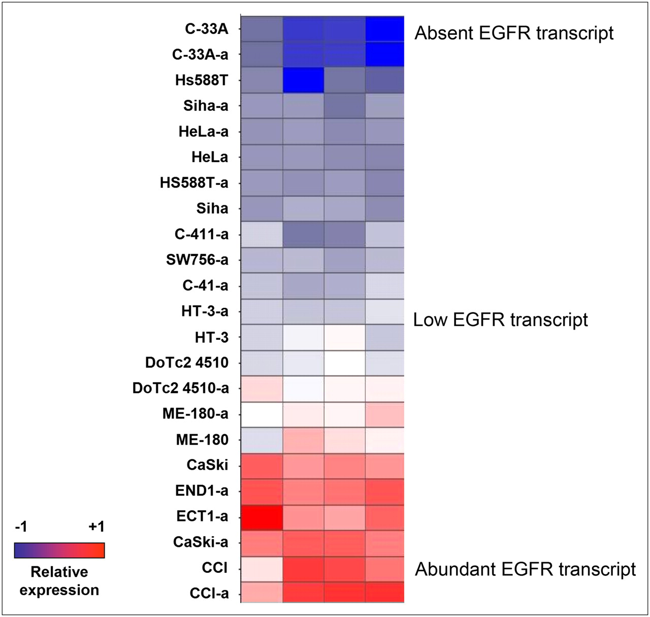
Twenty-three human cervical cancer cell lines (including several identical cell lines grown in different laboratories) were analyzed by gene expression microarray analysis. Among the approximately 54,000 probe sets represented on the array, 4 probe sets representing the EGFR transcript were analyzed to characterize EGFR transcript levels in each line (Fig. 1). Cell lines were classified according to their level of EGFR expression from the averaged probe set signals. In addition, cDNA templates from each of the cell lines were used to perform double-stranded resequencing of the entire EGFR coding region. None of the cell lines demonstrated previously characterized sequence alterations either within or external to the EGFR coding domain (data not shown).

Transcriptional profiling of cervical cancer cell lines with U133Plus2 GeneChip microarrays (Affymetrix). EGFR expression is based on 4 different oligonucleotide probe sets (each column shows response to 1 probe set). Cell lines are ordered based on their relative levels of EGFR probe signals. Cell lines with suffix designation “-a” were grown and harvested independently.
Five cervical cancer cell lines, covering the entire range of EGFR mRNA expression, were selected for further study: CaSki (high EGFR expression), ME-180 and DoTc2 4510 (both midrange EGFR expression), HeLa (low EGFR expression), and C-33A (negative for EGFR expression).
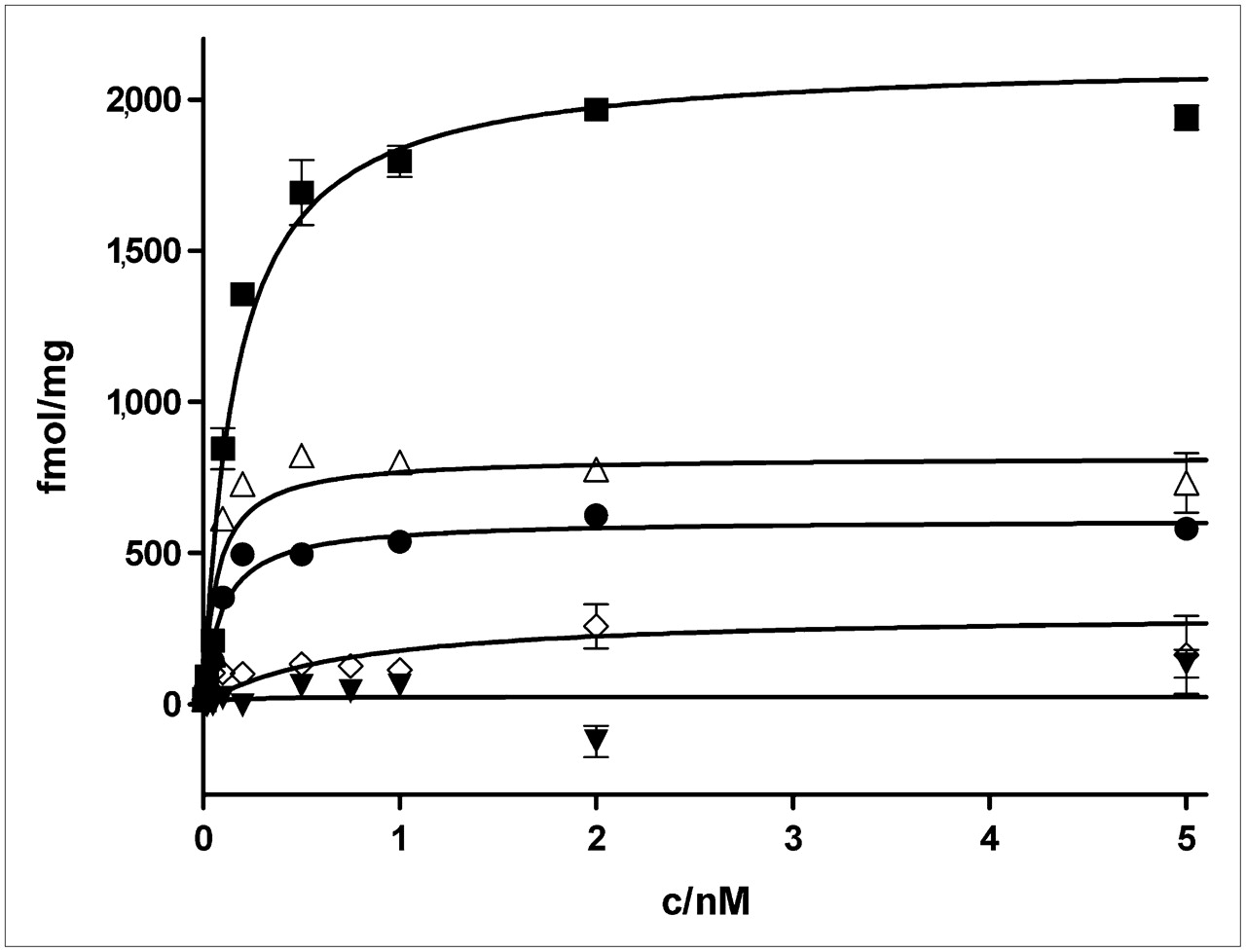
To determine the relationship between EGFR gene expression levels and cell-surface expression of the EGFR, saturation receptor binding of 64Cu-DOTA-cetuximab to the EGFR was performed. Binding studies were performed in live cells at 4°C to inhibit internalization and trafficking of cetuximab. The results are shown in Figure 2.

Saturation receptor binding of 64Cu-DOTA-cetuximab to 5 cervical cancer cell lines (n = 3). EGFR densities (Bmax in fmol/mg) on cell surface were, from high to low, CaSki (▪, 2,130 ± 60) > ME-180 (Δ, 820 ± 40) > DoTc2 4510 (•, 610 ± 20) > HeLa (⋄, 310 ± 110) > C-33A (▾, not applicable).
In Figure 2, 4 of the 5 cell lines exhibited saturable 64Cu-DOTA-cetuximab binding. The fifth line, C-33A, showed no specific 64Cu-DOTA-cetuximab binding. The affinity of 64Cu-DOTA-cetuximab binding for the EGFR was similar in the 4 EGFR-positive lines, varying from 0.1 to 0.7 nM. These values are consistent with those reported for the binding of unmodified cetuximab to the EGFR (dissociation constant, 0.1–0.4 nM) (18). Therefore, conjugation to DOTA and complexation with 64Cu did not affect the affinity of cetuximab toward its antigen. The Bmax for 64Cu-DOTA-cetuximab binding to these cell lines paralleled the levels of EGFR expression determined by microarray analysis (Fig. 2; Table 1).
Quantification of EGFR mRNA and Protein Levels in 5 Cervical Cancer Cell Lines
Internalization assays were next performed to determine whether EGFR number correlated with increased uptake of 64Cu-DOTA-cetuximab by receptor-mediated endocytosis. For these experiments, cells were incubated with 64Cu-DOTA-cetuximab at 37°C, and internalization was measured by determining the amount of cell-associated 64Cu-DOTA-cetuximab that was stable to acid wash (PBS) supplemented with 20 mM sodium acetate, pH 3.0. The data in Figure 3A show that all EGFR-positive cell lines internalized 64Cu-DOTA-cetuximab. Internalization was linear for the first 15–30 min but began to plateau after approximately 1 h. No uptake was observed in the EGFR-negative C-33A cells. The maximum level of internalization observed after 4 h paralleled receptor expression levels as determined by 64Cu-DOTA-cetuximab saturation binding (Table 1).
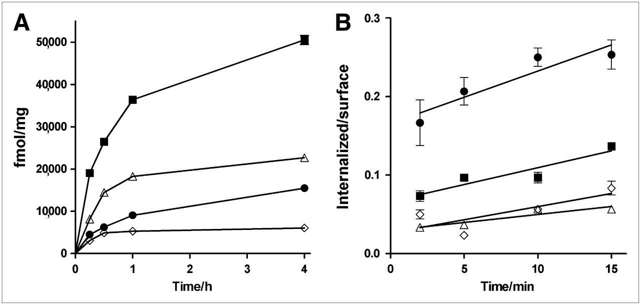
Internalization of 64Cu-DOTA-cetuximab (n = 3). (A) Internalization standardized to protein mass over 4 h displayed same relative order as seen by expression profiling and receptor binding: CaSki (▪) > ME-180 (Δ) > DoTc2 4510 (•) > HeLa (⋄). (B) Rate of internalization in first 15 min of exposure to 64Cu-DOTA-cetuximab, displayed as internalized to surface-bound activity.
To determine whether the difference in uptake was because of an increase in the rate of internalization, 64Cu-DOTA-cetuximab uptake was followed over a 15-min time course, and the data were plotted as the ratio of internalized 64Cu-DOTA-cetuximab to cell-surface 64Cu-DOTA-cetuximab versus time. This normalizes uptake to receptor number and allows assessment of the rate of internalization on a per-receptor basis. Figure 3B shows that the slopes of the InSur plots were similar for all 4 EGFR-positive cell lines, indicating that the rate of uptake was similar in all lines. Thus, the difference in the absolute amount of 64Cu-DOTA-cetuximab internalized can be attributed to differences in the number of cell-surface EGFRs.
In CaSki cells, the line that expressed the highest level of EGFRs, approximately 50,000 fmol of 64Cu-DOTA-cetuximab per milligram were internalized within 4 h. This corresponds to an internalized 64Cu-DOTA-cetuximab–to–receptor ratio of 24 (Table 1). This correspondence suggests that the EGFRs are continuously recycled to the cell surface rather than being targeted into the degradation pathway. The ratio of internalized 64Cu-DOTA-cetuximab to EGFR number was similar for the ME-180 (28), DoTc2 4510 (25), and HeLa (20) cells, indicating that all lines exhibited the same phenomenon of receptor recycling followed by cetuximab-induced receptor internalization.
To assess the interaction of 64Cu-DOTA-cetuximab with cervical cancer cells in vivo, 5 × 10⁶ CaSki cells were injected into female SCID mice. Three weeks later, palpable tumors were observed. 64Cu-DOTA-cetuximab was then injected into these tumor-bearing athymic nude mice (n = 5), and its biodistribution was assessed 24 h after injection (Fig. 4). Uptake into the cervical cancer cell tumor was high (13.2 ± 1.2 %ID/g). By contrast, uptake was low in muscle (3.4 ± 0.2 %ID/g) and bone (2.6 ± 0.2 %ID/g). However, uptake in other nontarget organs remained high at 24 h, especially in the blood (23.8 ± 0.9 %ID/g) and the spleen (25.6 ± 1.7 %ID/g). This biodistribution at 24 h indicates that optimal contrast is likely observed at time points greater than 24 h after injection, which is consistent with data from Cai et al. (14) and data from our group (15).

Biodistribution of 64Cu-DOTA-cetuximab in CaSki tumors implanted into SCID mice at 24 h after injection.
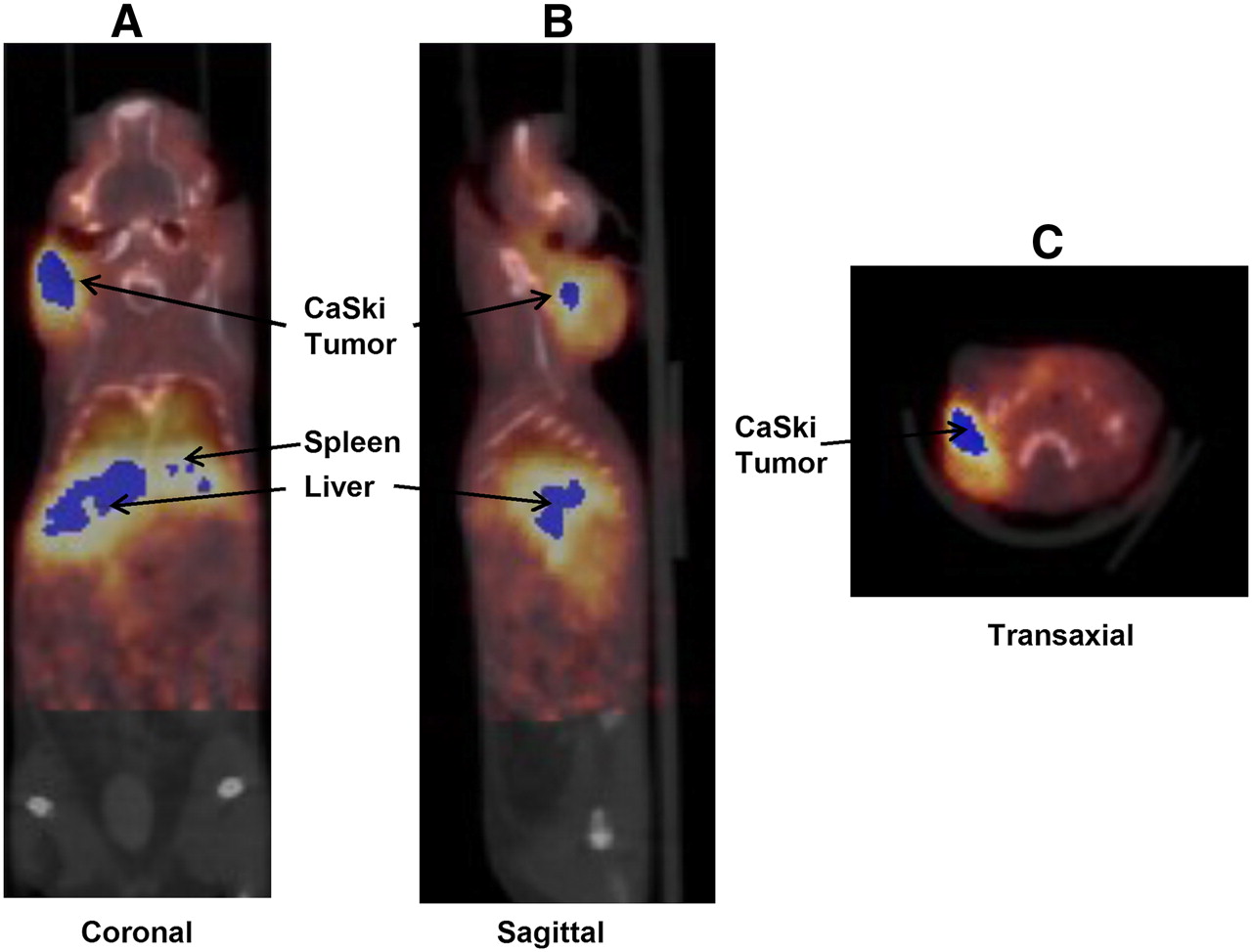
A preliminary small-animal PETCT experiment was performed in a CaSki tumor-bearing mouse that showed high 64Cu-DOTA-cetuximab uptake in the tumor mass but relatively low nontarget uptake 24 h after injection (Fig. 5). The specific uptake value for the tumor was 1.39, whereas the values for the liver and heart were 1.72 and 1.63, respectively. These data are consistent with the biodistribution data, indicating relatively high liver and blood accumulation, although the tumor is visible at 24 h.

Small-animal PET imaging of 64Cu-DOTA-cetuximab in CaSki tumor–bearing SCID mice at 24 h after injection: coronal (A), sagittal (B), and transaxial (C) views.
DISCUSSION
Despite widespread screening for cervical cancer, this disease continues to claim lives, particularly among medically underserved populations of women. Gardasil (Merck & Co., Inc.), a recombinant vaccine against human papilloma virus types 6, 11, 16, and 18, was approved by the Food and Drug Administration in 2006 and by the European Union in 2007. Although this is an important step in the battle against cervical cancer, the viral types for which this vaccine provides protection are responsible for only 70% of the cases of cervical cancer. Also, the vaccine will provide no protection for women who have already been exposed to the target strains (19). Therefore, the need for advances in the diagnosis and treatment of cervical cancer will persist.
Cetuximab has been approved in Europe and the United States for the clinical treatment of advanced metastatic colorectal cancer and advanced head and neck cancer. Like cervical cancer, these tumors are known to express elevated levels of EGFRs (20,21). The studies presented here were aimed at investigating the use of cetuximab in the diagnosis and subtyping of cervical cancers.
Microarray analysis of 23 human cervical cancer cell lines demonstrated significant differences among lines in the level of expression of EGFR mRNA. Thus, the sensitivity of these tumors to cetuximab-induced cell killing is likely to be highly variable. In particular, tumors that do not express EGFRs are not likely to be effectively targeted by cetuximab.
To determine the potential use of cetuximab in identifying EGFR-positive cell lines, we conjugated cetuximab with DOTA and radiolabeled it with 64Cu. This radionuclide (half life, 12.7 h; β+, 17.4%; β−, 41%) emits both β+ and β− radiation, allowing for the use of 64Cu radiopharmaceuticals with high tumor-to-background ratios for both PET imaging and targeted radiotherapy of cancer.
We used 64Cu-DOTA-cetuximab to quantify EGFR levels and found that the expression of EGFR mRNA correlated with the number of cell-surface EGFR in 5 human cervical cancer cell lines. In addition, the degree of 64Cu-DOTA-cetuximab internalization correlated with both EGFR mRNA and protein levels. CaSki cells, which showed the highest EGFR mRNA levels by microarray analysis, were also found to have the highest maximum density of EGFR per milligram of protein. In addition, they internalized more 64Cu-DOTA-cetuximab than did the other cell lines. At the other end of the spectrum, C-33A cells responded negatively to all oligonucleotide probe sets, and no EGFR could be detected by 64Cu-DOTA-cetuximab–binding assays. Thus, 64Cu-DOTA-cetuximab binding appears to be a useful probe for quantifying EGFR number and function.
Several recent reports have investigated whether the response of colorectal tumors to cetuximab therapy is correlated with EGFR mRNA copy number or EGFR protein levels. Moroni et al. (21) demonstrated a correlation between response to cetuximab and increased EGFR gene copy number in patients with metastatic colorectal cancer. By contrast, Vallböhmer et al. (22) failed to find a significant correlation between colorectal tumor EGFR mRNA levels and patient response to cetuximab, but this study was hampered by a small sample size. Nonetheless, it was observed that patients with higher levels of gene expression of the EGFR had a shorter overall survival. Despite these correlations, 1 study of patients with colorectal cancer with no detectable tumor levels of EGFR as determined by immunohistochemistry showed that approximately 25% of these patients respond to cetuximab (23). The data presented in these studies suggest that the quantification of EGFR by immunohistochemistry is unreliable and demonstrate the need for a more sensitive method for measuring EGFR for use in stratifying patients for potential response to cetuximab. Molecular imaging of EGFR with PET/CT and cetuximab labeled with a positron emitter, compared with traditional methods, may be a sensitive alternative measure of EGFR.
To assess the potential for 64Cu-DOTA-cetuximab as a PET/CT agent, a preliminary study was performed in which 64Cu-DOTA-cetuximab was injected into nude mice bearing tumors derived from CaSki cervical cancer cells. Biodistribution analysis demonstrated high uptake of 64Cu-DOTA-cetuximab in implanted CaSki tumors at 24 h after injection (13.2 ± 1.2 %ID/g). This result is comparable to the highest level of uptake of 64Cu-DOTA-cetuximab in a recent study of 7 tumor cell lines implanted in athymic nude mice (14). The uptake in the liver was comparable in both studies (14.0 ± 1.4 %ID/g at 16 h after injection vs. 15.5 ± 0.5 %ID/g at 24 h after injection in our study), but retention of radiopharmaceutical in the blood was significantly higher in our system (14). Copper can dissociate from the chelator DOTA, which may in part account for the observed uptake in nontarget organs. The use of bifunctional chelators that form more stable copper complexes, such as 4,11-bis(carboxymethyl)-1,4,8,11-tetraazabicyclo[6.6.2]hexadecane (CB-TE2A), can alleviate copper dissociation and reduce nonspecific copper uptake (24). However, CB-TE2A is labeled with 64Cu at 95°C and a pH greater than 8 (25). Antibodies are denatured under these conditions, and until suitable prelabeling procedures for 64Cu-CB-TE2A with subsequent conjugation to antibodies are available, it will be necessary to use chelators that can be radiolabeled under milder conditions. At any rate, that uptake of 64Cu-DOTA-cetuximab into CaSki tumors was readily detected in our studies and that its extent of uptake correlates with EGFR number suggests that further examination of this radiopharmaceutical with additional time points with the CaSki model, as well as imaging of EGFRs in other cervical cancer mouse models, is warranted.
CONCLUSION
We demonstrated that the expression of EGFR mRNA correlated well with protein levels of EGFR as determined by 64Cu-DOTA-cetuximab. In addition, the levels of internalization of 64Cu-DOTA-cetuximab in the 5 cervical cancer cell lines corresponded to the EGFR density, and the rates of internalization were similar. As several studies have demonstrated that EGFR densities in tumors do not typically correlate with response to cetuximab therapy, questions arise as to whether determination of EGFR concentration in tumors with 64Cu-DOTA-cetuximab and PET would be a more valuable tool than immunohistochemistry and less invasive than tumor biopsy followed by mRNA analysis. Data presented here strongly suggest that 64Cu-DOTA-cetuximab is an accurate biomarker of EGFR expression levels, and this agent with PET might be useful for patient selection and therapeutic monitoring with all types of EGFR inhibitors, including small-molecule and antibody-based, although further imaging studies are warranted.
Acknowledgments
We thank Susan Adams and Christopher Sherman for technical assistance, Jerrel Rutlin for small-animal PET/CT image analysis, and Wenping Li for helpful discussions. Cetuximab was provided by ImClone Systems. This research was supported by National Cancer Institute (NCI) grant P50CA9405604. The production of 64Cu at Washington University School of Medicine was supported by NCI grant R24 CA86307. Small-animal imaging at Washington University School of Medicine was supported by NIH grant 5 R24 CA83060.
Footnotes
-
COPYRIGHT © 2008 by the Society of Nuclear Medicine, Inc.
References
- Received for publication February 29, 2008.
- Accepted for publication May 12, 2008.


Jump to section
Related Articles
Cited By...
- 64Cu-Labeled Repebody Molecules for Imaging of Epidermal Growth Factor Receptor-Expressing Tumors
- Molecular Fluorescence Endoscopy Targeting Vascular Endothelial Growth Factor A for Improved Colorectal Polyp Detection
- Interrogating Tumor Metabolism and Tumor Microenvironments Using Molecular Positron Emission Tomography Imaging. Theranostic Approaches to Improve Therapeutics
- The Role of p53 in Combination Radioimmunotherapy with 64Cu-DOTA-Cetuximab and Cisplatin in a Mouse Model of Colorectal Cancer
- Rift Valley Fever Virus Strain MP-12 Enters Mammalian Host Cells via Caveola-Mediated Endocytosis
- Preparation, Biological Evaluation, and Pharmacokinetics of the Human Anti-HER1 Monoclonal Antibody Panitumumab Labeled with 86Y for Quantitative PET of Carcinoma
- Molecular Imaging: 18F-FDG PET and a Whole Lot More
- Small-Animal PET of Tumors with 64Cu-Labeled RGD-Bombesin Heterodimer
- PET of EGFR Antibody Distribution in Head and Neck Squamous Cell Carcinoma Models