


Abstract
To evaluate the possibility of radionuclide gene therapy and imaging in hepatocellular carcinoma cancer, we investigated the iodine accumulation of a human hepatocellular carcinoma cell line, SK-Hep1, by transfer of human sodium iodide symporter (hNIS) gene. By targeting NIS expression in SK-Hep1, we could also investigate whether these cells concentrate 99mTc-pertechnetate and 188Re-perrhenate as well as 125I in vitro and in vivo. Methods: The hNIS gene was transfected to human hepatocellular carcinoma SK-Hep1 cell lines using lipofectamine plus reagent. The uptake and efflux of 125I, 99mTc-pertechnetate, and 188Re-perrhenate were measured in the transfected and parental cells. Biodistribution was studied in nude mice bearing SK-Hep1 and SK-Hep1-NIS at 10 and 30 min and at 1, 2, 6, 16, and 23 h after injection of 125I, 99mTc- pertechnetate, or 188Re-perrhenate. In tumor imaging studies, the nude mice were intravenously injected with 188Re-perrhenate and imaged with a γ-camera equipped with a pinhole collimator at 30 and 60 min after injection. The survival rate (%) was determined by the clonogenic assay after 37 MBq/10 mL (1 mCi/10 mL) 131I and 188Re-perrhenate treatment. Results: SK-Hep1-NIS, stably expressing the NIS gene, accumulated 125I up 150 times higher than that of SK-Hep1. Iodine uptake of SK-Hep1-NIS is completely blocked by perchlorate. NIS gene transfection into SK-Hep1 also resulted in 112- and 87-fold increases of 99mTc-pertechnetate and 188Re-perrhenate uptake, respectively. Iodide efflux from SK-Hep1-NIS was relatively slow, with only 10% released during the initial 5 min, and 60% remained at 25 min. In the biodistribution study using SK-Hep1-NIS–xenographed mice, the tumor uptake of 125I, 188Re-perrhenate, and 99mTc-pertechnetate was 68.0 ± 15.0, 46.2 ± 9.1, and 59.6 ± 16.2 %ID/g (percentage injected dose per gram) at 2 h after injection, respectively. After 188Re-perrhenate injection in SK-Hep1 and SK-Hep1-NIS–xenographed nude mice, whole-body images clearly visualized the SK-Hep1-NIS tumor, whereas the control tumor was not visualized. The survival rate (%) of SK-Hep1-NIS was markedly reduced to 46.3% ± 10.1% and 28.9% ± 5.2% after 37 MBq/mL (1 mCi/10 mL) 131I and 188Re-perrhenate treatment compared with the survival rates of the parental cells. These results demonstrated that SK-Hep1-NIS could be selectively killed by the induced 131I and 188Re-perrhenate accumulation through NIS gene expression. Conclusion: NIS-based gene therapy using β-emitting radionuclides has the potential to be used in hepatocellular carcinoma management.
Sodium iodide symporter (NIS) is a specialized active iodide transporter that cotransports a sodium and an iodide ion. NIS is an essential glycoprotein located in the basolateral plasma membrane of thyroid follicular cells (1,2). The sodium gradient, generated by sodium-potassium adenosine triphosphatase (Na+, K+-ATPase), serves as the driving force for iodide uptake. Moreover, NIS-mediated iodide transport is inhibited by the Na+, K+-ATPase inhibitor ouabain and by the competitive inhibitors thiocyanate and perchlorate.
Iodine uptake by NIS is impaired in some cases of thyroid carcinoma (3). Approximately one third of differentiated thyroid cancers and all anaplastic thyroid cancers fail to concentrate radioiodine due to reduced NIS expression, which prevents the use of radioiodine for metastatic thyroid cancer therapy. The recent cloning and characterization of the NIS gene could lead to a novel gene strategy for radioiodine therapy in thyroid cancer (2,4). The transfer of NIS gene and the functional expression of NIS protein in cancer cells would enable these cells to concentrate iodide from plasma and enable radioiodine therapy (5).
By combining the targeting and the expression of the NIS gene, radioiodine treatment could also be used to treat nonthyroid malignant disease. Several investigators have shown that the gene transfer of NIS into a variety of cancer cells confers increased radioiodine uptake in nonthyroid cancers and in thyroid cancer (6–10). Radioiodine therapy with NIS gene transfer has been demonstrated by several researchers (7,11–14). To achieve therapeutic effectiveness, several problems remain to be solved. Of these, rapid washout after sufficient iodide uptake is probably the main problem, which is due to the defective organification of iodide. Cotransfection of the thyroid peroxidase gene has been examined in this context (15). Another approach involved the use of more powerful and suitable radionuclides transported by NIS, such as 188Re-perrhenate or 211At rather than 131I. 188Re-Perrhenate is a β-emitter and has a shorter half-life (t1/2) and is effective over a wider range than 131I (16,17). 211At is a high-energy α-emitter with a short physical t1/2 (18).
In this study, we transferred the human NIS (hNIS) gene into a human hepatocellular carcinoma cell, SK-Hep1. The established cell line, SK-Hep1-NIS, showed high uptake of 99mTc-pertechnetate and of 188Re-perrhenate in addition to 125I, which may make it suitable for radionuclide gene therapy.
MATERIALS AND METHODS
Cells, Vectors, and Chemicals
Human hepatocellular carcinoma cell line SK-Hep1 was obtained from the Korea Cell Bank and maintained as recommended. The human NIS-expressing vector, FL*-hNIS/pcDNA3, was kindly provided by Dr. Sissy Jhiang (Ohio State University, Columbus, OH) and controlled using a cytomegalovirus promoter. 125I in the form of sodium iodide was purchased from New England Nuclear, 188Re-perrhenate in the form of sodium perrhenate was eluted from a 188W/188Re generator (Oak Ridge National Laboratory), and 99mTc-pertechnetate was eluted from a 99Mo/99mTc generator (Amersham Pharmacia Biotech).
Establishment of Human Hepatocellular Carcinoma Cell Line Expressing hNIS
SK-Hep1 cells were grown as a monolayer in RPMI 1640 medium (Invitrogen) supplemented with 292 mg/mL glutamine, 100,000 IU/L penicillin, 100 mg/L streptomycin, and 10% fetal bovine serum (FBS). Purified expression vector was transfected into SK-Hep1 cells using lipofectamine plus reagent (Invitrogen) according to the manufacturer’s instructions. Stable transfectants were selected with 600 μg/mL geneticin in RPMI 1640 medium containing 10% FBS for 3 wk.
Radionuclide Uptake and Efflux Assay
Iodine uptake by cells in vitro was measured as described by Nakamoto et al. (10). Briefly, the cells were plated in 24-well plates and cultured with RPMI 1640 medium containing 10% FBS. When the cells reached confluence (∼1 × 10⁶ cells), they were incubated at 37°C for 5–120 min in 500 μL of Hanks’ balanced salt solution (HBSS) containing 0.5% bovine serum albumin and 10 mmol/L of the sodium salt of 2-(4-[2-hydroxyethyl]-1-piperazinyl)ethanesulfonic acid–NaOH, pH 7.4, with 18.5-kBq (0.5 μCi) carrier-free Na125I and 10 μmol/L NaI, to yield a specific activity of 740 MBq/mmol (20 mCi/mmol). The cells were then quickly washed with 2 mL of ice-cold HBSS, and the radioactivity of the detached cells was counted with a γ-counter (Canberra Industries). Unless otherwise stated, the iodide uptake was expressed as picomoles per 106 cells. To modulate the iodide uptake, the cells were incubated for 1 h in either Na125I medium or Na125I medium supplemented with 50 μmol/L sodium perchorate (Sigma) or with 100 μmol/L lithium chloride to block iodide uptake (19). For iodine efflux studies, cells were incubated with 10 μmol/L NaI and 3.7 kBq (0.1 μCi) Na125I in 500 μL of HBSS incubation buffer at 37°C for 1 h. The cells were washed 2 times with HBSS and added to HBSS at 37°C and further incubated. At the specified time points (0, 3, 6, 9, 12, 15, 21, and 27 min), the buffer was removed and its radioactivity was measured. After the last time point, the cells were collected to determine the residual radioactivity. All data points were measured in triplicate and are displayed as mean ± SEM.
Animal Model
To investigate the uptake of 125I, 188Re-perrhenate, or 99mTc-pertechnetate by NIS-expressing hepatocellular carcinoma tumors, a xenografted tumor model was used in nude mice. Five million tumor cells were transplanted subcutaneously into either the right (SK-Hep1-NIS) or the left (SK-Hep1) thigh of 6-wk-old BALB/c nu/nu mice. Experiments involving animals were performed with the approval of the Seoul National University Animal Research Committee.
Biodistribution
Biodistribution studies (n = 60) were performed when the tumor weighed ∼300 mg. At 10 and 30 min and at 1, 2, 6, 16, and 23 h after administering radionuclides, the animals were killed, and their organs and tumors were removed and weighed for radioactivity measurement. Results are expressed as a percentage of the injected dose per gram (%ID/g) of tissue.
Tumor Imaging
For imaging studies, which were performed under general anesthesia achieved by an intraperitoneal injection of 40 μL of ketamine and xylazine (4:1) solution before tracer injection, only animals bearing tumors with a size > 15 mm in diameter were accepted. Immediately after injecting 7.4 MBq (200 μCi) 188Re-perrhenate into the lateral tail vein, mice bearing tumors were placed in a spread prone position and scanned with a γ-ray camera (ON 410; Ohio Nuclear) equipped with a 4-mm pinhole collimator. The images were acquired for 5 min. The relative accumulations of radioactivity in the tumors of the right (SK-Hep1-NIS) and the left (SK-Hep1) thighs on the 30-min and 1-h images were then examined.
In Vitro Clonogenic Assay
The procedure was performed as described previously (7) with minor modifications. Briefly, cells were grown in a 75-cm2 flask and incubated for 7 h at 37°C with 5 mL HBSS containing 37 MBq/10 mL (1 mCi/10 mL) Na131I or 37 MBq/10 mL (1 mCi/10 mL) 188Re-perrhenate. The reaction was terminated by removing the radioisotope-containing medium and washing the cells twice with HBSS. The cells were then trypsinized, counted, and plated at densities of 250 or 1,000 cells per well with RPMI 1640 medium in 6-well plates. The cells were grown for 10 d, fixed with 3:1 methanol/acetic acid, and stained with crystal violet, and the number of macroscopic colonies was counted. Survival rate was calculated as the percentage of colony numbers in a plate treated with radionuclides compared with the number in a plate treated with HBSS only.
Statistical Analysis
All numeric data are expressed as the mean ± SD. Statistical significance of differences was determined by conducting a paired Student t test. P values < 0.05 were considered statistically significant.
RESULTS
Radioisotope Uptake
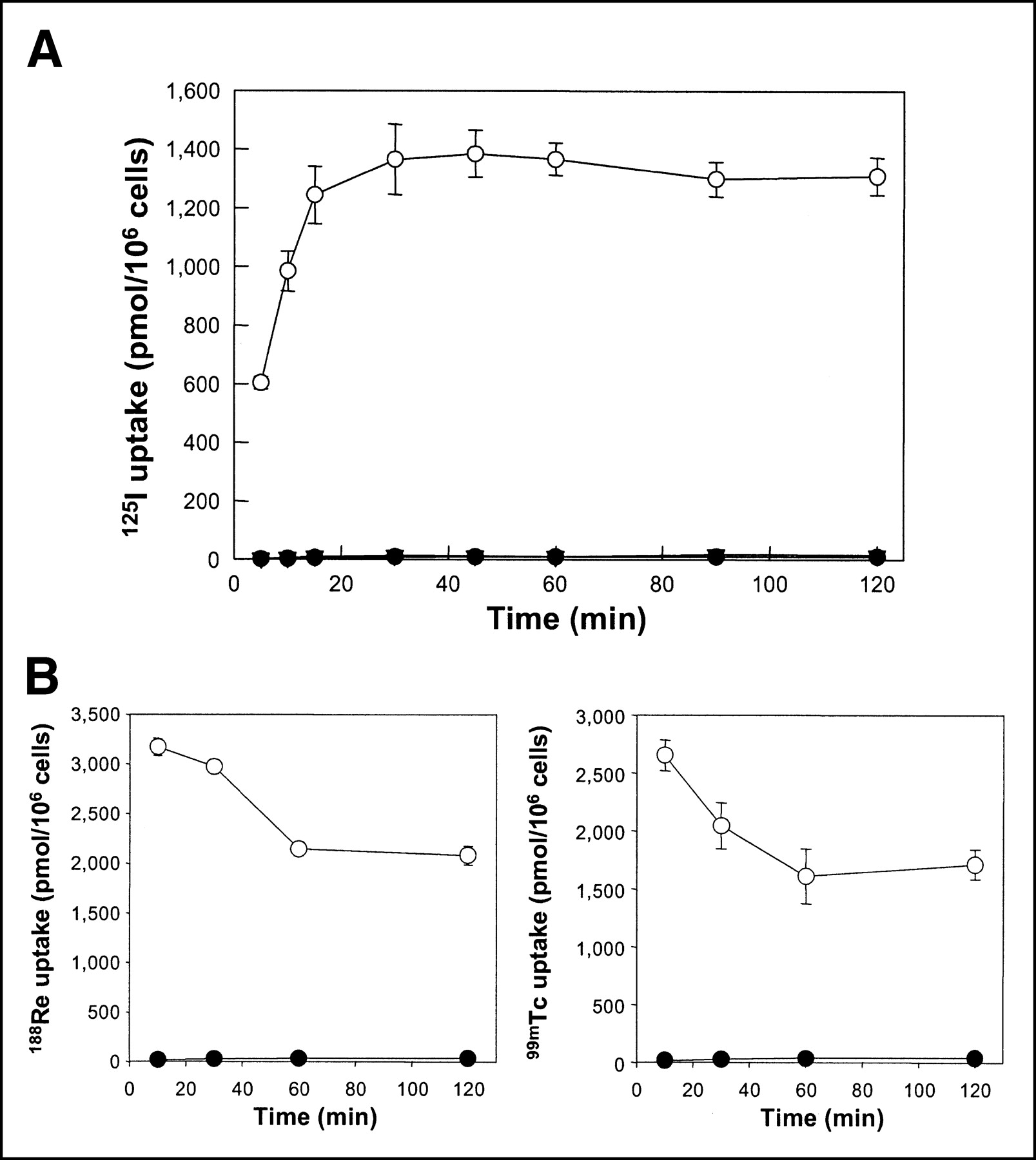
The iodide uptake by SK-Hep1-NIS cells reached a half-maximal level within 10 min and a plateau at 30 min, as shown in Figure 1A. Compared with the iodide uptake of the parental cell line at baseline, NIS gene transfection resulted in an ∼150-fold increase in iodide uptake. The perchlorate, specific NIS inhibitor, completely blocked the iodide uptake of SK-Hep1-NIS cells. SK-Hep1-NIS cells efficiently accumulated 188Re-perrhenate and 99mTc-pertechnetate as well as 125I. In the case of 99mTc-pertechnetate and 188Re-perrhenate, SK-Hep1-NIS cells took up 112 and 87 times more than that of the nontransfected cells, respectively (Fig. 1B).

(A) Time course of radioiodide uptake by SK-Hep1 cells (•), SK-Hep1-NIS cells without perchlorate (○), and SK-Hep1-NIS cells with 1 mmol/L perchlorate (▾). Cells in 24-well plates were incubated with 3.7 kBq (0.1 μCi) carrier-free Na125I and 10 μmol/L NaI at 37°C for 5–120 min. At the various time points shown, cells were quickly washed with buffer and their radioactivity was counted with a γ-counter. All data are expressed as means (pmol/106 cells) of triplicate wells. (B) Time course of 188Re-perrhenate (left) and 99mTc-pertechnetate (right) uptake by SK-Hep1 cells (•) and SK-Hep1-NIS cells (○). 188Re in the form of perrhenate and 99mTc-pertechnetate as pertechnetate were eluted from a 188W/188Re generator and a 99Mo/99mTc generator, respectively.
Iodide Efflux
SK-Hep1-NIS cells exhibited a relatively slow release; only 10% was released during the initial 5 min and 60% remained at 25 min. The t1/2 of the iodide efflux was about 35 min. However, in the presence of Li+ ion, iodide efflux was complete at 6 min, with a t1/2 of 2 min (Fig. 2).
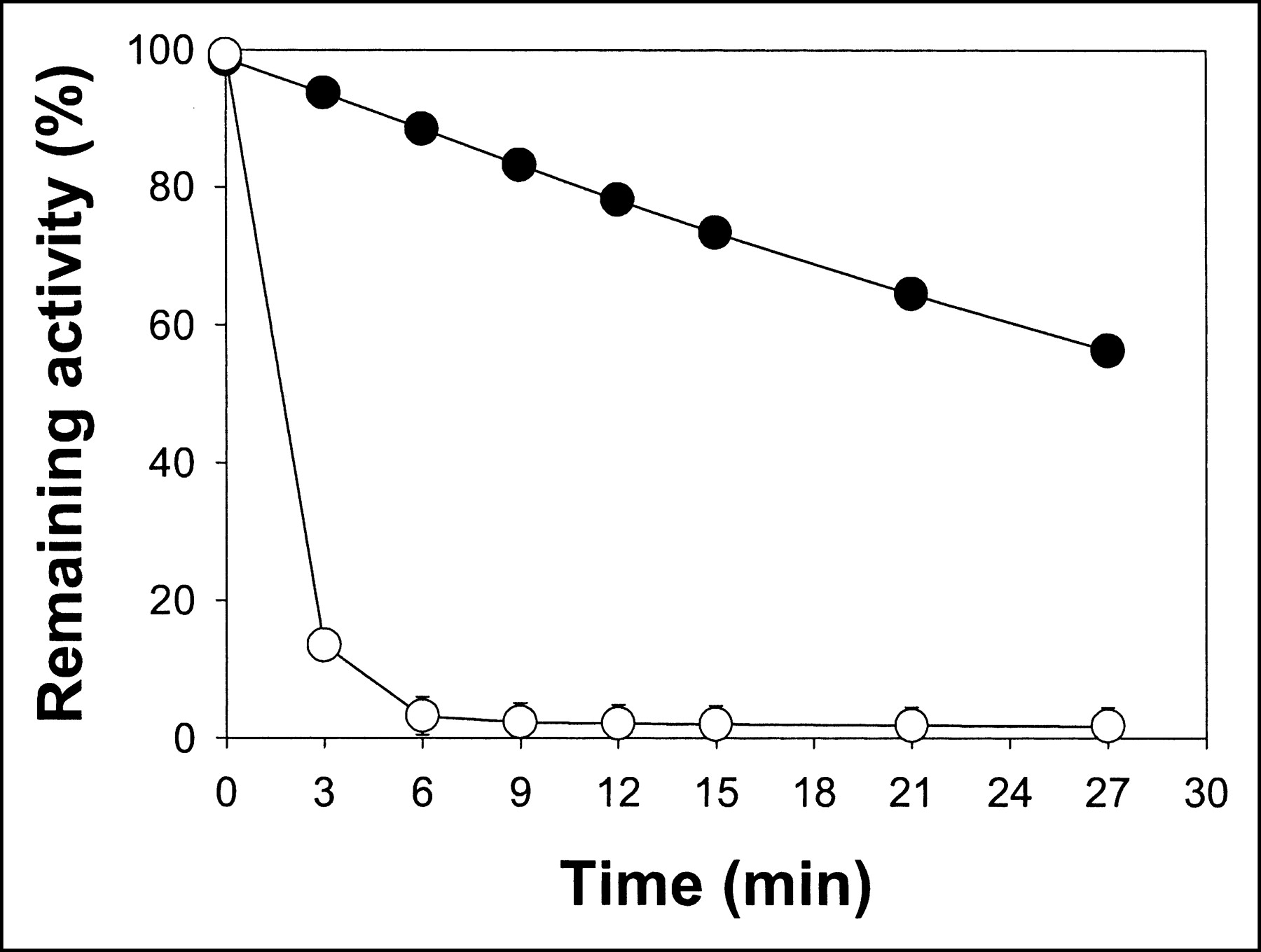
Iodide efflux from SK-Hep1-NIS cells after 1-h incubation with Na125I without (•) or with (○) medium containing 100 μmol/L Li+ ion in place of Na+ ion. After cells had been incubated with Na125I at 37°C for 1 h, the buffer, containing radioiodide, was removed at various time points and its radioactivity was determined. Data are expressed as means (remaining % of 125I) of triplicate wells.
Biodistribution Study
The biodistribution data for 125I, 188Re-perrhenate, and 99mTc-pertechnetate in SK-Hep1-NIS–and SK-Hep1–bearing nude mice are summarized in Figure 3. The NIS-expressing tumors exhibited increased uptake of 188Re-perrhenate, 99mTc-pertechnetate, and 125I versus the original SK-Hep1 tumors. SK-Hep1-NIS tumors accumulated 68.0 ± 15.0, 46.2 ± 9.1, and 59.6 ± 16.2 %ID/g of 125I, 188Re-perrhenate, and 99mTc-pertechnetate, respectively, 2 h after radionuclide injection. The ratios of SK-Hep1-NIS to blood of 125I, 188Re-perrhenate, and 99mTc-pertechnetate were 17.6 ± 2.9, 12.6 ± 3.6, and 7.2 ± 1.6, respectively, and the ratios of SK-Hep1-NIS to muscle were 21.8 ± 7.1, 59.1 ± 23.0, and 30.0 ± 6.7 at 2 h after injection, respectively (Table 1).

Biodistribution of 125I (A), 188Re-perrhenate (B), and of 99mTc-pertechnetate (C) in tumor-bearing BALB/c nude mice. Nude mice were injected subcutaneously with 5 × 10⁶ SK-Hep1 or SK-Hep1-NIS cells. Two weeks the injection, animals were injected with Na125I, 188Re-perrhenate, or 99mTc-pertechnetate via tail vein. At specified time points, animals were killed, their organs and tumors were removed and weighed, and radioactivities were measured. Data are expressed as %ID/g of tissue. SK = SK-Hep1; SK_NIS = SK-Hep1-NIS.
Tumor (SK-Hep1-NIS)–to-Blood, -Muscle, and -Tumor (SK-Hep1) Ratios of 99mTc, 125I, and 188Re in Tumor-Xenografted BALB/c Nude Mice
Tumor Imaging
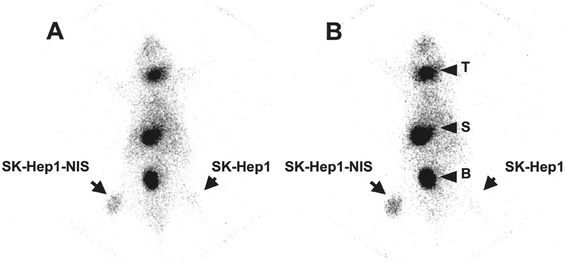
Thirty minutes and 1 h after the 188Re-perrhenate injection, whole-body planar scintigraphic images were obtained (Fig. 4). Normal NIS-expressing tissues, including those of the salivary gland, thyroid, and stomach, were visualized. NIS-transduced tumor sites were also clearly visible, whereas the control tumor was not.

Whole-body planar scintigraphic images of tumor-bearing nude mouse at 30 min (A) and at 1 h (B) after injection of 188Re-perrhenate. Mouse was subcutaneously transplanted with NIS-expressing cells (SK-Hep1-NIS) in right thigh and parental cells (SK-Hep1) in left thigh. T = thyroid; S = stomach; B = urinary bladder.
In Vitro Clonogenic Assay
Using an in vitro clonogenic assay (7), we investigated whether 131I and 188Re-perrhenate showed selective cytotoxic activity in NIS-transfected SK-Hep1 and SK-Hep1 cells. As shown in Figure 5, the survival rate of SK-Hep1-NIS cells, based on the clonogenic assay, was markedly reduced to 46.3% ± 10.1% and 28.9% ± 5.2% in response to 131I and 188Re-perrhenate compared with the survival rate of SK-Hep1 cells, respectively.
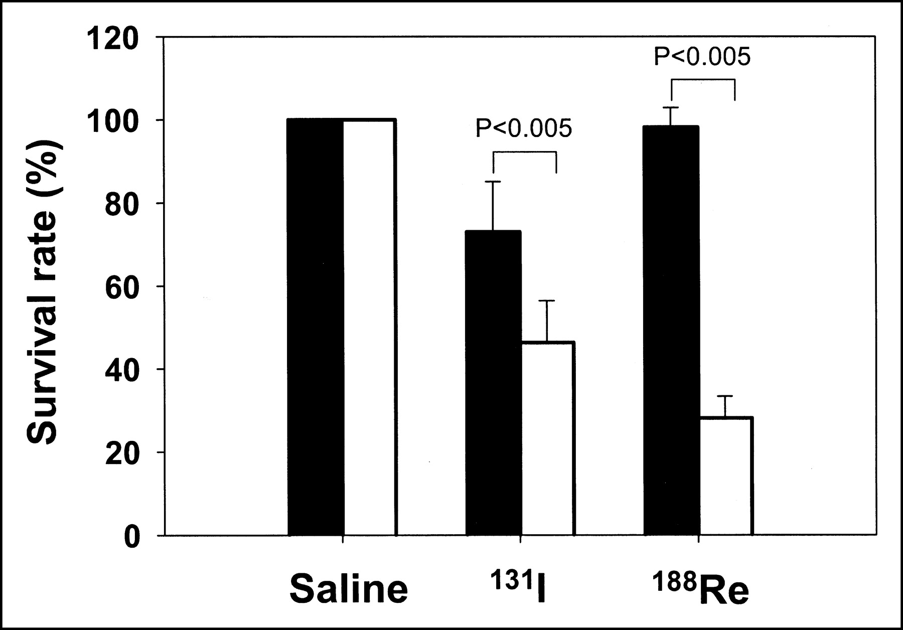
Survival rates (%) of SK-Hep1-NIS cells treated with 37 MBq/10 mL (1 mCi/10 mL) 131I or 188Re-perrhenate. SK-Hep1 and SK-Hep1-NIS cells were exposed to 37 MBq/10 mL (1 mCi/10 mL) 131I or 37 MBq/10 mL (1 mCi/10 mL) 188Re-perrhenate for 7 h at 37°C. Cells were then rinsed with buffer, counted, and plated at a density of 250 or 1,000 cells per well in 6-well plates. Cells were then grown for 10 d and stained with crystal violet, and numbers of colonies were counted. Survival rate was calculated by expressing number of colonies in plates treated with radionuclides as the percentage of number of colonies in plates containing buffer only. Black bars and white bars represent SK-Hep1 and SK-Hep1-NIS cells, respectively.
DISCUSSION
NIS is important for the biosynthesis of thyroid hormones as it mediates the accumulation of iodide into thyrocytes. NIS has a central role in the diagnosis and treatment of thyroid diseases and, above all, in the therapeutic management of thyroid carcinoma, comprising surgery and radioiodine therapy (20). By combining the transfer of the NIS gene with radioiodine therapy, it is possible to treat other cancers as well as differentiated thyroid carcinoma. To investigate the possibility of radiotherapy and of tumor-imaging of the NIS-transfected tumor cells, we established a hepatocellular carcinoma cell line stably expressing the NIS gene and evaluated its in vitro and in vivo characteristics.
The expression of NIS in an NIS-transfected human hepatocellular carcinoma cell line, SK-Hep1-NIS, resulted in higher uptakes of 99mTc-pertechnetate, 188Re-perrhenate, and 125I than that in the parental control cells. Radioiodine accumulation in SK-Hep1-NIS cells was blocked by perchlorate, which confirmed NIS-mediated iodide transport in these cells (2). Several researchers have reported that NIS gene transfer to various cancer cells shows increased radioiodine uptake up to 235-fold that of nontransfected control cells. For example, undifferentiated thyroid carcinoma cells transfected with NIS exhibited increased radioiodine uptake (6), and Haberkorn et al. (8) reported that the transfection of rat hepatocellular carcinoma with the hNIS gene using a retroviral vector was sufficient to induce iodide transport in hepatocellular carcinoma cells. The transfection of the NIS gene into nonthyroid cells, including breast cancer, prostate cancer, and glioma cells, was also found to increase iodine uptake (7,9,10,15).
In our biodistribution study, NIS-expressing tumors were found to efficiently accumulate 188Re-perrhenate, 99mTc- pertechnetate, and 125I (Fig. 3). In particular, the %ID/g of SK-Hep1-NIS–induced tumors was about 70% in terms of the 125I biodistribution, and this value was similar to that of the stomach, a physiologic NIS-expressing organ, in the same mice. SK-Hep1-NIS cells also accumulated 46.2 %ID/g of 188Re-perrhenate and 59.6 %ID/g of 99mTc-pertechnetate. To our knowledge, these are the highest values of radionuclide accumulation reported to date. Therefore, we believe that this system is suitable for the experimental study of NIS-based radionuclide gene therapy. Specific uptake was observed in the stomach and thyroid when endogenous NIS was expressed (21).
Radioiodide efflux from SK-Hep1-NIS cells seemed to be somewhat slower than that of other cells (6–10). However, when a large amount of Li+ ion was added to the media to inhibit NIS activity, the washout of iodide was rapid (t1/2 = 2 min), as occurs in other cells. Since the Li+ ion seems to block the reuptake of I− ion with the Na+ ion, this finding suggests that the reuptake of iodide in SK-Hep1-NIS cells was very active compared with that in SK-Hep1 cells and that this caused the slow washout. This finding also suggests that iodide is not organized in thyroid hormones, such as thyroglobulin. The short exposure of radioiodide from rapid efflux reveals a limitation of radio-concentrating gene therapy using the NIS gene. SK-Hep1-NIS cells offer some advantages in this context.
In this study, clonogenic survival of NIS-transfected hepatocellular carcinoma cells was reduced to 46% after 131I treatment versus that of the control cells. This finding is in accord with previous reports (6,7,9). However, Schipper et al. (14) reported a 99% reduction in the survival rate of NIS-transfected neuroendocrine tumor cells. This discrepancy may be due to the differences in experimental conditions or to the intrinsic radiosensitivity of the tumor cells used.
In addition to iodide, several anions, such as ClO4−, ReO4−, SCN−, ClO3−, and Br−, are transported by NIS (22). Monovalency and anion size are the determinators of NIS substrates. It is for these reasons that technetium pertechnetate and rhenium perrhenate are transported by NIS. It has been reported that NIS-expressing tissues or cells could concentrate pertechnetate (TcO4−), perrhenate (ReO4−), or astatide (At−). Moreover, some research groups have proposed that since 188Re-perrhenate is a powerful β-emitting radiometal (Eave = 0.764 MeV [Eave = average energy]) that it be used in the treatment of NIS-expressing tumors in place of 131I (16). 188Re-Perrhenate is conveniently obtained from a 188W/188Re generator (23) and has better emission characteristics and physical properties than 131I (16). In particular, it has a short t1/2, which makes it more suitable for the treatment of NIS-expressing tumors, because they show rapid radionuclide washout. Recently, Shen et al. reported that 188Re-perrhenate was more efficient than 131I to enhance the survival of rats bearing NIS-expressing glioma (17).
As shown in Figure 5, the cytotoxic effect of 188Re-perrhenate in SK-Hep1-NIS cells was much higher than that of 131I. In addition, its accumulation in NIS-transfected tumors can be evaluated by γ-scintigraphy (Fig. 4). These results suggest that radioisotope-concentrator tumor therapy based on 188Re-perrhenate is feasible for the control of NIS-expressing tumors—if these findings can be transferred to the in vivo situation.
CONCLUSION
We successfully applied radionuclide gene therapy to a human hepatocellular carcinoma model using NIS gene transfer. The NIS gene transfer in hepatocellular carcinoma cells is promising to tumor therapeutic strategies as a β-emitter and tumor imaging as 99mTc-pertechnetate.
Acknowledgments
This work was supported by a research grant from the Cancer Research Institute, Seoul National University, and by the BK21 Project for Medicine, Dentistry, and Pharmacy.
Footnotes
Received Nov. 20, 2003; revision accepted Mar. 16, 2004.
For correspondence contact: June-Key Chung, MD, PhD, Department of Nuclear Medicine, Seoul National University College of Medicine, 28 Yongon-dong, Chongno-gu, Seoul, 110-744, Korea.
E-mail address: jkchung{at}plaza.snu.ac.kr
REFERENCES
In this issue


Jump to section
Related Articles
Cited By...
- A Novel Orally Active Inverse Agonist of Estrogen-related Receptor Gamma (ERR{gamma}), DN200434, A Booster of NIS in Anaplastic Thyroid Cancer
- Detection of Increased 64Cu Uptake by Human Copper Transporter 1 Gene Overexpression Using PET with 64CuCl2 in Human Breast Cancer Xenograft Model
- Specific Activation of Sodium Iodide Symporter Gene in Hepatoma Using Alpha-fetoprotein Promoter Combined with Hepatitis B Virus Enhancer (EIIAPA)
- Combination Therapy and Noninvasive Imaging with a Dual Therapeutic Vector Expressing MDR1 Short Hairpin RNA and a Sodium Iodide Symporter
- Synergistic tumoricidal effect of combined hMUC1 vaccination and hNIS radioiodine gene therapy
- Development of a Dual Membrane Protein Reporter System Using Sodium Iodide Symporter and Mutant Dopamine D2 Receptor Transgenes
- A perspective view of sodium iodide symporter research and its clinical implications.
- Radioiodine Therapy of Hepatoma Using Targeted Transfer of the Human Sodium/Iodide Symporter Gene