


Abstract
Because of the development of gene knockout and transgenic technologies, small animals, such as mice and rats, have become the most widely used animals for cardiovascular imaging studies. Imaging can provide a method to serially evaluate the effect of a particular genetic mutation or pharmacologic therapy (1). In addition, imaging can be used as a noninvasive screening tool for particular cardiovascular phenotypes. Outcome measures of therapeutic efficacy, such as ejection fraction, left ventricular mass, and ventricular volume, can be determined noninvasively as well. Furthermore, small-animal imaging can be used to develop and test new molecular imaging probes (2,3). However, the small size of the heart and rapid heart rate of murine models create special challenges for cardiovascular imaging.
Although many challenges to small-animal cardiovascular imaging presently exist, the methods presented in this paper can be used as a noninvasive screening tool for particular cardiovascular phenotypes. In addition, small-animal imaging provides an economic platform for the rapid translation of new knowledge in cardiovascular medicine to the clinical arena.
CARDIAC MRI
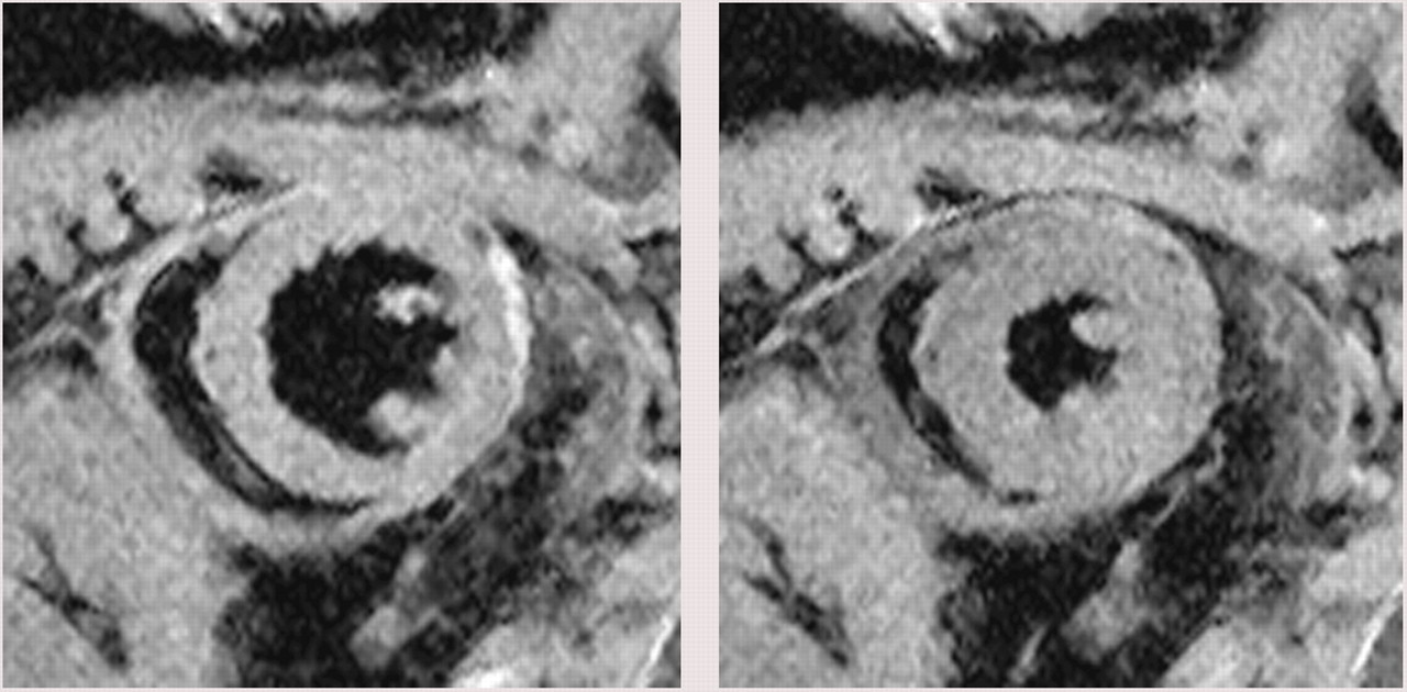
Because of the small heart size and rapid heart rates in small animals such as mice and rats, most small-animal cardiac MRI has been performed on scanners with field strengths greater than 4.7-T. To minimize the effects of anesthetics on cardiac contractility, inhalational anesthesia—in combination with methods to maintain the animal's core temperature at 38°C—is used for MRI and other small-animal imaging modalities. As in clinical scanners, blurring due to cardiac motion is avoided by gating image acquisition to the electrocardiogram (ECG) and deriving a single image from several heart cycles. However, image acquisition time for multiple short- and long-axis views can result in 1 to several hours per animal. Obviously, this image acquisition time is not compatible with high-throughput screening of genetically manipulated mouse models. Several groups are exploring methods to image multiple mice simultaneously (4–6). To image multiple animals simultaneously, receiver coil arrays can be positioned over each animal or individual coils for each animal can be designed (5,6). Although parallel imaging techniques can be used to take advantage of individual receiver coil sensitivities and increase the speed of imaging, these systems are not often available or the software is lacking to implement such routine techniques on commercially available high-field small-animal scanners. One approach to enable the rapid translation of high-field small-animal techniques to the clinical realm is the marriage of a high-field 7-T MRI scanner with the clinical user interface and a high number of receiver channels. Figure 1 shows an example of black-blood cardiac MR images (7) using such a system and demonstrates the exquisite soft-tissue detail that can be obtained in the mouse. Another alternative is to develop specialized gradient inserts and receiver coils for small animals on clinical 1.5- and 3-T imaging platforms (8). Another approach to shorten acquisition time in multiple-mouse imaging is to acquire images without cardiac gating and retrospectively reconstruct the images on the basis of a separately acquired navigator or the image data itself (4,9). Imaging times can be reduced from several hours to a few minutes per imaging slice while simultaneously imaging several animals using such a wireless ECG approach.

Representative short-axis end-diastolic (left) and end-systolic (right) images from 16–cardiac phase cine, black-blood imaging sequence in mouse using 7-T MRI scanner (spatial resolution of 0.1 × 0.1 × 1 mm) with a clinical user interface acquired in 4 min. Notice high contrast from ventricular cavity and myocardium that enables highly accurate measurement of left ventricular global function. Courtesy of Dr. Fred Epstein.
For cell tracking using superparamagnetic iron oxides or perfusion imaging, moving to the higher field strengths used in small-animal imaging may not always be advantageous because of increased field inhomogeneity and artifacts caused by magnetic susceptibility effects such as the lung–heart or blood–tissue interfaces (10). However, higher field strengths have been advantageous for enhanced spatial resolution of the components of atherosclerotic plaques in combination with targeted contrast agents (2,3). A recent review by Nahrendorf et al. (11) provides more detail on these novel MRI molecular targets for cardiovascular MRI.
OPTICAL IMAGING
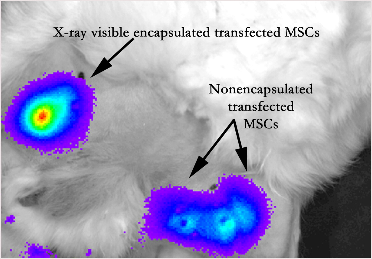
Optical imaging, like MRI, is beneficial because there is no exposure to ionizing radiation. Although techniques are under development for intravascular fluorescence imaging (12), most fluorescence imaging techniques have been performed ex vivo or as microscopic validation (13). One of the challenges of any optical imaging technique is the relatively shallow penetration of light-emitting probes due to photon scatter. As a result, these techniques are ideally suited for the imaging of small animals, where the light emission at the body surface can easily be detected. Anatomic imaging is provided by simple visible light camera systems. The introduction of commercially available systems for small-animal bioluminescence imaging (BLI) (14) that are relatively simple to operate has resulted in a rapid expansion of these techniques for studying angiogenesis and cell tracking (15,16). Small-animal imaging systems have been developed to probe deeper in the body with tomographic fluorescent imaging (17). Although the BLI signal tends to be lower than fluorescent signals, the lack of background luminescence signal, compared with native tissue fluorescence, provides a slight advantage to in vivo BLI (18). Optical imaging techniques often rely on creating transfected cells to express a nonmammalian bioluminescence probe or introducing a probe that emits light when cleaved in the presence of a particular substrate, such as vascular cell adhesion molecule-1 or matrix metalloproteinases, which are both currently targets in the atherosclerotic plaque (3,13,19). Recently, Min et al. have demonstrated the BLI of cord blood mesenchymal stem cells transfected with a reporter gene expressing firefly luciferase to study the effects of immunosuppressive regimes on transplanted cell survival in the rat myocardium (16). One of the major benefits of a reporter gene technique for cell tracking is that only viable cells will be detected. We have recently demonstrated the combination of radiographic fluoroscopic imaging for the detection of encapsulated cells with BLI for the assessment of cell viability in a rabbit model of peripheral arterial disease (Fig. 2) (20).

BLI after intramuscular injection in medial thigh of rabbit model of peripheral arterial disease provides ability to assess cell viability in vivo in radiography-visible encapsulated MSCs similar to that in nonencapsulated MSCs. Bioluminescence image was acquired in 60 s and overlaid on light image for anatomic delineation. MSCs = mesenchymal stem cells.
CT
CT offers the ability to perform whole-body imaging with high anatomic detail. However, to achieve an image quality with CT comparable to that with a clinical system for in vivo small-animal imaging, a resolution of approximately 100 μm is required (21,22). Achieving this resolution requires some combination of increased dose or decreased ability to distinguish soft tissues (i.e., a decrease in contrast resolution), creating a unique challenge for small-animal micro-CT systems.
Micro-CT systems have been designed to rotate the radiographic or detector system around the animal, as is typically done in clinical systems. Most commercial micro-CT systems consist of a low-power radiographic tube of approximately 85 kVp and a flat-panel detector that acquires 2-dimensional (2D) projection images from multiple views around the animal. When imaging small live animals, these systems normally operate with an acquisition time on the order of several minutes and provide 3-dimensional (3D) volume images with a resolution of 100–200 μm. High-speed micro-CT for small-animal cardiovascular imaging is possible with a special high-power microfocus radiographic tube and a slip-ring gantry similar to that on human CT systems. This kind of system provides fast (∼1–2 s) cardiovascular imaging at somewhat reduced resolutions (∼200 μm) and increased radiation dose (23).
As with cardiac MRI, ECG gating strategies to freeze cardiac motion are used with micro-CT in cardiovascular imaging. In addition, traditional radiopaque contrast agents that are injected intravascularly to enhance contrast rapidly wash out of the blood pool and, thus, must be modified for micro-CT applications. A specialized lipid-emulsion contrast agent that has delayed uptake by hepatocytes (Fenestra VC; ART Advanced Research Technologies Inc.) and, therefore, remains intravascular for several hours has been developed for cardiovascular applications in micro-CT (24,25). Delayed-enhancement micro-CT has also been used to image myocardial infarction (26). However, most micro-CT studies use CT for anatomic localization typically in combination with radionuclide studies (Fig. 3).

SPECT/CT rendering of 111In-oxine radiolabeled MSCs (∼2,886 kBq [∼78 μCi] total activity) delivered intravenously to rat with doxyrubicine cardiotoxicity demonstrates initial high lung uptake (blue green on SPECT) immediately after injection (left), followed by redistribution to other organs at 24 h (right). MSCs = mesenchymal stem cells.
RADIONUCLIDE IMAGING
Radionuclide imaging, one of the traditional imaging modalities, has received renewed interest as an important molecular imaging technique. By injecting a trace amount of biomarkers labeled with radioisotopes, radionuclide imaging allows the study of various myocardial functions and related diseases, including ejection fraction, regional wall motion abnormalities, congestive heart failure, perfusion, viability, oxygen consumption, and glucose and fatty acid metabolism (27–29). Also, coronary artery functions and related diseases, such as ischemia, infarction, and atherosclerosis, can be investigated (30–32). In addition, because of its exceptional target specificity of radiotracers, radionuclide imaging allows imaging at the molecular level, such as receptor imaging (33), which cannot be accomplished by other imaging techniques.
Conventional radionuclide imaging uses a position-sensitive radiation detector, such as a scintillation or γ-camera, to detect the γ-ray photons emitting from the 3D distribution of radioactivity of the radiolabeled biomarker in vivo and form a 2D projection image. SPECT and PET apply image-reconstruction methods, which are based on mathematic formulations, to 2D projection images from multiple views and generate 3D images that represent the distribution of radioactivity in vivo in much higher image contrast and clarity than the 2D projection images.
During the past decade, significant advances have been made in detector technologies that provide much improved intrinsic resolution. The new technologies have been applied to clinical and small-animal SPECT and PET systems. In SPECT, using pinhole imaging geometry with magnification, both high resolution (on the order of 1 mm or less with small pinhole aperture and low photon energy) and high detection efficiency (on the order of 10−3 with a full-ring detector geometry and submillimeter resolution) can be achieved (34,35). For example, a unique line of multipinhole SPECT systems for small-animal imaging (U-SPECT-I and its commercial successor, U-SPECT-II) provide unprecedented resolution in mice of 350 μm for 0.35-mm pinholes and 450 μm for 0.6-mm pinholes. This system allows for the visualization of tracer uptake and retention in minute detail in the myocardium of the mouse, including the papillary muscle (Fig. 4) (35,36). Another advantage of small-animal SPECT is its ability to image multiple radiotracers that emit different energy photons simultaneously (37). By taking advantage of radionuclides with relatively long half-lives, small-animal SPECT has been used to track the migration of radiolabeled mesenchymal stem cells to myocardial infarction (38). Also, quantitative SPECT image-reconstruction methods with compensation for collimator-detector response, photon attenuation, and scatter that have been successfully applied to clinical SPECT (39,40) are becoming available to small-animal SPECT, for further improvement in image quality and quantitative accuracy (41–43).
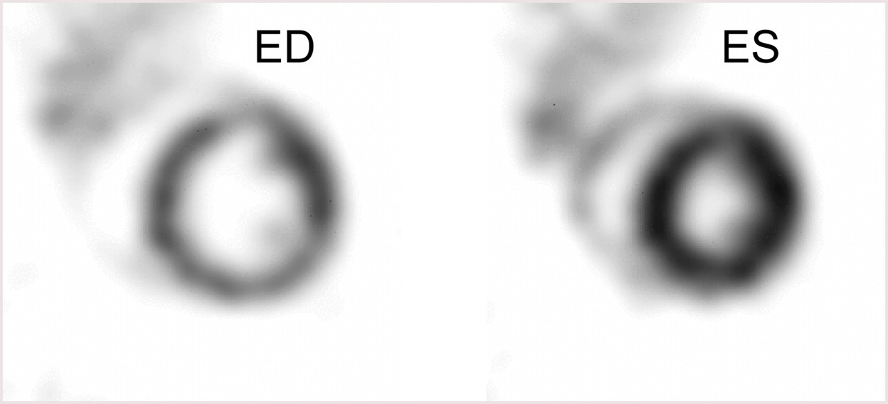
Sample ultra-high-resolution 99mTc-tetrofosmin SPECT images of heart of mouse in end-diastole (ED) and end-systole (ES), showing myocardial perfusion in minute detail in papillary muscles and right ventricular wall. Male C57BL/6 mouse (30 g) was injected intravenously with 190 MBq of 99mTc-tetrofosmin and anesthetized using ketamine, medetomidine, and atropine. At 45 min after injection, mouse was imaged for 1 h using U-SPECT-II system with 0.6-mm-diameter pinhole inserts. During image acquisition, an ECG trigger signal was acquired (BioVet; m2m Imaging) and incorporated in list-mode data. A 16-gate reconstruction was performed. Image data courtesy of Freek J. Beekman.
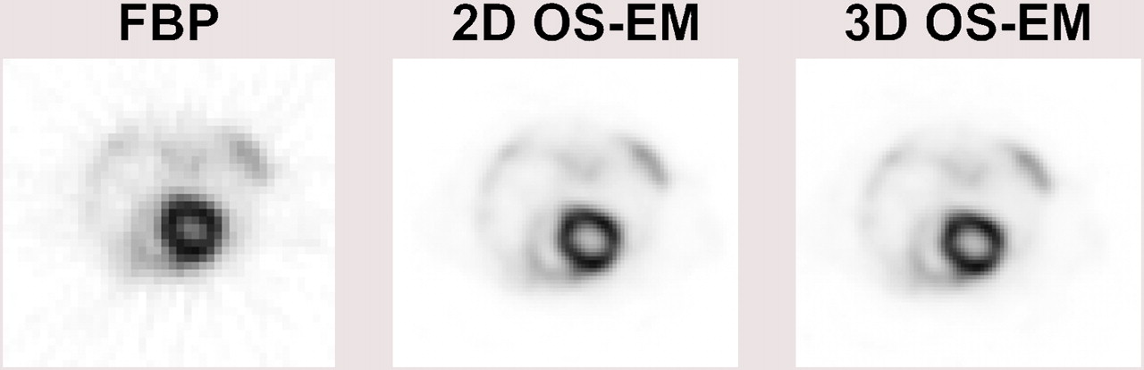
The 511-keV high-energy photons, effective range of energetic positrons (e.g., from 0.2 mm for 18F to ∼2.6 mm for 82Rb), and small noncolinearity between the 2 annihilation 511-keV photons become limiting factors for high-resolution small-animal PET (44,45). Current small-animal PET systems have a spatial resolution of 1.5 mm and detection efficiency on the order of a few percent (46,47). The main advantages of PET include its higher detection efficiency and the availability of positron-emitting radionuclides, including 11C, 13N, 15O, and 18F, which allows the labeling of many physiologically and biochemically interesting biomarkers that are involved in health and disease (48). Three-dimensional quantitative image-reconstruction methods with compensation for detector response, photon attenuation, and scatter have also been applied to small-animal PET for further improvement in image quality of quantitative accuracy (Fig. 5) (49,50).

Same single whole-body transaxial section from 18F-FDG mouse study (26-g mouse, ∼11.1 MBq [0.3 mCi] injected). Data were acquired using commercial small-animal PET system over 30 min, about 1 h after injection, and reconstructed with 3 different methods: Fourier rebinning (FORE)/filtered backprojection, FORE/2D ordered-subset expectation maximization (OS-EM), and 3D OS-EM. RV myocardium and intensely labeled LV myocardium are seen in all 3 18F-FDG reconstructions. FBP = filtered backprojection.
Although small-animal SPECT and PET images offer unique functional information at the molecular level, they are often difficult to interpret because of the lack of correlation with anatomic structures or biologic landmarks. Because CT images provide excellent anatomic information, multimodality SPECT/CT and PET/CT have become standard clinical and small-animal molecular imaging systems (51). More recently, trimodality preclinical small-animal systems with SPECT/PET/CT are becoming commercially available. Also, because of the anatomic information, soft-tissue differentiation, and additional functional information offered by coregistered MR images, dual-modality clinical and preclinical small-animal SPECT/MRI (52) and PET/MRI (53) systems are under active research and development.
Because of the availability of commercial radiolabeled tracers, preclinical molecular imaging using radionuclide techniques has enjoyed much progress. At the same time, fueled by advances in the development of new radiomarkers and radiopharmaceuticals, radionuclide imaging including SPECT and PET has become increasingly important in the preclinical molecular imaging of cardiovascular functions and diseases. Because success in preclinical molecular radionuclide imaging techniques can be directly translated to clinical studies, these methods are particularly important in drug development and translation medicine from in vitro to clinical practice.
CONCLUSION
Although many challenges to small-animal cardiovascular imaging presently exist, whole-body imaging allows the testing of new therapies in relevant disease models to study the safety and efficacy with outcome measures of promising agents similar to those that will be found in future clinical trials. In addition, genetically modified animals allow us to probe the underlying mechanisms of disease development and response to specific treatments that could enable the identification of potential new therapies or molecular imaging probes. Thus, small-animal imaging provides an economic platform for the rapid translation of new knowledge in cardiovascular medicine to the clinical arena.
Acknowledgments
This work was supported in part by the National Institutes of Health under grants R01-EB01558, U24-CA092871, R21-HL89029, R01-EB007825, and 2008 MSCRFII-0399-00 (MD Stem Cell Research Fund).
Footnotes
-
COPYRIGHT © 2009 by the Society of Nuclear Medicine, Inc.
References
- Received for publication November 24, 2008.
- Accepted for publication February 24, 2009.


