


Abstract
Adenosine modulates brain activity through 4 G protein-coupled receptors, primarily adenosine A1 receptors (A1ARs). A1ARs are heterogeneously distributed throughout the brain and participate in many physiologic processes—for example, the induction of sleep and feedback inhibition of excitatory neurotransmission. There is also evidence that A1ARs are involved in brain pathologies, including cerebral ischemia, epilepsy, and neurodegeneration. Therefore, measuring A1ARs in the living brain has been a long-standing goal. This report describes the preclinical evaluation of 18F-8-cyclopentyl-3-(3-fluoropropyl)-1-propylxanthine (18F-CPFPX), a novel A1AR PET ligand. Methods: CPFPX, a xanthine-based A1AR antagonist, was labeled with either 18F or 3H, maintaining identical chemical structures, and evaluated in rats as a putative radioligand for in vivo or in vitro imaging of brain A1ARs by quantitative receptor autoradiography and the combination of high-resolution small animal PET and MRI. Results: 3H-CPFPX bound with nanomolar affinity (Kd, 4.4 nmol/L) to A1ARs and showed a distribution typical of cerebral A1ARs. In extensive in vitro competition studies, 3H-CPFPX proved to be a highly selective and specific A1AR radioligand. Neither the nonxanthine-type adenosine A2A receptor antagonist ZM 241385 nor multiple cholinergic, serotoninergic, and glutamatergic receptor compounds competed for 3H-CPFPX below the micromolar level. In vivo animal PET and ex vivo autoradiographic experiments measured radioactivity in discrete brain regions after intravenous injection of 18F-CPFPX. 18F-CPFPX had excellent in vivo stability and penetrated the blood-brain barrier immediately after injection due to its high lipophilicity. Brain uptake was rapid and particularly high in gray matter regions. Retention of 18F-CPFPX was highest in the cerebellum, thalamus, and neocortex with evidence of saturable binding. Low binding potentials were found in the midbrain. In vivo displacement PET experiments with the A1AR antagonist 8-cyclopentyl-1,3-dipropylxanthine showed a 72% ± 8% displacement of 18F-CPFPX. Conclusion: 18F-CPFPX is a highly selective and specific ligand for A1ARs and a suitable radioligand for noninvasive PET imaging of A1ARs in the living brain. These studies also support the application of high-resolution animal PET as an effective in vivo imaging tool in the evaluation process of new radioligands.
Adenosine, an endogenous purine, exerts its physiologic actions via G protein-coupled receptors, 4 of which (A1, A2A, A2B, A3) have been pharmacologically characterized and cloned in several species, including rats and humans (1–3). Each subtype of adenosine receptor is heterogeneously distributed throughout the body, particularly in excitable tissues such as heart and brain. The A1AR subtype is the most abundant adenosine receptor in the brain and is distributed in a heterogeneous pattern in neocortical and allocortical regions, as well as in the basal ganglia, thalamus, and cerebellum (4–6). The A1AR seems to have an inhibitory net effect on neuronal tissue, although this is still a matter of debate (7). There is evidence that adenosine contributes to the induction and maintenance of sleep and the regulation of arousal (8–10), functions as a retrograde synaptic messenger (11), and plays a role in the autoregulation of cerebral blood flow (12). A1ARs are involved in multiple pathologic conditions of the brain—for example, epilepsy (13) and neurodegenerative diseases, including Alzheimer’s dementia (14–16).
Given the importance of A1ARs both in physiology and in brain pathology, several attempts have been made to image A1ARs in vivo. So far, 3 A1AR antagonists, labeled with 11C, have been described: 1-propyl-11C-8-dicyclopropylmethyl-1,3-dipropylxanthine (11C-KF15372) (17,18) and its 11C-ethyl- and 11C-methyl- derivatives 1-ethyl-11C-8-dicyclopropylmethyl-1,3-dipropyl-xanthine (11C-EPDX) (19) and 1-methyl-11C-8-dicyclopropylmethyl-1,3-dipropylxanthine (11C-MPDX) (20), respectively. 11C-KF15372 has been evaluated as a potential candidate for A1AR PET imaging. Although primary data looked quite promising, serious limitations to the routine application of 11C-KF15372 occurred. Due to the multistep and time-consuming radiosynthesis in combination with the short physical half-life of 11C (20.4 min), the radiochemical yield of 11C-KF15372 was relatively low (∼5%) and specific activity (As) was in the range of 10–56 GBq/μmol (0.3–1.5 Ci/μmol). Brain uptake in mice after intravenous injection was 1.9% of injected dose per gram after 5 min and fell thereafter (17). The content of radioligand was high in hippocampus, cerebral and cerebellar cortex, and caudate-putamen. The administration of unlabeled ligand reduced the content of the radioligand to about 45% of the control level (18). Most important, however, the low rates in both radiochemical yield and As impose severe restrictions to the routine application of 11C-KF15372. The evaluation of 11C-MPDX and 11C-EPDX revealed different affinities to A1ARs in the following order: 11C-EPDX > 11C-KF15372 > 11C-MPDX. In mice, the highest initial brain uptake was found with 11C-MPDX, followed by 11C-EPDX and 11C-KF15372. The washout of 11C-MPDX, however, was relatively fast in comparison with the other compounds (19). 11C-KF15372 and 11C-MPDX have since been tested in monkey studies. Both tracers showed the same brain distribution. Brain uptake of 11C-MPDX (inhibition constant [Ki], 4.2 nmol/L) was much higher and washout was faster than that of 11C-KF15372 (Ki, 3.0 nmol/L). Cerebral binding was blocked by carrier loading or displacement by an A1AR antagonist. The regional cerebral binding pattern as evaluated with kinetic analysis was consistent with data reported from previous in vitro studies (20,21). Up to now, there is no report that any of these 11C-labeled radioligands have reached the stage of human studies.
This report describes the preclinical evaluation of the novel xanthine-based A1AR antagonist 8-cyclopentyl-3-(3-fluoropropyl)-1-propylxanthine (CPFPX). The ligand is an analog of 8-cyclopentyl-1,3-dipropylxanthine (DPCPX), which, due to its high selectivity and affinity for A1ARs, is the prototypical A1AR antagonist (22–24). The synthesis and pharmacologic in vitro characterization of CPFPX as well as the method for the radiosynthesis of noncarrier-added 18F-CPFPX have recently been published (25). 18F-CPFPX is routinely produced and reliably obtained ready for injection in about 55-min overall synthesis time with a radiochemical yield of 45% ± 7% (7.5 ± 0.5 GBq [200 ± 20 mCi]; n = 22), a radiochemical purity of >98%, and an As of >270 GBq/μmol (>7.5 Ci/μmol) at the end of synthesis. 18F-CPFPX shows excellent in vitro stability at room temperature in aqueous solution without any indication of disintegration products up to 6 h after preparation. The octanol-water partition coefficient (log P value) is 2.1 ± 0.15, indicating that 18F-CPFPX is sufficiently lipophilic to cross the blood-brain barrier.
In rat striatal membranes, 18F-CPFPX showed a nanomolar affinity for A1ARs and a 1,200-fold selectivity for A1ARs over A2AARs. In mice, brain uptake was high, and specific binding accounted for 70%–80% of the radioactivity. The radioligand was stable after penetrating the blood-brain barrier but underwent degradation into at least 2 polar metabolites in blood (25).
In this article we discuss in vitro autoradiographic competition studies, in vivo high-resolution small animal PET, and ex vivo dissection techniques to further evaluate 18F-CPFPX for its future use as a PET ligand in nonhuman primates and in humans.
MATERIALS AND METHODS
Radiopharmaceuticals and Chemicals
CPFPX was synthesized in house as previously described (25). Noncarrier-added radiolabeling of CPFPX with 18F has also been published (26). The target compound 18F-CPFPX was obtained with a radiochemical yield of 45% ± 7% and a radiochemical purity of >98%. The As of the product was in the range of >270 GBq/μmol (>7.5 Ci/μmol). 18F-CPFPX was formulated in 0.9% NaCl/7% ethanol for the purpose of intravenous injection.
3H-CPFPX (As, 2,179 GBq/mmol [58.9 Ci/mmol]) was synthesized as recently described (27). The radiochemical purity was >98% as determined by high-pressure liquid chromatography. The 18F- and 3H-labeled probes of CPFPX had identical chemical and pharmacologic properties.
3H-DPCPX (As, 4,129 GBq/mmol [111.6 Ci/mmol]) and 3H-4-(2-[7-amino-2-(2-furyl)[1,2,4]triazolo[2,3-a][1,3,5]triazin-5-ylamino]ethyl)phenol (3H-ZM 241385; As, 629 GBq/mmol [17 Ci/mmol]) were purchased from New England Nuclear.
Adenosine deaminase, (R−)-N6-(2-phenylisopropyl)adenosine (R−)-PIA), caffeine, dopamine, phentolamine, nicotine, carbachol, l-glutamate, and γ-aminobutyric acid were purchased from Sigma-Aldrich Co. 8-Cyclopentyl-1,3-dipropylxanthine (DPCPX) and ZM 241385 were purchased from Tocris Cookson Ltd. All other chemicals were of reagent grade and obtained commercially.
Animals
Adult male Wistar rats (bred in house; 230–250 g body weight) were used. All animals were kept under a natural light/dark cycle and had unlimited access to water and food. The local government approved all procedures according to the German Law on the Protection of Animals. Animal experiments were also approved by the Animal Research Committee of the Scientific and Technical Advisory Board of the Research Center Jülich.
Autoradiographic In Vitro Studies
Animals were decapitated between 6 and 9 am. Whole brains were rapidly removed, blotted free of excess blood, and immediately frozen in 2-methylbutane (−50°C). Subsequently, the brains were cut in a cryostat microtome (CM 3050; Leica; section thickness, 20 μm) at −20°C. Sections were thaw-mounted onto silica-coated object slides, stored in a desiccator containing silica gel, dried overnight at 4°C, and stored adjacently at −80°C in plastic bags until use.
Incubation conditions for 3H-CPFPX and 3H-DPCPX were identical and similar to those previously described by other groups (4,28). All incubations were performed at 22°C in a TRIS-HCl buffer (170 mmol/L, pH 7.4) containing 2 international units (IU)/L adenosine deaminase and MgCl2 (1 mmol/L). Sections were preincubated in buffer for 20 min and subsequently incubated with 4.4 nmol/L 3H-CPFPX or 1.0 nmol/L 3H-DPCPX for 120 min with or without displacer, respectively. They were washed twice in ice-cold buffer (2 × 5 min), rapidly rinsed in ice-cold distilled water, and finally placed under a stream of dry air to facilitate rapid drying. Nonspecific binding was defined as the residual activity in the presence of 100 μmol/L (R−)-PIA. Specific binding was calculated as the difference between total and nonspecific binding.
3H-ZM 241385 was incubated in a TRIS-HCl buffer (170 mmol/L, pH 7.4). Sections were preincubated for 30 min at 37°C in buffer containing ethylenediaminetetraacetic acid (1 mmol/L) and 2 IU/L adenosine deaminase. Sections were washed twice for 10 min at 22°C in incubation buffer containing 10 mmol/L MgCl2 and subsequently incubated with 0.4 nmol/L 3H-ZM 241385 for 120 min at 22°C in buffer containing 2 IU/L adenosine deaminase and MgCl2 (10 mmol/L). Nonspecific binding was defined as the activity remaining in the presence of 2-chloroadenosine (20 μmol/L). Sections were finally washed twice for 5 min in ice-cold buffer, briefly dipped into distilled water, and subsequently dried under a stream of dry air.
A series of adjacent sections of all autoradiographs was stained by Nissl’s method and used for cytoarchitectural identification of brain nuclei and cortical regions.
Saturation Studies
Saturation experiments with 3H-CPFPX and 3H-DPCPX were performed to evaluate the respective dissociation constants, which define the ligand concentration of half-maximum receptor occupation and allow an absolute quantification of receptor binding according to the equation: RT = RL × ([Kd + F]/F), where RT is total receptor concentration, RL is specific binding, Kd is dissociation constant, and F is free ligand concentration.
For saturation experiments, 4 quadruplicates of randomized coronal sections were chosen from different parts of the brain (caudate-putamen and dorsal hippocampus planes, respectively) and incubated in a series with rising concentrations of 3H-ligands and a parallel series with the addition of (R−)-PIA (100 μmol/L) to determine nonspecific binding.
High-Resolution Small Animal PET Studies
PET imaging of in vivo blockade and displacement experiments with 18F-CPFPX was performed on a prototype of a dedicated high-resolution small animal PET scanner (TierPET), which has previously been described (29,30). The TierPET scanner (Fig. 1) consists of 2 orthogonal pairs of detectors, which are mounted on linear feeds to adjust detector-detector distances individually (range, 16–58 cm; present experiment, 32 cm). Feeds are mounted on an aluminum wheel, which is rotated by 90° for the acquisition of complete projections (7.5° angular steps, step-and-shoot mode). Each detector module consists of a 20 × 20 array of 2 × 2 × 15 mm yttrium aluminum perovskite crystals, which are polished and optically isolated by reflective layers. The arrays are coupled to a Hamamatsu R2487 position-sensitive photomultiplier. The TierPET scanner runs in 3-dimensional mode and acquires data in list mode. List mode data are converted into 2-dimensional sinograms using a multiple-slice rebinning method (31). Two-dimensional sinograms are subsequently reconstructed using an iterative reconstruction based on the maximum-likelihood expectation maximization algorithm. Reconstructed image resolution is 2.1 mm (full width at half maximum), which is homogeneously maintained throughout the entire field of view. To reduce scatter-related perturbations, the neck and body of the animal were shielded with a hollow lead sleeve perfused with water heated to 37°C to maintain a constant body temperature. A precise anatomic identification of rat brain regions was achieved by coregistration with structural MR images (Siemens Magnetom VISION; 1.5 T, equipped with a dedicated transmit/receive radiofrequency coil and using a scanning sequence developed in house) and TierPET datasets. For this purpose, animals were placed in a transportable stereotactic head holder along with 3 fiducial markers, which served as spatial landmarks for PET and MRI. They were alternately filled with 18F for PET or cod liver oil for MRI. The head holder was screwed to an object tablet, which could be moved along the x-, y- and z-axes by computer-controlled step motors, thus permitting a precise positioning of the animals within the field of view. Regions of interest (ROIs) were delineated on fusion images of MRI and TierPET datasets with dedicated software (Multi-Purpose-Imaging-Tool; ATV) (32).
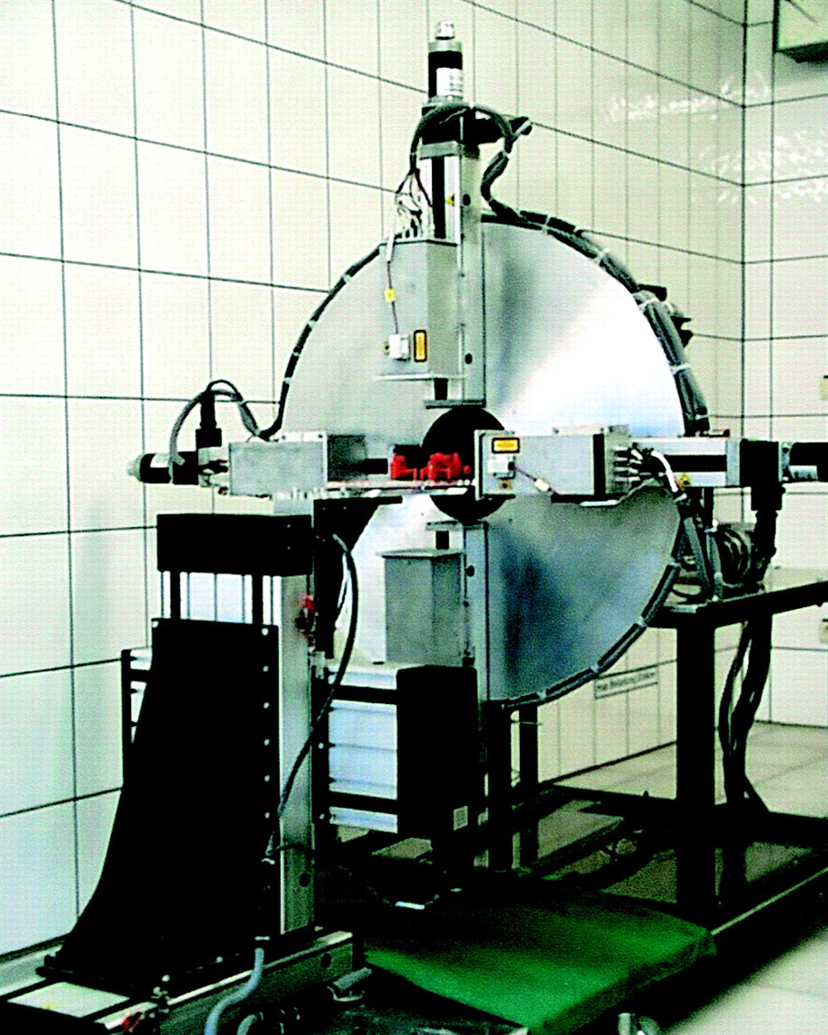
Photograph of TierPET system, a dedicated high-resolution small animal PET scanner developed at Research Center Jülich, Jülich, Germany.
Animals were sedated in an isoflurane atmosphere (2%–5%) and anesthetized with ketamine (100 mg/kg)/xylazine (10 mg/kg). After placing a jugular vein catheter, animals received a bolus injection of 0.3 mL 18F-CPFPX (37 MBq/mL [1 mCi] dissolved in 0.9% NaCl/7% ethanol). TierPET data were acquired for 60 min after injection in 10 frames of 6-min duration. Blood samples (∼0.1 mL) were collected at 10, 20, 40, and 60 min after ligand administration to determine radioactivity and metabolites. Metabolite analysis was performed as previously described (25). In brief, venous plasma was obtained from heparinized whole blood samples (0.1 mL), diluted with methanol/dichloromethane (80:20, v/v, 0.2 mL), vortexed for 20 s at room temperature, and centrifuged (20,000g, 1 min). Aliquots (0.1 mL) of the supernatants were withdrawn, CPFPX (20 ng) was added, the mixtures were vortexed for 10 s, and samples (10–15 μL) were spotted onto thin-layer chromatography plates (SIL G; Macherey-Nagel), which were developed with ethyl acetate/hexane (50:50, v/v) and analyzed using a high-performance image plate reader (InstantImager; Canberra Packard). In vivo distribution and displacement studies were performed in identical animals by intravenous application of 1 mg/kg DPCPX 12 min after the injection of 18F-CPFPX.
High-Resolution MRI
All animals underwent MRI scanning for anatomic comparison. Images were obtained using a whole-body MRI scanner (Siemens Magnetom VISION; 1.5 T) equipped with actively shielded gradient coils (maximum gradient strength, 25 mT/m; slew rate, 83 T/m/s). The animals were positioned, either individually or in pairs, inside a 22-cm-diameter transmit/receive radiofrequency coil. Anatomic images were obtained with a 3-dimensional gradient echo pulse sequence with a slab selection (slab thickness, 17 cm; effective slice thickness, 1 mm) along the sagittal orientation. Other acquisition parameters were field of view, 180 mm; matrix size, 256 × 256 with an in-planar resolution of 0.7 × 0.7 mm; number of excitations, 1. The total acquisition time was about 14 min. The final imaging resolution was 0.7 × 0.7 × 1.0 mm.
Autoradiographic Ex Vivo Studies
To confirm reliability of the in vivo measurements, 18F-CPFPX PET studies were calibrated by ex vivo experiments. Animals were killed by cervical dislocation on completion of the scan. Brains were removed immediately, frozen in 2-methylbutane at −50°C, and cut in coronal sections (thickness, 20 μm) with a cryostat microtome (CM 3050; Leica).
Image Processing and Statistical Analysis
Sections from autoradiographic experiments were placed on phosphor imaging plates (BAS-SR 2025; Raytest-Fuji) along with in-house-made and calibrated 18F brain paste standards for 18F studies or industrial 3H activity standards (Microscales; Amersham Biosciences) for 3H studies, respectively. Upon exposure, the imaging plates were scanned with a high-performance imaging plate reader (BAS 5000 BioImage Analyzer; Raytest-Fuji) providing a spatial resolution of 50 μm.
The evaluation of digital receptor autoradiography was processed according to standard image analysis software (AIDA 2.31; Raytest). Competition and saturation curves were approximated numerically and calculated with standardized in-house-developed software and commercially available curve-fitting programs (Prism 3.0; GraphPad Software). Ex vivo autoradiographic data were corrected for injected dose, weight, decay, exposure time, and film efficacy before being analyzed with dedicated software (AIDA 2.31). Similarity of receptor densities as determined by PET and ex vivo autoradiography was analyzed with linear regression analysis using the same software.
RESULTS
CPFPX was labeled with 18F or 3H at the positions indicated in Figure 2. The 2 ligands are chemically identical.

Structures of 18F-CPFPX (A) and 3H-CPFPX (B).
In Vitro Distribution Studies
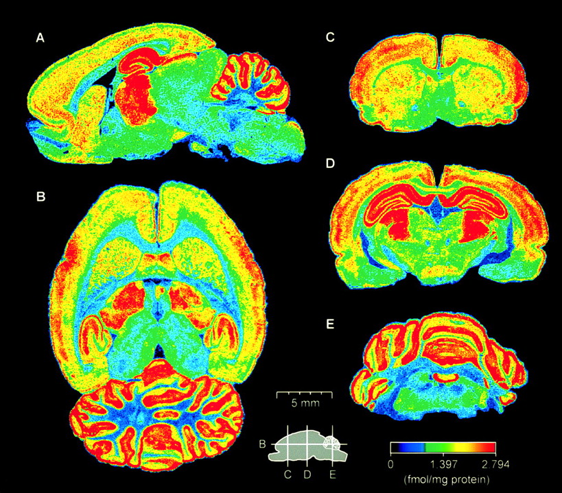
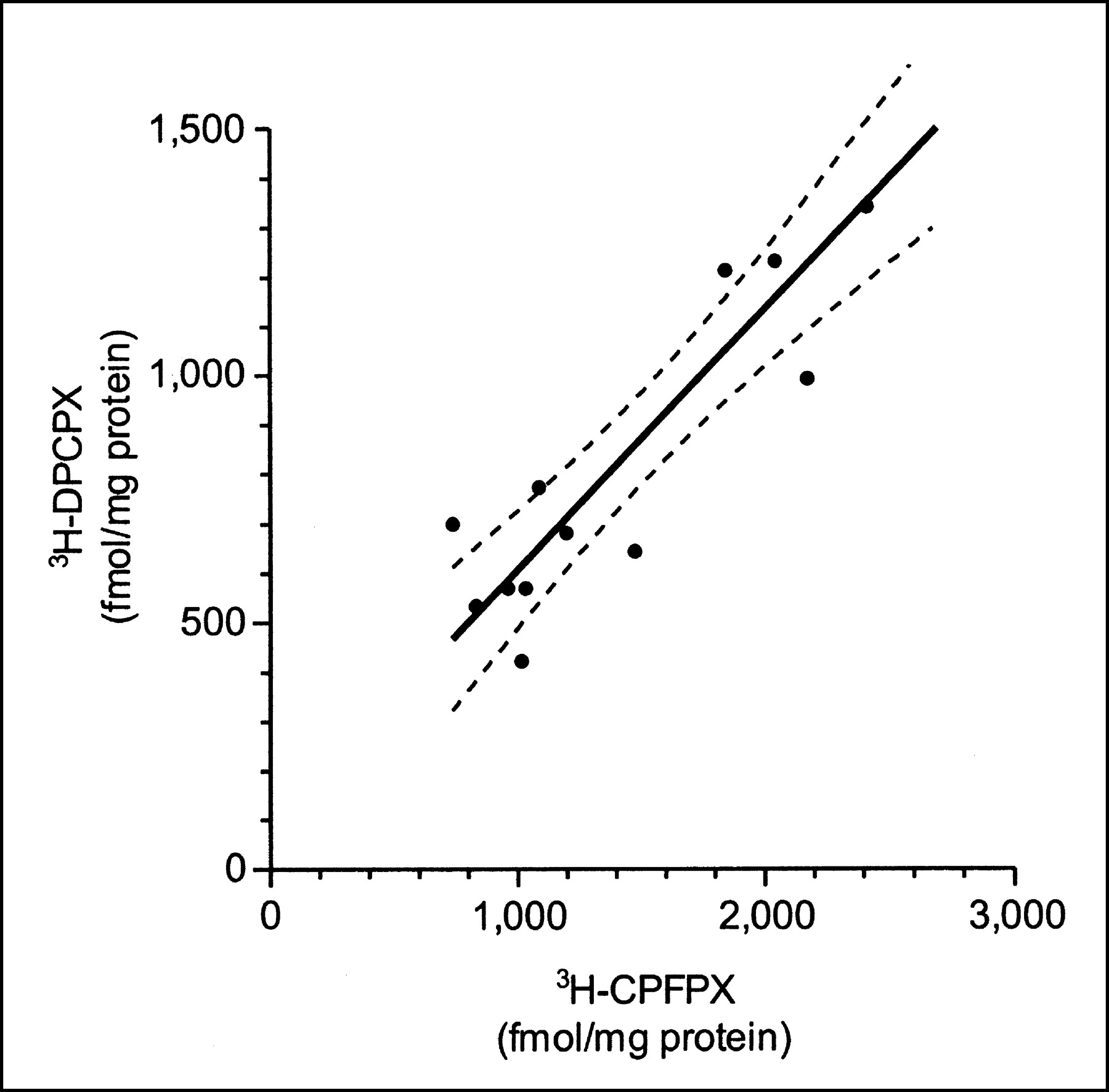
Anatomic distribution of 3H-CPFPX was evaluated with quantitative receptor autoradiography. We chose 12 ROIs to calculate absolute receptor binding. In vitro autoradiographic binding of 3H-CPFPX in rat demonstrated a typical pattern of cerebral A1AR distribution with a low background and a high specific binding, particularly in gray matter regions. The highest receptor densities were found in the cerebellum, neocortex, and thalamus (Fig. 3). Nonspecific binding in rat brain amounted to a maximum of only 7% of total binding. A comparison of 3H-CPFPX and 3H-DPCPX binding established from adjacent serial sections revealed a highly significant similarity of the binding profiles of 3H-CPFPX and 3H-DPCPX (Fig. 4).

Representative autoradiographs of sagittal (A), horizontal (B), and coronal (C–E) sections of rat brain. Sagittal scout on bottom indicates coronal and horizontal planes of transsection. Absolute receptor density is indicated according to color scale at bottom right. Note high accumulation of 3H-CPFPX in thalamus (A, B, and D) and cerebellum (A, B, and E). Neocortical regions exhibit region- and layer-specific receptor binding. Low accumulation of radioligand is found in midbrain (A and D) and brain stem (A, D, and E). For direct reading of absolute receptor density, see color scale at bottom right.

Autoradiographic data from parallel sections of 12 rat brain regions (n = 5) labeled with 3H-CPFPX and 3H-DPCPX, respectively. Similarity, as calculated with linear regression analysis, is highly significant (P < 0.0001; r2, 0.8539; dashed lines indicate 95% confidence intervals).
In Vitro Competition Studies
3H-CPFPX binding in rat brain was saturable, and Scatchard analysis revealed a Kd of 4.4 nmol/L (Fig. 5A). Competition studies were performed at ligand concentrations close to the Kd value. Competitor concentrations were chosen between 1 pmol/L and 10 μmol/L. The affinity to 3H-CPFPX binding sites was negligible (Ki, >1 μmol/L) for various competitors from representative neurotransmitter systems, in particular amino acid systems (γ-aminobutyric acid, l-glutamate), monoaminergic transmitter systems (5-hydroxytryptamine, dopamine), and cholinergic systems (carbachol, nicotine). In contrast, the A1AR antagonists CPFPX (Ki, 1.21 nmol/L), DPCPX (Ki, 6.29 nmol/L), and N-0840 (Ki, 11 nmol/L) competed for 3H-CPFPX binding sites in the nanomolar range. The unselective adenosine agonist (R−)-PIA (Ki, 39 nmol/L) and the antagonist caffeine (Ki, 216 nmol/L) were less effective in competing with 3H-CPFPX. ZM 241385, an A2AAR-selective antagonist, displaced 3H-CPFPX in the high micromolar range (Ki, 15 μmol/L), underscoring the A1AR selectivity of CPFPX (Fig. 5B).
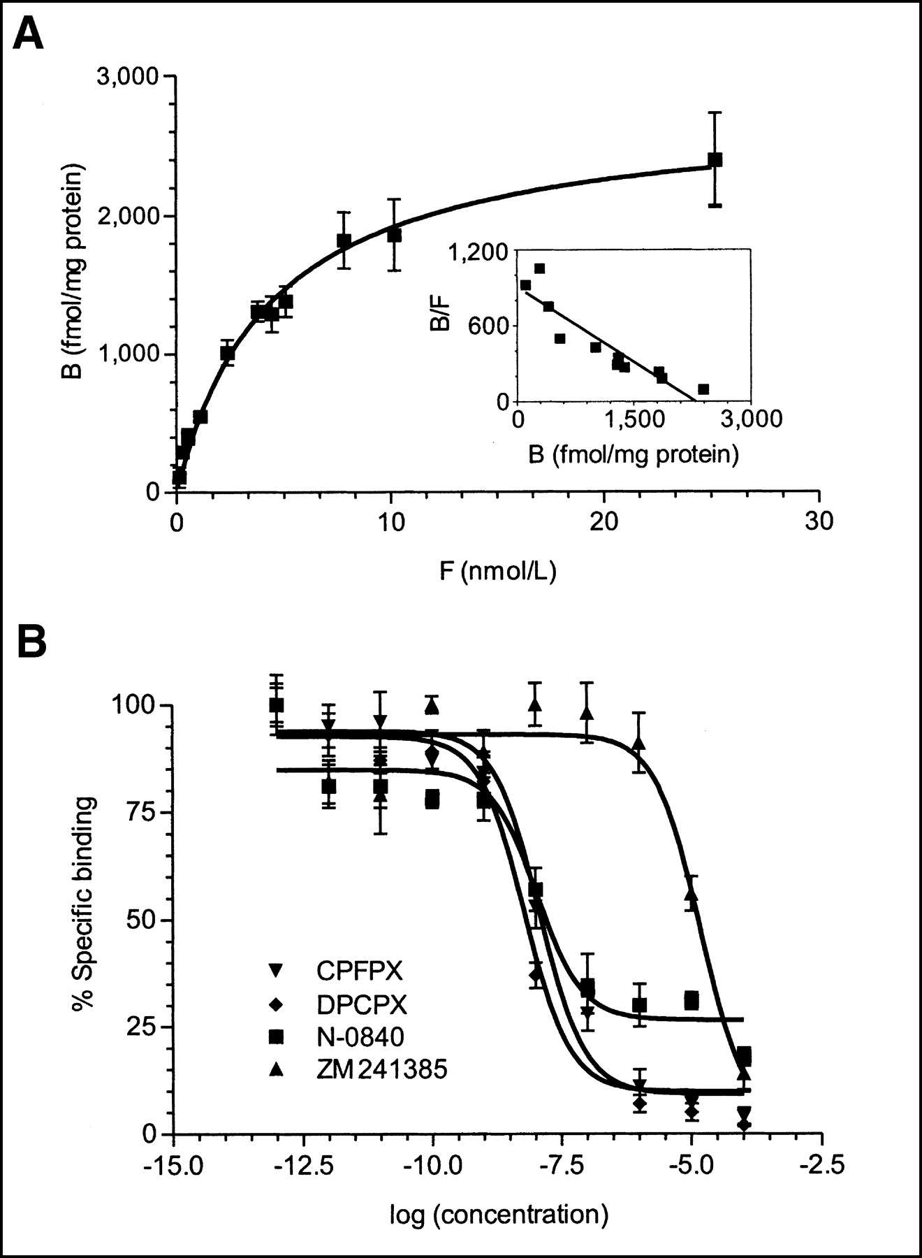
(A) Representative saturation curve and Scatchard plot (insert) of 3H-CPFPX (Kd, 4.4 nmol/L; maximum number of binding sites [Bmax], 2,746 fmol/mg protein; r2 [saturation curve], 0.989). Curve fitting is consistent with 3H-CPFPX binding to single high-affinity site. B = number of binding sites; F = free ligand concentration. (B) Representative competition curves of selective adenosine A1 (CPFPX, DPCPX, N-0840) and A2A (ZM 241385) antagonists vs. 3H-CPFPX, which demonstrate selectivity of 3H-CPFPX for A1AR.
In Vivo Animal PET and MRI Studies
High-resolution brain MRI scans obtained on all investigated animals served as an anatomic reference for a reliable identification of the brain. Inspection of the horizontal, coronal, and sagittal planes identified all major cranial and cerebral structures. The brain was clearly distinguishable from other cranial structures and was contoured accordingly on the MR datasets. The brain contours served as an overlay that was transferred to individually coregistered TierPET datasets.
After intravenous application, brain uptake of 18F-CPFPX was rapid (Fig. 6). Upon completion of the first frame (6 min after injection), the maximum of intracerebral uptake was already reached, followed by a gradual washout down to 67% of the maximum accumulation at the end of the scan (Fig. 6). Accumulation of radioactivity was high throughout the brain and extracranial soft tissues, particularly in the masseter muscles and the submandibular and harderian glands. Figure 7B is a representative example of the TierPET images. During the scanning procedure, 18F-CPFPX was metabolized into 2 polar metabolites to the extent of 54% ± 8% within 5 min.

Time-activity curves of baseline (n = 3) and displacement studies (n = 3) with 18F-CPFPX in absence and presence of DPCPX, respectively. DPCPX was injected 12 min after start of scanning (arrow). Whole rat brain was taken as ROI. Data ± SD were normalized to injected dose.

Representative example of in vivo displacement study of DPCPX vs. 18F-CPFPX measured by TierPET shows MRI (A) and coregistered TierPET images (B and C) of horizontal (left column), coronal (middle column), and sagittal (right column) sections. Entire brain was chosen as ROI. (A) Red trace outlines brain based on individual MRI datasets, which were subsequently superimposed onto TierPET datasets (B and C, brain outlined in white). (B) Summed TierPET images of frames 1 and 2 (in total, 12 min) before application of displacer. (C) Summed TierPET images of frames 3–10 (in total, 48 min) after intravenous application of DPCPX (1 mg/kg). Note significant displacement of 18F-CPFPX in brain after DPCPX was applied, whereas activity is not significantly reduced in other parts of head.
To test the displaceability of 18F-CPFPX in vivo, 1 mg/kg DPCPX was injected 12 min after tracer administration. Figures 7B and 7C illustrate horizontal, coronal, and sagittal TierPET images of a representative animal after 18F-CPFPX (Fig. 7B) and after intravenous injection of DPCPX (Fig. 7C). DPCPX caused a rapid and significant displacement of cerebral 18F-CPFPX (62% ± 8% decrease of activity after 6 min; n = 3), indicating the specific and reversible binding of 18F-CPFPX (Fig. 6). In contrast, other cranial structures, particularly the masseter muscles and the submandibular glands, retain a considerable amount of activity, pointing to a nonspecific extracerebral ligand accumulation (Figs. 7B and 7C). In vivo PET experiments were completed by ex vivo dissection of the rat brains. Displacement of 18F-CPFPX by DPCPX amounted to 76% ± 5% in ex vivo measurements.
DISCUSSION
There is a substantial amount of evidence from in vitro studies that the neuromodulator adenosine and its receptors are involved in numerous processes of normal brain function as well as neuropathologic conditions, including epilepsy, Alzheimer’s disease, and cerebral ischemia. Given the importance of the adenosine system, in vivo imaging of the A1AR, the most abundant cerebral adenosine receptor, has been a long-standing goal. This study pursues the evaluation process of 18F-CPFPX, of which precursor synthesis, radiochemistry, basic ex vivo pharmacokinetics, and ex vivo distribution studies in mice have previously been reported (25).
CPFPX was labeled with either 3H or 18F. Both ligands had identical chemical and binding properties. 3H-CPFPX was produced to facilitate and enhance the evaluation process, because 3H-ligands are comparatively stable tracers (half-life, 12.3 y) and, thus, easier to handle in incubation protocols. An additional advantage is that 3H gives a substantially higher resolution than 18F in autoradiographic studies. A high resolution is required to identify layer- or nucleus-specific distribution patterns, as shown in Figure 3, for example.
3H-CPFPX demonstrated an A1AR typical binding pattern in the rat brain. The labeled receptors had a highly heterogeneous distribution and were mainly concentrated in the cerebellar cortex, hippocampus, some thalamic nuclei, and cerebral cortex. In contrast, the hypothalamus and brain stem were sparse in binding. These results agree well with data from previous autoradiographic (5) and immunohistochemical (33,34) studies on the distribution of A1ARs in the rat brain. They are also in excellent accordance with previous reports on the distribution of the A1AR gold standard 3H-DPCPX (35,36). A direct comparison of both ligands demonstrated a highly significant correlation in cerebral ligand distribution. Saturation studies revealed that 3H-CPFPX bound with nanomolar affinity to a single and saturable binding site.
In numerous competition studies, it was clearly shown that 3H-CPFPX is not displaceable by ligands that have high affinities for other receptor systems having brain distributions like those of the A1AR or by a selective A2AAR antagonist. The latter is of special relevance for the high binding capacity of the caudate-putamen, the main brain region coexpressing A1ARs and A2AARs. In contrast, 3H-CPFPX is displaceable in vitro and in vivo by DPCPX. All of these results provide substantial evidence that CPFPX is a highly specific and selective A1AR ligand.
Thus far, high-resolution small animal PET has not been applied widely in the initial stages of ligand development, although its demand and potential benefit have repeatedly been emphasized (37,38). This study includes the application of in vivo imaging studies with a dedicated high-resolution small animal PET scanner to test the feasibility and the usefulness of this technique in the early process of ligand development.
Analysis of 18F-CPFPX PET time-activity curves revealed a rapid cerebral uptake of the ligand followed by a gradual washout over the 60-min scan time. The whole brain was defined as an ROI for further data processing. The kinetic data of 18F-CPFPX agreed with previous results of ex vivo mice experiments (25). To test the specificity of 18F-CPFPX binding, the A1AR standard antagonist DPCPX was used as an unlabeled displacer, injected 12 min after scan onset. Within 6 min, the brain activity diminished to 62% ± 8% of the baseline radioactivity. These data indicate that 18F-CPFPX is reversibly bound to cerebral A1ARs and, thus, may be used to provide a direct measurement of drug-induced occupancy of A1ARs in vivo. In contrast, other sites of ligand accumulation outside the rat cranium—for example, muscles of mastication and salivary glands—showed persistent activity after the administration of DPCPX, which is in accordance with the nonspecific accumulation of 18F-CPFPX and its metabolites. In summary, in vivo experiments support the view that 18F-CPFPX is a suitable PET ligand for the detection of A1ARs.
CONCLUSION
This study demonstrates that 18F-CPFPX is a suitable radioligand for the in vivo imaging of A1ARs with PET. It penetrates the blood-brain barrier rapidly, binds specifically and highly selectively to A1ARs, and is displaceable in vivo. Additionally, this study demonstrates the effectiveness of high-resolution small animal PET as a tool in the early evaluation process of radioligands.
Acknowledgments
The authors gratefully acknowledge the helpful comments of Dirk Bier, Institute of Nuclear Chemistry; and the excellent technical assistance of Sabine Wilms, Tobias Rustige, and Ute Meisert (laboratory staff) as well as Maria-Liisa Grosse-Ruyken (MRI staff) of the Institute of Medicine; and Markus Lang, Bettina Palm, and Erika Wabbals of the Cyclotron and Radiosynthesis Staff of the Institute of Nuclear Chemistry. Both institutes are part of the Research Center Jülich. This work was supported by grants from the Hermann von Helmholtz-Gemeinschaft Deutscher Forschungszentren (Strategiefonds), the American Heart Association (Cardiovascular Research, University of South Florida), and the Deutsche Forschungsgemeinschaft (SFB 575).
Footnotes
Received Feb. 20, 2003; revision accepted May 22, 2003.
For correspondence contact: Andreas Bauer, MD, Institute of Medicine, Research Center Jülich, 52425 Jülich, Germany.
E-mail: an.bauer{at}fz-juelich.de
REFERENCES
In this issue


Jump to section
Related Articles
Cited By...
- Effect of Acute Hypoxia Exposure on the Availability of A1 Adenosine Receptors and Perfusion in the Human Brain
- Caffeine Occupancy of Human Cerebral A1 Adenosine Receptors: In Vivo Quantification with 18F-CPFPX and PET
- Activation of Adenosine1 Receptors Induces Antidepressant-Like, Anti-Impulsive Effects on Differential Reinforcement of Low-Rate 72-s Behavior in Rats
- 18F-CPFPX PET Identifies Changes in Cerebral A1 Adenosine Receptor Density Caused by Glioma Invasion