


Abstract
242315
Introduction: Nuclear medicine plays a key role in the diagnosis and differentiation of thyroid pathologies. Currently, in addition to subjective 99mTc-Pertechtenate scintigraphy image assessments, physicians might utilize the absolute counts of thyroid uptake, based on the uptake values derived from pre- and post-injection of syringe, injection site, thyroid, and background images, for diagnosis ranging from normal tissue to thyroiditis and nodular goiter. In this study, we aimed to provide an AI-assisted model in conjunction with radiomic features to distinguish pathological from healthy cases, using only thyroid images.
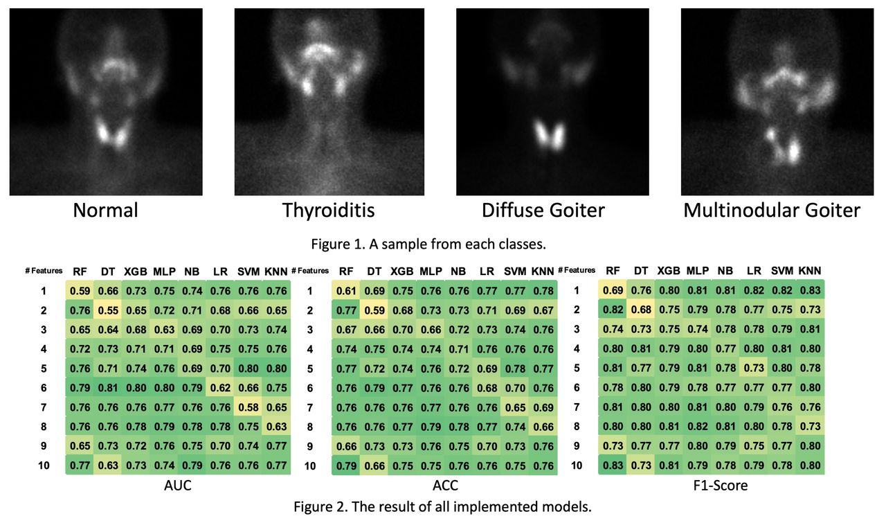
Methods: We retrospectively collected thyroid scintigraphy imaging data from 191 patients. The ground truth was based on thyroid uptake used by nuclear medicine physician for clinical diagnosis (50 diffuse goiter, 29 multinodular goiter, 41 thyroiditis, and 71 normal). One representative sample of each class can be seen in Figure 1. A total of 100 radiomic features (shape, intensity, and second-/high-order texture features from Gray Level Dependence Matrix (GLDM), Gray Level Co-occurrence Matrix (GLCM), Gray Level Run Length Matrix (GLRLM), Gray Level Size Zone Matrix (GLSZM), and Neighboring Gray Tone Difference Matrix (NGTDM)) were extracted using the Pyradiomics python library from segmented thyroids in antero-posterior scintigraphic images. Using nested cross-validation (outer and inner loops of 5 and 4, respectively), one to ten of the most predictive feature combinations were selected iteratively via the Minimum Redundancy Maximum Relevance (MRMR) algorithm. In each fold, the feature(s) were subsequently fed to machine learning (ML) models for binary classification task (normal vs. abnormal). We utilized 8 ML algorithms, including (Random Forest (RF), Naive Bayesian (NB), Decision Tree (DT), eXtreme Gradient Boosting (XGB), Multilayer Perceptron (MLP), Logistic Regression (LR), Support Vector Machine (SVM), and K-Nearest Neighbors (KNN)). Metrics including area under the receiver operating characteristic curve (AUC-ROC), accuracy (ACC), and F1-score were reported.
Results: Our results demonstrated that the top 5 most selected features in the entire feature selection process were Coarseness and Strength (from NGTDM features), Maximum_2D_Diameter_Column (from shape features), Informational Measure of Correlation 2 (Imc2) (from GLCM features), and Gray_Level_Variance (from GLSZM features). The results of all models are illustrated in Figure 2. Among ML classifiers, the best-performing model according to AUC was DT (AUC=0.809 ± 0.081, ACC=0.785 ± 0.091, F1=0.802 ± 0.092, using most predictive combination of 6 features), and the best accuracy and F1-score were achieved by RF (ACC=0.790 ± 0.077, F1=0.835 ± 0.059, AUC=0.773 ± 0.084, using most predictive combination of 10 features).
Conclusions: Our study demonstrated that ML algorithms coupled with radiomic features are reasonably capable of differentiating healthy cases from patients with thyroid disease only from thyroid images without the need for calculating uptake values, which can be time-saving and effectively contribute to daily clinical practice. Moreover, the streamlined process eliminates the need for pre- and post-injection imaging of syringe, injection site imaging, and background imaging, thereby decreasing the workload and radiation exposure to staff.

In this issue


Jump to section
Related Articles
Cited By...
- No citing articles found.