


Abstract
242067
Introduction: Prostate cancer (PCa) is a diagnostic challenge due to its heterogeneity and varying degrees of aggressiveness. Previous studies have established a correlation between disease aggressiveness and tumor perfusion, and it is therefore possible that tumor perfusion may be valuable not only for prognostic purposes but also for evaluating treatment response. In this study, we aimed to evaluate long axial field-of-view (LAFOV) [15O]H2O PET as a tool for whole-body quantitative tumor perfusion imaging in metastatic PCa.
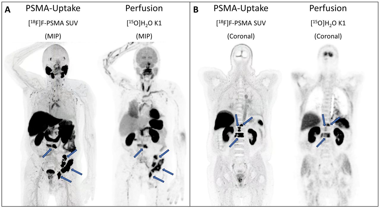
Methods: Six patients with metastatic PCa underwent dynamic LAFOV PET scans (Siemens, Quadra). The imaging protocol consisted of a dynamic 5-minute [15O]H2O PET followed by a static [18F]PSMA-1007 PET. The primary prostate lesion and metastatic sites in bones and lymph nodes were identified and semi-automatically delineated on the [18F]PSMA-1007 images using a fixed SUV threshold of 4 (Affinity Viewer, Hermes). These delineations were then transferred to the [15O]H2O PET images, and time-activity curves were extracted. K1 (mL/min/mL) was used as the metric of perfusion and was estimated using a single-tissue compartment model and a cardiac image-derived input function. Parametric images of perfusion were calculated using the basis-function method with initial voxel-wise estimation of delay using a leading-edge approach.
Results: On the [18F]PSMA-1007 PET scans, 5 primary tumor lesions, 63 lymph nodes, and 33 bone metastases were detected. The median perfusion of the primary prostate tumor was 0.20 mL/min/mL, with a range of 0.08-0.35 mL/min/mL. The lymph node metastases had a median perfusion value of 0.16 mL/min/mL, ranging from 0.03-0.58 mL/min/mL. Additionally, for bone metastases, the median perfusion was found to be 0.29 mL/min/mL, with a range of 0.06-0.98 mL/min/mL.
Conclusions: Long axial field-of-view PET/CT imaging using [15O]H2O allows for the estimation of whole-body tumor perfusion. This approach provides valuable insights into the distribution and quantification of perfusion values, not only in the primary tumor but also in metastatic sites. The ability to assess whole-body tumor perfusion could be valuable for evaluating disease severity, monitoring treatment response, and characterization of the different metastatic sites in patients with PCa.

In this issue


Jump to section
Related Articles
Cited By...
- No citing articles found.