


Abstract
242440
Introduction: Fumarate hydratase - deficient renal cell carcinoma (FHRCC) is a rare renal cancer, associated with the germline / somatic gene mutation of fumarate hydratase. 18F-FDG PET/CT can be used for staging and restaging for FHRCC due to the activation of glycolytic pathway. 68Ga-FAPI PET/CT can reflect the expression of fibroblasts associated protein (FAP) on cancer associated fibroblasts and was a useful tool for diagnosing RCC. However, FHRCC has not been reported yet. Therefore, the aim of this study is to compare the efficiency of imaging of 18F-FDG PET/CT and 68Ga-FAPI PET/CT for FHRCC.
Methods: Patients with FHRCC confirmed by pathology were prospectively enrolled to performed for 68Ga-FAPI PET/CT and 18F-FDG PET/CT from May 2022 to December 2023 (ChiCTR2300072818). Two senior nuclear medicine physicians interpreted the PET/CT images based on lesion features, enhanced CT or MRI results and clinical data. The maximum standardized uptake (SUVmax) and tumor to liver ratio (TLR) of two tracers were compared for Wilcoxon signed-rank test.
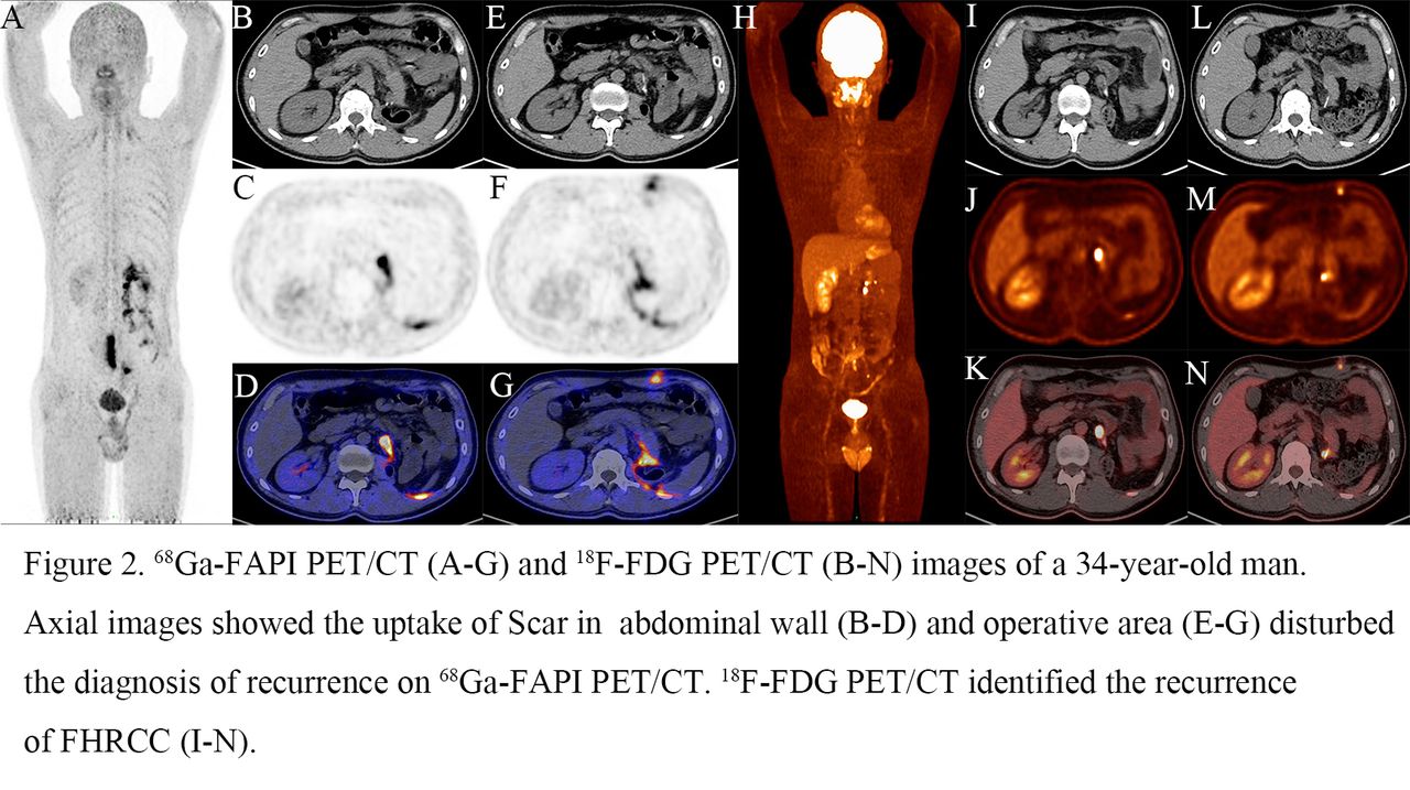
Results: Totally, 11 patients (median age: 42 years old) with 85 lesions were enrolled in the study. 92.9% (79/85) of lesions were detected on 18F-FDG PET/CT and 84.7% of lesions were detected on 68Ga-FAPI PET/CT. The rate of 18F-FDG PET/CT in detecting FHRCC-related lesions were higher than 68Ga-FAPI PET/CT (primary lesions: 62.5% vs 50.0%; lymph nodes: 94.9% vs 87.2%; bone lesions: 100.0% vs 95.2%; visceral lesions: 94.1% vs 82.3%). Bizarrely, there were two patients with three primary lesions not detected by 18F-FDG PET/CT and 68Ga-FAPI PET/CT. Besides, false-positive lesions of liver hyperplasia were found on 68Ga-FAPI PET/CT and the uptake of scar on 68Ga-FAPI PET/CT disturbed the diagnosis of metastases of abdominal wall. In semi-quantitative analysis, the median SUVmax of primary and metastatic lesions on 18F-FDG PET/CT were comparable to 68Ga-FAPI PET/CT (primary lesions: 14.72 vs 16.35, P = 0.730; lymph nodes: 10.04 vs 8.86, P = 0.306; bone lesions: 13.49 vs 11.05, P = 0.273; visceral lesions: 7.51 vs 4.20, P = 0.070). However, the median TLR of primary and metastatic lesions on 68Ga-FAPI PET/CTwere higher than 18F-FDG PET/CT (primary lesions: 30.44 vs 5.86, P = 0.016; lymph nodes: 16.68 vs 3.95, P = 0.000; bone lesions: 16.43 vs 5.21, P = 0.000; visceral lesions: 9.26 vs 3.22, P = 0.001).
Conclusions: Overall, 18F-FDG PET/CT detected more FHRCC-lesions than 68Ga-FAPI PET/CT. However, the high TLR of FHRCC on 68Ga-FAPI PET/CT may indicate the therapeutic potential of targeting FAP for FHRCC.


In this issue


Jump to section
Related Articles
Cited By...
- No citing articles found.