


Abstract
241100
Introduction: Positron emission tomography with magnetic resonance imaging (PET/MRI) has the potential to improve diagnostic accuracy and provide noninvasive molecular characterization of breast cancer. However, clinician or patient reluctance may be limiting widespread use due to concerns of current radiation dose from the radiopharmaceutical. The overall goal of this research is to reduce the radiation dose from 2-deoxy-2-[18F]fluoro-D-glucose (18F-FDG) breast PET/MRI by 90%, which would result in a radiation dose similar to full-field digital mammography, while preserving image quality. The specific aim of this reader study was to evaluate the diagnostic image quality of simulated low dose 18F-FDG breast PET/MRI with and without a vendor-provided, deep learning-based, denoising algorithm.
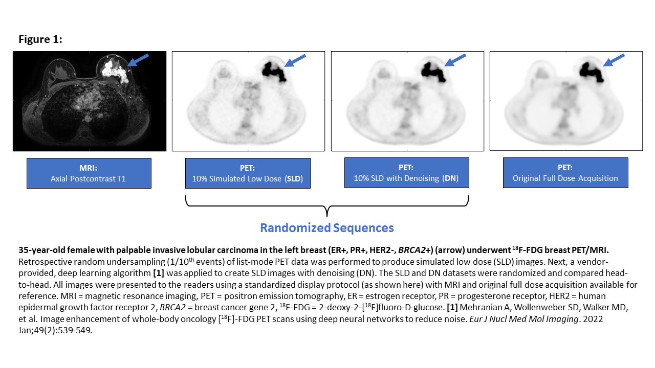
Methods: The data used for this reader study was obtained from a single-institution prospective study, which included women with biopsy-proven invasive breast cancer undergoing preoperative breast MRI from 2016 to 2018. Fasting participants were injected with 10 mCi (370 MBq) of 18F-FDG followed by an uptake period of 85 minutes. Prone breast PET/MRI was accomplished using standard breast MRI sequences with simultaneous PET acquisition for 30 minutes. Retrospective random undersampling (1/10th events) of the list-mode PET data was used to produce simulated low dose PET images. Three reconstructed PET datasets were generated: 1) simulated low dose images without denoising (SLD), 2) simulated low dose images with denoising (DN), and 3) original full dose acquisition (Figure 1). The SLD and DN datasets were displayed in blinded, randomized order and independently reviewed by three board-certified radiologists and/or nuclear medicine physicians. Using a 5-point Likert scale, the readers evaluated SLD and DN for artifacts, signal-to-noise ratio, image sharpness/resolution, and overall image quality. The readers also rated the diagnostic image quality and lesion detectability in SLD and DN compared to the full dose acquisition as a measure of clinical acceptability. Post-contrast MRIs were provided for anatomic reference. Reader-averaged scores were compared between datasets using the Wilcoxon signed rank test.
Results: Twenty-three women (mean age 50 years; range 33-70) were included in the study. Pathologic findings were comprised of 16 invasive ductal carcinomas, six invasive lobular carcinomas, one mucinous carcinoma, and one invasive mammary carcinoma with lobular features. The average tumor size was 38 mm (range 11-88 mm) measured on MRI. Compared to SLD, the DN datasets scored better for artifacts (p<0.001), signal-to-noise ratio (p<0.001), image sharpness/resolution (p<0.001), and overall image quality (p<0.001) (Table 1). Compared to the full dose acquisition, DN scored better than SLD for diagnostic image quality (p<0.001) and lesion detectability (p=0.002) (Table 1). For diagnostic image quality, DN was rated as equivalent or slightly inferior to full dose images in 89.9% (62/69) of the reads. Overall, the readers preferred DN (76.8%; 53/69) to SLD (4.3%; 3/69), and both datasets were preferred equally in 10.1% (7/69) of the reads.
Conclusions: The results demonstrate that acceptable imaging quality can be achieved using lower doses with denoising reconstruction algorithms. Larger, multi-institution trials with the denoising algorithm are needed in addition to prospective evaluations with 10% lower injected activity to validate these findings.


In this issue


Jump to section
Related Articles
Cited By...
- No citing articles found.