


Abstract
242585
Introduction: To compare the clinical evaluation of 18F-AlF-LNC1007 PET/CT in primary and metastatic lesions of Breast cancer with that of 18F-FDG and 18F-FAPI-04 PET/CT.
Methods: 33 patients with highly suspected breast cancer were collected from July 2023 to December 2023, all of their pathological data were obtained .Thirty patients were diagnosed with invasive breast cancer (including 8 patients with triple-negative breast cancer), 2 patients with breast fibroadenoma, and 1 patient with breast cancer combined with kidney cancer. Among the 30 patients, 29, 22 and 9 patients underwent 18F-AlF-LNC1007, 18F-FDG and 18F-FAPI-04 PET/CT,respectively. SUVmax was measured in background tissue, primary tumor and metastasis, and the ratio of SUVmax in primary tumor and metastasis to background SUVmax (TBR) was calculated. Collect ER, PR, HER2 and Ki-67 data. The difference between 18F-AlF-LNC1007 and 18F-FDG /18F-FAPI-04 PET/CT groups was analyzed by Wilcoxon test. The difference between the three groups was analyzed by Kruskal-Wallis test. The correlation was measured by Spearman correlation coefficient. The diagnostic efficiency of metastatic lymph nodes was analyzed by receiver operating characteristic curve (ROC). The relationship between some immunohistochemical indexes and lesion SUVmax was analyzed by optimal scaling regression (CATREG). All tests were considered statistically significant with a P-value less than 0.05.
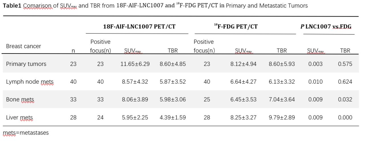
Results: In the 18F-AlF-LNC1007and 18F-FDG PET/CT groups,the18F-AlF-LNC1007 PET/CT group had higher SUVmax levels of primary tumors, lymph nodes and bone metastases than those of 18F-FDG group(P≤0.01), the TBR of bone metastasis was lower than 18F-FDG (5.98±3.06 vs. 7.04±3.64,P=0.032). The SUVmax and TBR in liver metastasis were higher in 18F-FDG (P <0.01)(Table1).
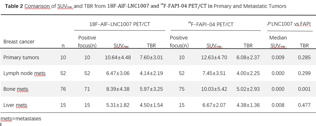
In the18F-AlF-LNC1007 and 18F-FAPI-04 PET/CT groups,the 18F-FAPI-04 PET/CT group had higher SUVmax levels of primary tumors, lymph nodes and bone metastases than those of 18F-AlF-LNC1007 (P≤0.01), the TBR of bone metastasis was lower than 18F-AlF-LNC1007 PET/CT (5.02±2.93 vs.5.97±3.25,P=0.032), and there was no significant difference among other lesions (all P >0.05) (Table2).
In bone metastasis with MTV less than 1, the SUVmax value was 8.73±5.04 (18F-FAPI-04), 6.38±2.69 (18F-AlF-LNC1007) and 4.42±1.59 (18F-FDG) from high to low (P=0.006), respectively. Moreover, in these small bone metastases, there was a moderate to strong correlation between SUVmax and MTV in the 18F-FDG group (r=0.605,P=0.008), and a weak correlation in the [18F]AlF-LNC1007 group (r=0.304,P=0.011), no correlation in the 18F-FAPI-04 group (r=0.187,P=0.171). In 18F-AlF-LNC1007 PET/CT group, there was a positive relationship between HER2 level and SUVmax (Beta=0.413,P=0.009). In 18F-FDG PET/CT group, PR and SUVmax had a reverse relationship (Beta=-0.830,P=0.017). The SUVmax cutoff values of 18F-AlF-LNC1007 and 18F-FDG PET/CT in diagnosis of lymph node metastasis were 2.62 and 3.90, respectively. Their AUC, Youden index, Sensitivity and Specificity were 0.986 and 0.790, 0.952 and 0.516, 95.2% and 66.67, 100% and 85.00(all P<0.01) respectively.
Conclusions: In the primary breast cancer, lymph nodes and bone metastases, all the uptake of 18F-FDG, 18F-AlF-LNC1007 and 18F-FDG decreased successively. In the liver metastases were 18F-FDG, 18F-FAPI-04, 18F-AlF-LNC1007 decreased successively. In the diagnosis of small bone metastases, the sensitivity of 18F-FDG, 18F-AlF-LNC1007, and 18F-FDG decreased successively. In the identification of metastatic lymph nodes, 18F-AlF-LNC1007 had better diagnostic efficacy than 18F-FDG.


In this issue


Jump to section
Related Articles
Cited By...
- No citing articles found.