


Abstract
241061
Introduction: Significantly increased sensitivity and high timing resolution of long axial field of view (LAFOV) PET scanners have enabled significant reductions in injected radiopharmaceuticals and sparked growing interest in ultra-low-dose PET imaging. Nevertheless, the advantages of low-dose PET examinations with such scanners can be hindered by the radiation dose linked to the CT scans conducted for PET attenuation correction. Recently, methods have been developed to utilize the background radiation from Lutetium 176 (Lu176) found in some PET scintillators as transmission source (LSO-TX) and combine this information with joint reconstruction- and deep learning-based methodologies to generate CT-free attenuation maps [1,2]. In this work, we evaluate the performance of LSO-TX-based attenuation correction methods in ultra-low dose PET scans using a LAFOV PET scanner where the patients were administered less than 10 MBq 18F-FDG.
Methods: Four subjects were administered a radiotracer dose of 7.9 ± 1.0 MBq (approximately 0.09 MBq/kg) and underwent a 90-minute-long PET scan, starting 60 minutes post-injection, using Biograph Vision Quadra (Siemens Healthineers) LAFOV PET scanner. The patients also received low-dose CT scans (voltage: 100 kV, reference tube current: 45 mA). LSO-TX data were also separately acquired for 5 minutes using a wide-energy window acquisition. LSO-TX based attenuation maps were generated using a combination of maximum likelihood for tomographic reconstruction (MLTR) and maximum-likelihood reconstruction of attenuation and activity (MLAA) based reconstruction methods and were enhanced using a deep learning-based method [1,2]. No ultra-low-dose datasets were included in the training of the deep learning model. CT-based and LSO-transmission-based attenuation maps were generated and used to reconstruct PET images. PET images were reconstructed using PSF-TOF algorithm with 4 iterations and 5 subsets and a 2 mm FWHM Gaussian filter. Percentage non-absolute and absolute relative change (%RC) maps were calculated using CT-based attenuation maps and PET images reconstructed with CT-based attenuation maps as reference images. Mean %RC values in different tissue types were reported. Furthermore, percentage SUV differences in ultra-low-dose PET images reconstructed with CT-based and LSO-TX-based attenuation maps are presented.
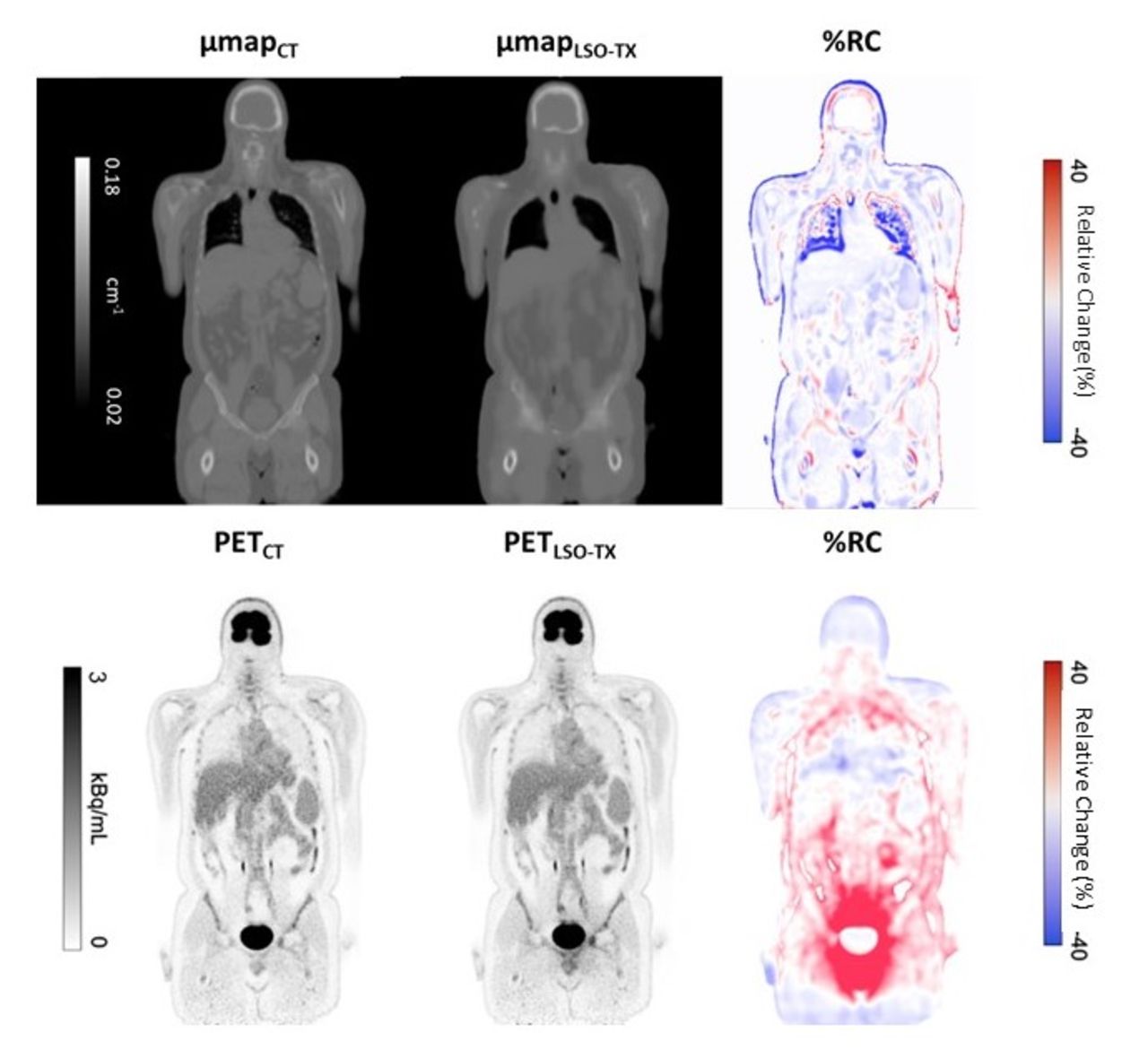
Results: The average effective dose received by the subjects was 0.153 ± 0.017 mSv for PET scans and 0.340 ± 0.104 mSv for CT scans. Figure 1 shows attenuation maps generated using CT-, and LSO-TX methods with their corresponding %RC map for a representative subject. PET images reconstructed using each attenuation map with the %RC map is also shown in the same figure. A very close visual resemblance were observed between CT- and LSO-TX-based attenuation maps as well as between PET images reconstructed with these attenuation maps. For the attenuation maps, the average %RC was 0.4 ± 1.1% for water-based soft tissue, -2.5 ± 0.8% for fat-based soft tissue and -5.3 ± 1.8% for bones. For the reconstructed PET images, the average %RC was -0.2 ± 1.8% for water-based soft tissue, -1.8±0.6% for fat-based soft tissue and -3.2 ± 1.3 for bones. The average differences in organ SUV values were -3.0 ± 1.9% in cerebral gray matter, -1.2 ± 3.3% in cerebral white matter, 3.3 ± 4.4% in liver, -4.5 ± 3.0% in the descending aorta and 2.4 ± 4.2% in spleen.
Conclusions: Results of this preliminary work indicate that an LSO-TX based attenuation correction method can be used to eliminate the radiation dose introduced by CT scans if they are solely performed for PET data corrections in ultra-low-dose PET examinations. Using LSO-TX-based attenuation correction, the total effective dose was reduced to ~0.15 mSV in these scans while preserving good quantitative accuracy.

In this issue


Jump to section
Related Articles
Cited By...
- No citing articles found.