


Article Figures & Data
Figures
- FIGURE 1.
Example of melanin-binding molecules, showing chemical interactions between benzopyrazine, picolinamide, and benzamide derivatives and melanin fragment.
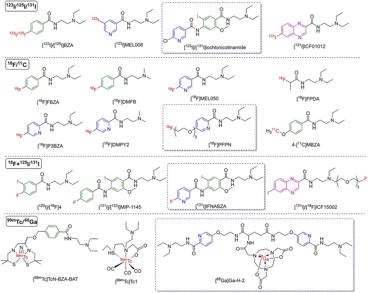
- FIGURE 2.
Structures of representative melanin-targeting molecules for nuclear medicine imaging. Isotopes used to label molecules are indicated in red. Benzene ring structure is shown in green. Nicotinamide and picolinamide ring structures are indicated in blue. Benzopyrazine is shown in peach. Molecules reported in Chinese studies are marked with blue boxes. [123I]MEL008 = N-(2-(diethylamino)ethyl)-5-[123I]iodonicotinamide; [125I]/[18F]4 = 125I- and 18F-labeled N-[2-(diethylamino)ethyl]-4-fluoro-3-iodobenzamide; [18F]DMFB = N-(2-(dimethylamino)ethyl)-4-[18F]fluorobenzamide; [18F]DMPY2 = N-(2-(dimethylamino)ethyl)-5-[18F]fluoropicolinamide; [18F]FPDA = N-(2-(diethylamino)ethyl)-2-[18F]fluoropropanamide; [18F]MEL050 = N-[2-(diethylamino)ethyl]-6-[18F]fluoronicotinamide; 4-[11C]MBZA = 4-11C-methoxy N-(2-diethylaminoethyl) benzamide; BZA-BAT = N-diethylaminoethyl-4-[8-methyl-3-(3-methyl-3-thio-1-azabutyl)-8-thio-2,6-oxoazanonyl]benzamide; ICF15002 = N-(12-ethyl-1-fluoro-3,6,9-trioxa-12-azatetradecan-14-yl)-6-iodoquinoxaline-2-carboxamide; IFNABZA = iodofluoronicotinamide benzamide.
- FIGURE 3.
Representative images of radiolabeled melanin-targeting molecules. PET/SPECT imaging shows mice bearing melanoma at different time points after injection of 18F-labeled tracers (A) (12,37,45), 125/131/123I-labeled tracers (B) (42,52,80), and 68Ga-labeled tracers (C) (60–62). Red arrows indicate tumors. [123I]MEL008 = N-(2(diethylamino)ethyl)-5-[123I]iodonicotinamide; [125I]40 = N-[2-[ethyl(4-fluorobut-2-ynyl)amino]ethyl]-6-[125I]iodoquinoxaline-2-carboxamide; [18F]DMPY2 = N-(2-(dimethylamino)ethyl)-5-[18F]fluoropicolinamide; 5-[131I]IPN = N-(2-(diethylamino)ethyl)-5-[131I]iodopicolinamide; PCA = procainamide. (Reprinted with permission of (12,45,52,60–62,80) and from (37,42).)
- FIGURE 4.
Representative images of [18F]PFPN and [18F]P3BZA PET in healthy volunteers and melanoma patients (72,73). Maximum-intensity projection (MIP) images were acquired with [18F]PFPN (A) and [18F]P3BZA (B) PET from healthy female volunteers. Moderate [18F]P3BZA uptake was observed in regional obsolete lymph nodes that showed calcification in chest. (C) Man who had surgical resection of choroidal melanoma underwent [18F]PFPN PET/MR and [18F]FDG PET/CT imaging. [18F]PFPN PET demonstrated capability to detect more lesions (indicated by arrowheads and arrows) than [18F]FDG PET. (D) MIP images of melanoma patient showed higher [18F]P3BZA uptake than [18F]FDG in melanoma lesions (arrows) at 60 min after injection. (Reprinted from (72,73).)
- FIGURE 5.
Representative [18F]PFPN PET and CT images of pigmented epithelial adenomas and clear cell sarcoma. (A) Patient who reported worsening vision and eye pain for more than 3 mo. MRI revealed 4.1 × 3.7 × 5.0 mm lesion in corpus ciliare with slightly reduced T1-weighted signals. [18F]PFPN showed high activity (SUVmax, 7.1), suggesting melanin expression. Subsequent pathology confirmed pigmented epithelial adenoma (74). (B) Patient with history of clear cell sarcoma surgery underwent [18F]PFPN PET scan for recurrence detection. [18F]PFPN PET found hepatic metastatic lesions < 1.0 cm missed with [18F]FDG (75). T1WI/FS = T1-weighted imaging/fat-suppressed; T2WI/FS = T2-weighted imaging/fat-suppressed. (Reprinted from (74,75).)
- FIGURE 6.
(A) Scheme of tetracycline-controlled transactivator tyrosinase (TYR) reporter system and molecular imaging applications. [18F]P3BZA PET (B), MRI (C), and photoacoustic (D) 3-modality imaging was used to monitor survival, distribution, and function duration of transfected stem cells in myocardial infarction area (14). Dopa = 3,4-dihydroxyphenylalanine; Dox = doxycycline; MSCs = mesenchymal stem cells; PDT = photodynamic therapy; PTT = photothermal therapy; TetIIP = tetracycline-inducible promoter; Ubi-TetR = ubiquitin promoter–tetracycline repressor. (Reprinted with permission of (14).)


Tables
Representative probe LogP Radiochemical yield (%) Radiochemical purity (%) Specific activity (GBq/μmol) Time point (h)* Highest tumor uptake (%ID/g) Reference [125I]BZA — 45 >95 1.33 × 10⁻³ 1 6.75 ± 0.67 (26) [125I]11 — 85–95 — — 1 5.21 ± 0.51 (33) [123I]IMBA 1.419 80–98 — — 1 6.7 (34) [125I]PAB — 78–94 — 66.6 1 3.87 ± 0.47 (32) [123I]MEL008 1.55 ± 0.03 30–80 >95 >2,000 1 7.8 ± 1.7 (42) 5-[131I]IPN 0.03 ± 0.03 55 ± 5 >98 5.45–6.55 1 16.37 ± 3.32 (80) [18F]FBZA 1.7† 50 >95 132–166 1 6.47 ± 2.16 (37) [18F]DMFB — 15–30 — >5.5 1 13.00 ± 3.90 (13) [18F]AlF-NOTA-BZA −1.96 20–35 >95 60–80 0.25 6.67 ± 0.91 (39) 4-[18F]FEBZA — 53 ± 14 >99 321.9 ± 40.7 1 8.66 ± 1.02 (38) [18F]FPBZA −0.48 40–50 >97 30–40 0.5 10.37 ± 1.13 (40) [18F]MEL050 1.1 35–45 >99 150–220 2 9.4 ± 1.6 (43) [18F]P3BZA −0.17 ± 0.03‡ 9.5 ± 1.9 >95 100–150 2 16.97 ± 3.28 (45) [18F]DMPY2 — 15–20 — >7.6 1 24.86 ± 2.30 (12) [18F]PFPN −0.69 ± 0.02 44.68 ± 5.99 >99 120–195 1 19.52 ± 1.69 (48) [18F]FPDA — 79.8 ± 13.5 >99 20–40 1 4.39 ± 0.51 (41) [18F]4 — 5 >99 8–14 1 6.77 ± 1.90 (50) [125I]4 — 73 >99 4–12 1 9.7 ± 2.6 (50) [18F]ICF15002 — 21 >99 45–53 1 14.05 ± 1.42 (51) [125I]40 — 8 96.3 5.2 × 10⁻³ 24 23.5 ± 1.4 (52) [99mTc]TcN-BZA-BAT§ −0.3 >40 >92 — 1 0.43 ± 0.16 (54) [99mTc]TcO-Cf 0.64 >95 — — 3 1.34 ± 0.46 (55) [99mTc]Tc-12 — 70 — — 6 4.35 ± 0.92 (56) [99mTc]4b — 28.4 99 — 1 3.39 ± 0.5 (57) [99mTc]2 3.5 60–98 — — 1 4.95 ± 1 (59) [99mTc]Tc1 0.53 ± 0.01 >95 — — 1 2.17 ± 0.42 (58) [68Ga]Ga-SCN-NOTA-BZA −3.25 ± 0.05 80–85 >97 10 1 1.57 ± 0.66 (61) [68Ga]Ga-SCN-DOTA-PCA −3.7 >80 >97 8.9 1 2.51 ± 0.5 (60) [68Ga]Ga-H-2 −2.37 ± 0.07 98 ± 2 >95 4.32 0.5 4.19 ± 1.79 (62) ↵* Time point (h) of highest tumor uptake.
↵† Values were obtained from Garg et al. (36).
↵‡ Values were obtained from Xu et al. (48).
↵§ The syn isomer.
— = no value mentioned in article; [125I]11 = N-(2-ethylaminoethyl)-4-[125I]iodobenzamide; [123I]IMBA = N-(2-diethylaminoethyl)-3-[123I]iodo-4-methoxybenzamide; [125I]PAB = N-(2-(piperidin-1-yl)ethyl)-4-[125I]iodobenzamide; [123I]MEL008 = N-(2-(diethylamino)ethyl)-5-[123I]iodonicotinamide; 5-[131I]IPN = N-(2-(diethylamino)ethyl)-5-[131I]iodopicolinamide; [18F]DMFB = N-(2-(dimethylamino)ethyl)-4-[18F]fluorobenzamide; 4-[18F]FEBZA = N-(2-diethylaminoethyl)-4-[18F]fluoroethoxybenzamide; [18F]FPBZA = N-(2-diethylaminoethyl)-4-[2-(2-(2-[18F]fluoroethoxy)ethoxy)ethoxy]benzamide; [18F]MEL050 = N-[2-(diethylamino)ethyl]-6-[18F]fluoronicotinamide; [18F]DMPY2 = N-(2-(dimethylamino)ethyl)-5-[18F]fluoropicolinamide; [18F]FPDA = N-(2-(diethylamino)ethyl)-2-[18F]fluoropropanamide; [18F]4 = 18F-labeled N-[2-(diethylamino)ethyl]-4-fluoro-3-iodobenzamide; [125I]4 = 125I-labeled N-[2-(diethylamino)ethyl]-4-fluoro-3-iodobenzamide; ICF15002 = N-(12-ethyl-1-fluoro-3,6,9-trioxa-12-azatetradecan-14-yl)-6-iodoquinoxaline-2-carboxamide; [125I]40 = N-[2-[ethyl(4-fluorobut-2-ynyl)amino]ethyl]-6-[125I]iodoquinoxaline-2-carboxamide; BZA-BAT = N-diethylaminoethyl-4-[8-methyl-3-(3-methyl-3-thio-1-azabutyl)-8-thio-2,6-oxoazanonyl]benzamide; PCA = procainamide.
Detailed information and structures of molecules are shown in Supplemental Fig. 1 and Supplemental Table 1.
Year Phase, NCT no. Purpose Tracer Dose Time point Population Patients (n) Country Reference 1993, 1998 Phase II DEE [123I]BZA 130 MBq 20–24 h Cutaneous and ocular melanoma, ocular melanoma 110, 48 France (68,69) 1994 — DEE (S)-[123I]IBZM 205 MBq 2, 4, and 24 h Melanoma 11 Italy (64) 1997 — DEE [123I]IMBA 200–300 MBq 2, 4, and 22 h Diagnosed or suspected metastatic melanoma 3 Germany (34) 1997 — DEE [123I]BZA 170 MBq 4–5 h Suspected ocular melanoma and ocular naevi 14 Belgium (65) 1998 — * [123I]IBZM 185 MBq 2–5 h and 1 d Melanoma 21 Germany (66) 1998 — DEE [123I]/[131I]MTB 135–560 MBq 1–120 h Melanoma 11 U.K. (67) 2002, 2004 Phase II DEE [123I]BZA2 130–185 MBq 2, 4, and 6 h Cutaneous melanoma, ocular melanoma 25, 40 France (31,70) 2013 Phase III DEE [123I]BZA2 2 MBq/kg 4 h Cutaneous and ocular melanoma 87 France (71) 2019 IIT, NCT03033485 Safety and dosimetry evaluation, DEE [18F]P3BZA 3.1 MBq/kg 10 min and 1 h Healthy volunteers, patients with suspected melanoma 6, 5 China (72) 2022 IIT, NCT04747561 Safety and dosimetry evaluation, DEE [18F]PFPN 3.0–5.4 MBq/kg 1 and 3 h Healthy volunteers, patients with suspected or confirmed melanoma 5, 21 China (73) 2023 IIT, NCT05645484 Prognosis evaluation [18F]PFPN 3.0–5.4 MBq/kg 2–3 h Melanoma 76 China (47) 2023 IIT, NCT05963035 DEE [18F]PFPN 3.0–5.4 MBq/kg — Clear cell sarcoma 3 China (75) 2013 Phase Ia and Ib, NCT00399113 and NCT00734188 Safety and dosimetry evaluation, TEE [188Re]Re-6D2 370 MBq, 740–2,220 MBq† — Stage III or IV metastatic melanoma 13 and 7† Israel (88) 2014 — Safety and dosimetry evaluation, TEE [123I]/[131I]BA52 235 ± 62 MBq, 3,900 ± 2,000 MBq‡ 10 min; 4, 24, and 48 h§ Metastatic, treatment-resistant melanoma 26, 9 for therapy Germany (27) ↵* Aim of study was to examine mechanisms of IBZM accumulation clinically and histologically.
↵† 13 patients were enrolled in phase Ia study, and 7 patients were enrolled in phase Ib study.
↵‡ Diagnostic dose, therapeutic dose.
↵§ Imaging time point of [123I]BA52.
NCT = National Clinical Trial; DEE = diagnosis efficiency evaluation; — = not available; IBZM = 2-hydroxy-3-iodo-6-methoxy-N-[(1-ethyl-2-pyrrolidinyl)methyl]benzamide; [123I]IMBA = N-(2-diethylaminoethyl)-3-[123I]iodo-4-methoxybenzamide; MTB = methylene blue; [123I]BZA2 = N-(2-diethylaminoethyl)-2-[123I]iodobenzamide; IIT = investigator-initiated trial; TEE = therapy efficacy evaluation; [123I]/[131I]BA52 = benzo(1,3)dioxolo-5-carboxylicacid(4-(2-diethylaminoethylcarbamoyl)-2-[123I]/[131I]iodo-5-methoxyphenyl)amide.
Supplemental Data
Files in this Data Supplement:


