


Article Figures & Data
Figures
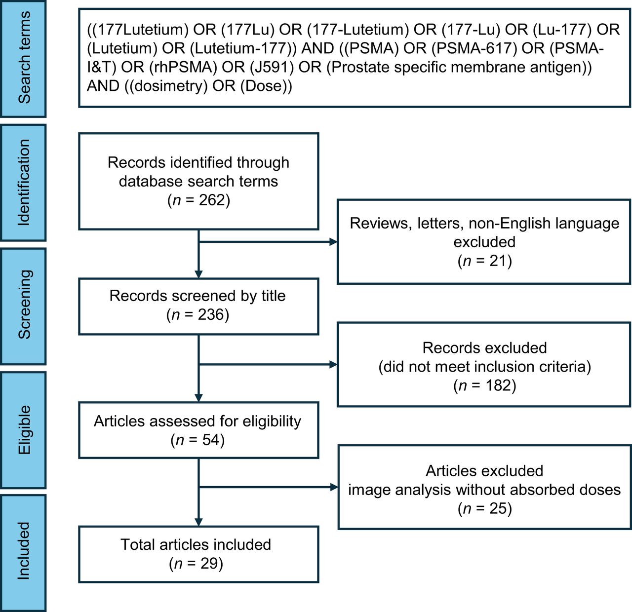
- FIGURE 1.
Study selection for metaanalysis. Using PRISMA criteria, we selected 29 studies for this metaanalysis. Only conference abstracts or fully published articles that were in English and reported absorbed dose to organs at risk or tumors were included.
- FIGURE 2.
Reported absorbed doses (Gy/cycle) to kidney from selected studies, allowing for computation of pooled average absorbed dose. *As reported in VISION trial. †As reported in SPLASH trial.
- FIGURE 3.
Reported absorbed doses (Gy/cycle) to tumor lesions from selected studies that were used to compute pooled average absorbed dose. Individual study estimates: gray box with black bar, each study's effect estimate (mean) is represented by gray box, with size of box proportional to study's weight in metaanalysis; black bar through box indicates CI for estimate; white cross in gray box, studies with very precise estimates; white cross indicates point estimate (mean), with gray box showing narrow CI; pooled estimate (diamond): Diamond Shape: The pooled estimate, or overall effect estimate, of the meta-analysis is shown as a diamond at the bottom of the plot. Width of the Diamond: The width of the diamond represents the confidence interval for the pooled estimate. The left and right tips of the diamond correspond to the lower and upper bounds of the CI, respectively. Center of the Diamond: The center of the diamond represents the overall effect estimate (mean) calculated from combining the individual study estimates. *Lesion location reported as soft tissue. †Lesion location reported as visceral tissue. ‡Lesion location reported within liver. §Lesion location reported within lung. ‖Tumor dosimetry reported from VISION trial.


Tables
Author Isotope Injected activity (GBq) Kidney (Gy/GBq) Parotid (Gy/GBq) Submandibular (Gy/GBq) Marrow (Gy/GBq) Liver (Gy/GBq) Lacrimal (Gy/GBq) Kabasakal (39) 617 0.2 0.88 ± 0.4 1.17 ± 0.31 – 0.03 ± 0.01 0.28 ± 0.09 – Delker (15) 617 3.6 0.6 ± 0.18 1.4 ± 0.53 – 0.01 ± 0.01 0.11 ± 0.06 – Fendler (10) 617 3.7 0.55 ± 0.25 1 ± 0.6 – 0.1 < 0.1 – Hohberg (40) 617 5.5 0.53 ± 0.17 0.72 ± 0.14 – – – 2.82 ± 0.76 Kratochwil (33) 617 3.0 0.75 ± 0.19 1.28 ± 0.4 1.48 ± 0.37 0.03 ± 0.01 – – Yadav (12) 617 2.5 0.99 ± 0.31 1.24 ± 0.27 – 0.05 ± 0.06 0.36 ± 0.11 – Scarpa (19) 617 6.1 0.6 ± 0.36 0.56 ± 0.25 0.5 ± 0.15 0.04 ± 0.03 – 1.01 ± 0.69 Gosewisch (41) 617 3.7 – – – 0.01 (0.01–0.02) – – Gosewisch (42) 617 5.2 – – – 0.012 – – Sarnelli (43) 617 5.0 0.67 ± 0.27 0.81 ± 0.74 – 0.04 ± 0.02 0.16 ± 0.15 – Violet (20) 617 7.8 0.39 ± 0.15 0.58 ± 0.43 0.44 ± 0.36 0.11 ± 0.1 0.1 ± 0.05 0.36 ± 0.18 Paganelli (44) 617 4.4 0.41 ± 0.19 1.04 ± 0.82 0.67 ± 0.36 0.04 ± 0.02 0.18 ± 0.14 2.06 ± 1.24 Mix (34) 617 6.0 0.67 ± 0.24 – – – – – Privé/Peters (8,9) 617 3.0 0.49 ± 0.11 0.39 ± 0.17 – 0.02 ± 0.01 0.09 ± 0.01 – Rosar (11) 617 6.4 0.54 ± 0.28 0.81 ± 0.34 0.72 ± 0.39 – 0.1 ± 0.05 – Völter (45) 617 6.0 – – – – – – Kamaldeep (16) 617 4.4 0.49 ± 0.17 0.53 ± 0.2 – 0.03 ± 0.02 0.07 ± 0.04 1.23 ± 0.7 Schuchardt (46) 617 6.5 0.8 0.5 – – – 5.1 Herrmann/Krause (47,48) 617 7.4 0.43 ± 0.16 0.63 ± 0.36 – 0.04 ± 0.02 – 2.1 ± 0.47 Uijen (22) 617 3.0 0.49 (0.34–0.66) – – – – – Okamoto (35) I&T 7.4 0.72 ± 0.21 0.55 ± 0.14 0.64 ± 0.4 – 0.12 ± 0.06 3.8 ± 1.4 Baum (17) I&T 5.8 0.8 (0.2–1.9) 1.3 (0.3–9.5) – 0.03 (0.01–0.04) – – Barna (13) I&T 7.4 0.71 ± 0.24 0.77 – – 0.27 – Chatachot (14) I&T 6.7 0.81 ± 0.24 0.21 ± 0.14 – 0.02 ± 0.01 0.13 ± 0.10 3.62 ± 1.78 Schuchardt (46) I&T 6.1 0.9 0.5 – – – 3.7 Kelk (49) I&T 7.4 0.305 0.11 0.24 – 0.03 0.8 Feuerecker (18) I&T 7.3 0.73 ± 0.18 0.8 ± 0.41 – 0.28 ± 0.2 0.07 ± 0.03 – Beauregard (50) I&T 6.8 0.73 ± 0.33 0.34 ± 0.27 – 0.03 ± 0.02 0.05 ± 0.04 1.2 ± 1.2 Uijen (22) I&T 7.4 0.73 (0.42–1.31) – – – – – Resch (51) I&T 7.4 2 (1.2–2.4) – – – – – Hohberg (21) I&T 7.2 0.53 ± 0.21 – – – – – Bander/Vallabhajosula (27,52) J591 2.8 1.41 ± 0.35 – – 0.32 ± 0.01 2.1 ± 0.6 – Data are reported as mean ± SD or as median followed by range in parentheses.
Author Isotope Injected activity (GBq) Unspecified/single-study exploration (Gy/GBq) Skeletal lesion (Gy/GBq) Nodal lesion (Gy/GBq) Liver lesion (Gy/GBq) Delker (15) 617 3.6 2.1 ± 0.8* 5.3 ± 3.7 4.2 ± 5.3 – Fendler (10) 617 3.7 2.16 ± 0.85* 4.92 ± 3.54 11.64 ± 5.44 – Scarpa (19) 617 6.1 – 3.4 ± 1.9 2.6 ± 0.4 2.4 ± 0.8 Violet (20) 617 7.8 – 5.28 ± 2.46 3.91 ± 3.93 – Paganelli (44) 617 4.4 – 4.7 (0.74–55.86) 3.64 (0.25–15.10) – Privé/Peters (8,9) 617 3.0 3.25 ± 3.19 1.1 (0.3–3.1) 3.1 (0.6–13) – Rosar (11) 617 6.4 – 1.68 ± 1.32 – – Volter (45) 617 6.0 – 4.7 ± 3.9 7.7 ± 9.7 – Schuchardt (46) 617 6.5 – 6 7.1 – Herrmann/Krause (47,48) 617 7.4 – 14.6 ± 29.8 12.5 ± 15.9 – Okamoto (35) I&T 7.4 1.75 ± 0.92† 3.4 ± 2.7 3.2 ± 2.2 1.2 ± 0.67 Baum (17) I&T 5.8 – 3 (0.2–40) 4 (0.14–78) – Barna (13) I&T 7.4 – 4.38 5.47 4.95 Schuchardt (46) I&T 6.1 – 5.9 6.9 – Feuerecker (18) I&T 7.3 – 1.7 ± 1.13 4.51 ± 2.69 – Hohberg (21) I&T 7.2 – 3.47 ± 2 3.73 ± 1.65 – Resch (51) I&T 7.5 – 5.8 ± 3.1 7.7 ± 4.5 – 617 I&T J591 Organ or group Gy/GBq Gy/7.4-GBq cycle Gy/GBq Gy/6.8-GBq cycle Gy/GBq Gy/5.6-GBq cycle 617 vs. I&T P Overall P Kidney 0.58 4.04 0.71 4.70 1.41 3.95 0.10 0.06 Parotid 0.84 5.85 0.43 2.62 – – <0.01 – Submandibular 0.74 5.15 0.64 4.35 – – 0.56 – Bone marrow 0.03 0.24 0.03 0.19 0.32 0.90 0.31 <0.01 Liver 0.16 1.11 0.09 0.56 2.10 5.88 0.05 <0.01 Lacrimal glands 1.58 11.03 2.83 19.23 – – 0.20 – Tumor lesion, bone only 3.57 26.43 4.10 27.87 – – 0.38 – Tumor lesion, soft-tissue only 4.19 31.00 2.94 19.98 – – 0.23 – Data are summary of pooled doses for organs at risk and tumor lesions from different 177Lu-based molecules shown as average (CI can be seen for kidney and tumors in Figs. 2 and 3 and for rest of organs in Supplemental Figs. 1–5).
Supplemental Data
Files in this Data Supplement:


