
Main menu

  • Home
  • Content
    • Current
    • Ahead of print
    • Past Issues
    • JNM Supplement
    • SNMMI Annual Meeting Abstracts
    • Continuing Education
    • JNM Podcasts
  • Subscriptions
    • Subscribers
    • Institutional and Non-member
    • Rates
    • Journal Claims
    • Corporate & Special Sales
  • Authors
    • Submit to JNM
    • Information for Authors
    • Assignment of Copyright
    • AQARA requirements
  • Info
    • Reviewers
    • Permissions
    • Advertisers
  • About
    • About Us
    • Editorial Board
    • Contact Information
  • More
    • Alerts
    • Feedback
    • Help
    • SNMMI Journals
  • SNMMI
    • JNM
    • JNMT
    • SNMMI Journals
    • SNMMI

User menu

  • Subscribe
  • My alerts
  • Log in
  • My Cart

Search

  • Advanced search
Journal of Nuclear Medicine
  • SNMMI
    • JNM
    • JNMT
    • SNMMI Journals
    • SNMMI
  • Subscribe
  • My alerts
  • Log in
  • My Cart
Journal of Nuclear Medicine

Advanced Search

  • Home
  • Content
    • Current
    • Ahead of print
    • Past Issues
    • JNM Supplement
    • SNMMI Annual Meeting Abstracts
    • Continuing Education
    • JNM Podcasts
  • Subscriptions
    • Subscribers
    • Institutional and Non-member
    • Rates
    • Journal Claims
    • Corporate & Special Sales
  • Authors
    • Submit to JNM
    • Information for Authors
    • Assignment of Copyright
    • AQARA requirements
  • Info
    • Reviewers
    • Permissions
    • Advertisers
  • About
    • About Us
    • Editorial Board
    • Contact Information
  • More
    • Alerts
    • Feedback
    • Help
    • SNMMI Journals
  • View or Listen to JNM Podcast
  • Visit JNM on Facebook
  • Join JNM on LinkedIn
  • Follow JNM on Twitter
  • Subscribe to our RSS feeds
Research ArticleClinical Investigation

Detecting High-Dose Methotrexate–Induced Brain Changes in Pediatric and Young Adult Cancer Survivors Using [18F]FDG PET/MRI: A Pilot Study

Lucia Baratto, Shashi B. Singh, Sharon E. Williams, Sheri L. Spunt, Jarrett Rosenberg, Lisa Adams, Vidyani Suryadevara, Michael Iv and Heike Daldrup-Link
Journal of Nuclear Medicine June 2024, 65 (6) 864-871; DOI: https://doi.org/10.2967/jnumed.123.266760
Lucia Baratto
1Division of Pediatric Radiology, Department of Radiology, Stanford University School of Medicine, Stanford, California;
  • Find this author on Google Scholar
  • Find this author on PubMed
  • Search for this author on this site
Shashi B. Singh
1Division of Pediatric Radiology, Department of Radiology, Stanford University School of Medicine, Stanford, California;
  • Find this author on Google Scholar
  • Find this author on PubMed
  • Search for this author on this site
Sharon E. Williams
2Child and Adolescent Psychiatry Clinic, Department of Psychiatry and Behavioral Sciences–Child and Adolescent Psychiatry and Child Development, Stanford University, Stanford, California;
  • Find this author on Google Scholar
  • Find this author on PubMed
  • Search for this author on this site
Sheri L. Spunt
3Department of Pediatrics–Hematology/Oncology, Lucile Packard Children’s Hospital, Stanford University, Stanford, California;
  • Find this author on Google Scholar
  • Find this author on PubMed
  • Search for this author on this site
Jarrett Rosenberg
4Department of Radiology, Stanford University School of Medicine, Stanford University, Stanford, California; and
  • Find this author on Google Scholar
  • Find this author on PubMed
  • Search for this author on this site
Lisa Adams
1Division of Pediatric Radiology, Department of Radiology, Stanford University School of Medicine, Stanford, California;
  • Find this author on Google Scholar
  • Find this author on PubMed
  • Search for this author on this site
Vidyani Suryadevara
1Division of Pediatric Radiology, Department of Radiology, Stanford University School of Medicine, Stanford, California;
  • Find this author on Google Scholar
  • Find this author on PubMed
  • Search for this author on this site
Michael Iv
5Neuroimaging Division, Radiology Department, Stanford Health Care, Stanford University, Stanford, California
  • Find this author on Google Scholar
  • Find this author on PubMed
  • Search for this author on this site
Heike Daldrup-Link
1Division of Pediatric Radiology, Department of Radiology, Stanford University School of Medicine, Stanford, California;
3Department of Pediatrics–Hematology/Oncology, Lucile Packard Children’s Hospital, Stanford University, Stanford, California;
  • Find this author on Google Scholar
  • Find this author on PubMed
  • Search for this author on this site
  • Article
  • Figures & Data
  • Supplemental
  • Info & Metrics
  • PDF
Loading

Article Figures & Data

Figures

  • Tables
  • Additional Files
  • Figure
    • Download figure
    • Open in new tab
    • Download powerpoint
  • FIGURE 1.
    • Download figure
    • Open in new tab
    • Download powerpoint
    FIGURE 1.

    Flowchart showing selection of study participants.

  • FIGURE 2.
    • Download figure
    • Open in new tab
    • Download powerpoint
    FIGURE 2.

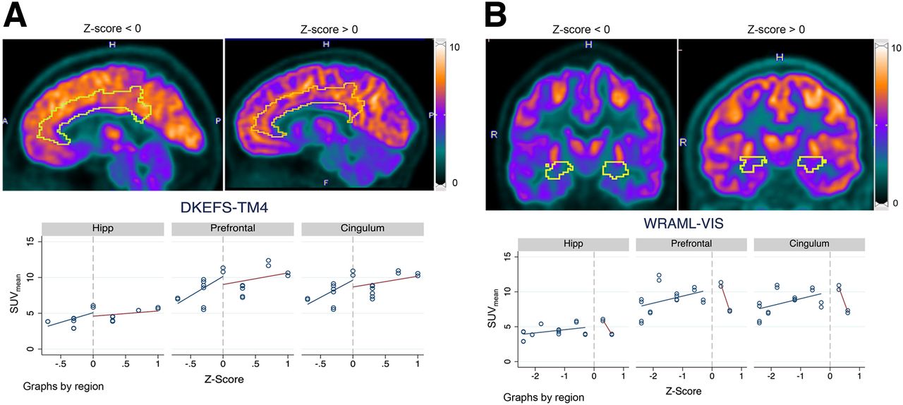
    Comparison of [18F]FDG uptake in cingulum and hippocampus (Hipp) for patients with low (z score < 0) vs. high (z score > 0) performance on DKEFS-TM4 (A) and WRAML-VIS (B), respectively. (A) Regression analysis showed significant correlation between SUVmean and DKEFS-TM4 for prefrontal cortex and cingulum (P = 0.003 and P = 0.012, respectively) but not for hippocampus (P = 0.111). (B) There was no significant correlation between SUVmean and WRAML-VIS for prefrontal cortex, cingulum, or hippocampus.

  • FIGURE 3.
    • Download figure
    • Open in new tab
    • Download powerpoint
    FIGURE 3.

    Comparison of CBFmean in cingulum and hippocampus (Hipp) for patients with low (z score < 0) vs. high (z score > 0) performance on DKEFS-TM4 (Fig. 3A) and WRAML-VIS (Fig. 3B), respectively. (A) Regression analysis showed significant correlation between CBFmean and DKEFS-TM4 for cingulum, hippocampus, and prefrontal cortex (all P < 0.001). (B) CBFmean for hippocampus and WRAML-VIS was inversely correlated (P = 0.003). However, there was no correlation between CBFmean and WRAML-VIS for cingulum (P = 0.071) and prefrontal cortex (P = 0.052).

Tables

  • Figures
  • Additional Files
    • View popup
    TABLE 1.

    Brain [18F]FDG PET/MRI Parameters

    ModalityParameterSpecification
    [18F]FDG PETImage protocol30-min static acquisition
    Image planeAxial
    Slice thickness (mm)2.78
    Field of view (cm)60
    Matrix size (mm)192 × 192
    Reconstruction algorithmTime of flight (28 subsets, 8 iterations)
    Glucose uptake (mg/dL)85.9 ± 10.16 (mean ± SD)
    Dose (MBq/kg)176.49 ± 42.55 (mean ± SD)
    Uptake time (min)43.50 ± 6.67 (mean ± SD)
    MRI*3D inversion recovery fast SPGR
     Image planeAxial
     Slice thickness (mm)1
     Field of view (cm)27
     Matrix size (mm)256 × 256
     Echo time (ms)3.1
     Repetition time (ms)7,664
     Flip angle11°
     Number of excitations1.00
    2D diffusion-weighted imaging†
     Image planeAxial
     Slice thickness (mm)5
     Field of view (cm)24
     Matrix size (mm)128 × 128
     Echo time (ms)76.5
     Repetition time (ms)5,000
     Flip angle90°
     Number of excitations3.00
    3D arterial spin labeling†
     Image planeAxial
     Slice thickness (mm)4
     Field of view (cm)24
     Matrix size (mm)512 × 8
     Echo time (ms)10.7
     Repetition time (ms)4,854
     Flip angle111°
     Number of excitations3.00
    • ↵* 3D T2 fluid-attenuated inversion recovery and 3D multiple-echo gradient-echo (QSM/R2*) sequences were also acquired.

    • ↵† Diffusion-weighted images were acquired with 2 diffusion weightings (b = 0 and 1,000 s/mm2). Apparent diffusion coefficient maps were automatically generated by software. Postprocessed arterial spin labeling imaging was performed by automated reconstruction script that sent CBF images directly to PACS.

    • View popup
    TABLE 2.

    Neurocognitive Tests

    FunctionMeasureAverage rangeAnatomic regionBroadman area
    Executive functionDKEFS10 ± 3Prefrontal cortexMiddle frontal gyrus and gyrus rectus (9 and 10/11)
    CingulumAnterior cingulate gyrus (24, 32, 33); posterior cingulate gyrus (23, 26, 29, 30, and 31)
    Intellectual quotientWASI100 ± 10Prefrontal cortex9 and 10/11
    Verbal and nonverbal memoryWRAML100 ± 10Hippocampus27, 28, 34, 35, 36, and 48
    • Each neurocognitive test comprised 2 or more battery sets: Delis Kaplan Executive Function System (DKEFS)-sequential tracking (TM4), DKEFS–design/nonverbal tasks, DKEFS–inhibition, Wechsler Abbreviated Scale of Intelligence (WASI)–full-scale intellectual quotient, WASI–verbal comprehension index, WASI–perceptual reasoning index, Wide Range Assessment of Memory and Learning (WRAML)–screening memory, WRMAL–verbal memory, and WRMAL-VIS.

    • View popup
    TABLE 3.

    Model Estimates of Marginal Effects of Negative z Score on Imaging Measures

    HippocampusCingulumPrefrontal cortex
    Neurocognitive testPET/MRI measureSlopePSlopePSlopeP
    WRAML-VIS (n = 16)SUVmean0.460.2851.020.1431.010.173
    CBFmean−6.040.003−5.970.071−5.930.052
    DKEFS-TM4 (n = 10)SUVmean2.700.1114.820.0125.410.003
    CBFmean8.68<0.00119.6<0.00110.9<0.001
    • n = number of measurements.

Additional Files

  • Figures
  • Tables
  • Supplemental Data

    Files in this Data Supplement:

    • Supplemental Data
PreviousNext
Back to top

In this issue

Journal of Nuclear Medicine: 65 (6)
Journal of Nuclear Medicine
Vol. 65, Issue 6
June 1, 2024
  • Table of Contents
  • Table of Contents (PDF)
  • About the Cover
  • Index by author
  • Complete Issue (PDF)
Print
Download PDF
Article Alerts
Sign In to Email Alerts with your Email Address
Email Article

Thank you for your interest in spreading the word on Journal of Nuclear Medicine.

NOTE: We only request your email address so that the person you are recommending the page to knows that you wanted them to see it, and that it is not junk mail. We do not capture any email address.

Enter multiple addresses on separate lines or separate them with commas.
Detecting High-Dose Methotrexate–Induced Brain Changes in Pediatric and Young Adult Cancer Survivors Using [18F]FDG PET/MRI: A Pilot Study
(Your Name) has sent you a message from Journal of Nuclear Medicine
(Your Name) thought you would like to see the Journal of Nuclear Medicine web site.
Citation Tools
Detecting High-Dose Methotrexate–Induced Brain Changes in Pediatric and Young Adult Cancer Survivors Using [18F]FDG PET/MRI: A Pilot Study
Lucia Baratto, Shashi B. Singh, Sharon E. Williams, Sheri L. Spunt, Jarrett Rosenberg, Lisa Adams, Vidyani Suryadevara, Michael Iv, Heike Daldrup-Link
Journal of Nuclear Medicine Jun 2024, 65 (6) 864-871; DOI: 10.2967/jnumed.123.266760

Citation Manager Formats

  • BibTeX
  • Bookends
  • EasyBib
  • EndNote (tagged)
  • EndNote 8 (xml)
  • Medlars
  • Mendeley
  • Papers
  • RefWorks Tagged
  • Ref Manager
  • RIS
  • Zotero
Share
Detecting High-Dose Methotrexate–Induced Brain Changes in Pediatric and Young Adult Cancer Survivors Using [18F]FDG PET/MRI: A Pilot Study
Lucia Baratto, Shashi B. Singh, Sharon E. Williams, Sheri L. Spunt, Jarrett Rosenberg, Lisa Adams, Vidyani Suryadevara, Michael Iv, Heike Daldrup-Link
Journal of Nuclear Medicine Jun 2024, 65 (6) 864-871; DOI: 10.2967/jnumed.123.266760
Twitter logo Facebook logo LinkedIn logo Mendeley logo
  • Tweet Widget
  • Facebook Like
  • Google Plus One
Bookmark this article

Jump to section

  • Article
    • Visual Abstract
    • Abstract
    • MATERIALS AND METHODS
    • RESULTS
    • DISCUSSION
    • CONCLUSION
    • DISCLOSURE
    • Footnotes
    • REFERENCES
  • Figures & Data
  • Supplemental
  • Info & Metrics
  • PDF

Related Articles

  • PubMed
  • Google Scholar

Cited By...

  • No citing articles found.
  • Google Scholar

More in this TOC Section

  • SNMMI Procedure Standard/EANM Practice Guideline for Brain [18F]FDG PET Imaging, Version 2.0
  • Meeting Upcoming Clinical and Diagnostic Needs in Oncologic Imaging: A Structured Reporting System for Fibroblast-Activation-Protein–Targeted Imaging—FAP-RADS Version 1.0
  • Imaging Efficacy of [18F]CTT1057 PET for the Detection of PSMA-Positive Tumors Using Histopathology as Standard of Truth: Results from the GuideView Phase 2/3 Prospective Multicenter Study
Show more Clinical Investigation

Similar Articles

Keywords

  • cancer survivors
  • methotrexate
  • neurocognitive test
  • neurotoxicity
  • PET
SNMMI

© 2025 SNMMI

Powered by HighWire