


Abstract
P711
Introduction: Spatial resolution plays a key role in cerebral PET imaging to resolve small structures of the human brain. We report the performance characteristics of the Ultra-High-Resolution (UHR) PET scanner, pursuing the physical limit of spatial resolution to produce PET images of the human brain with an unparalleled level of detail.
Methods: The basic LabPET II detector unit of the UHR scanner is a 4 x 8 array of 1.125 x 1.125 x 12 mm³ LYSO:Ce crystal pixels, independently coupled to a 4 x 8 monolithic array of 1.1 x 1.1 mm² APDs at a 1.2 mm pitch. Detection modules are made from assembling four of these basic detector units in a 2 x 2 configuration. The 128-channel front-end electronic modules include two 64-channel mixed analog/digital ASICs to digitize the signals using a time-over-threshold technique. The UHR consists of 56 radially inserted cassettes, each holding 18 detection modules along the axial direction, for a total of 129,024 independent pixel detectors arranged as a cylinder measuring 398 mm in diameter by 235 mm in length (fig. 1). Ethernet links transfer single events, physiological and motion data to the acquisition computer where the list-mode events are merged and sorted in real-time by a software coincidence engine. Image reconstruction is done using 3D-OSEM algorithm implementing analytical PSF modeling.
The performance characterization of the UHR was done using a mix of the NEMA NU 4-2008 and NU 2-2018 standards, as a brain scanner finds itself somewhere between preclinical and whole-body clinical tomographs in terms of size. The sensitivity, count rate capabilities, spatial resolution and imaging performance in phantoms and humans were all measured using a 350-650 keV energy window and a 5.3 ns time window, unless otherwise specified.
Results: The reconstructed images of the Mini Deluxe phantom (fig. 2) exhibit a 74% resolvability for the smallest 1.2 mm rods at a radial position of 0 cm, and a 100% resolvability for the other sectors (1.6, 2.4, 3.2, 4.0 and 4.8 mm). At a radial position of 5 cm, 1.2 mm rods have a 58% resolvability, which goes up to 95% for the 1.6 mm rods.
These resolvability results are coherent with the spatial resolution of 1.28 mm measured with a point source at the center of the scanner. This preliminary value obtained with a suboptimal single-ray Siddon projector was measured by adding a uniform warm background to the source (10:11 ratio) to avoid artificial enhancement of the spatial resolution.
Images of the smaller Ultra-Micro Hot Spot phantom, of the NEMA NU 4-2008 IQ phantom (fig. 3) and of a modified Jaszczak clinical phantom with 12 hot spheres of 2 to 37 mm diameter will also be analyzed and presented, as well as the count rates performance.
The sensitivity peaks at 1.1% at the axial center of the scanner and increases to 2.3% using a wider 200-650 keV energy window (with rejection of all inter-crystal scatter events).
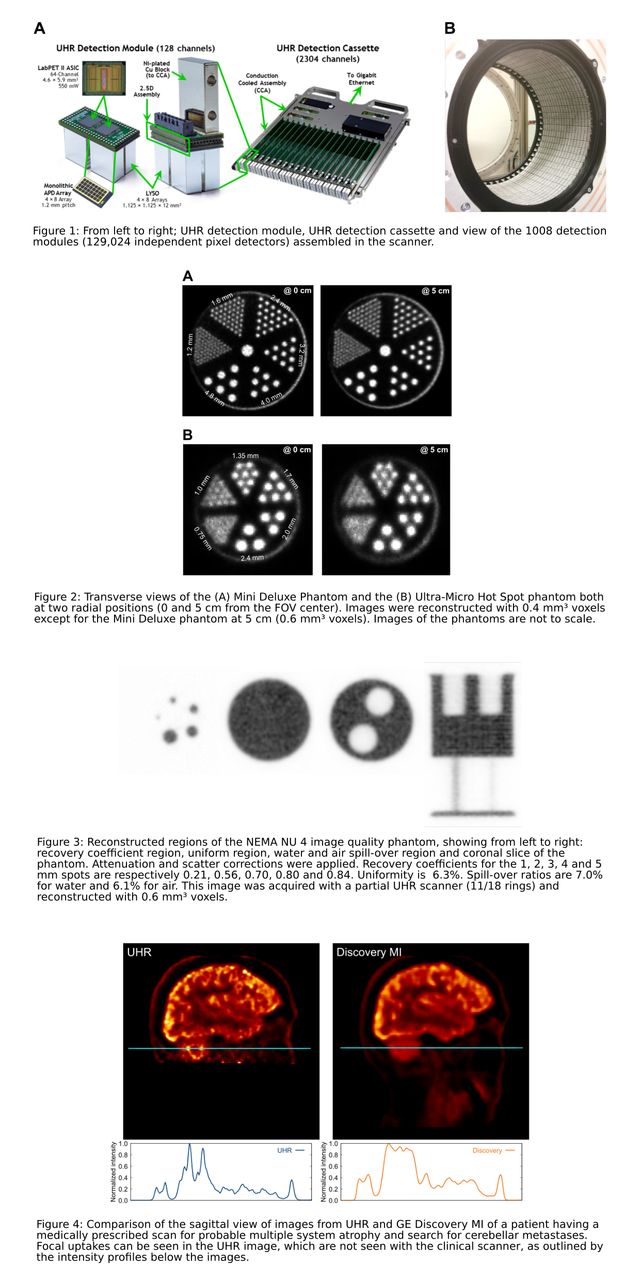
Human brain images obtained with the UHR show very well resolved cortex circumvolutions and higher contrast for several cortical and subcortical structures usually not resolved in PET images. For a patient having a medically prescribed scan on a GE Discovery MI PET/CT scanner for probable multiple system atrophy and search for cerebellar metastases, the UHR images revealed focal uptakes that were not seen with the clinical scanner (fig. 4).
Conclusions: The target resolution performance of the UHR brain PET scanner was confirmed by physical measurements and imaging data. Resolving smaller structures with a lower partial-volume effect enhances contrast and could enable detecting neurological diseases closer to their true developmental onset. The sensitivity remains a limiting factor due to the truly pixelated detectors and the rejection of inter-crystal scatter events. However, recovery of triple events is possible and undergoing investigation. Further improvement of image accuracy is expected with the implementation of real-time tracking and correction for patient motion.

In this issue


Jump to section
Related Articles
Cited By...
- No citing articles found.