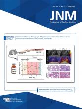
A 75-y-old man who was scheduled for resection of a lipomatous lesion of the left upper back underwent preoperative PET/CT with 68Ga-fibroblast activation protein inhibitor-46 (68Ga-FAPI-46) as part of a prospective study (NCT04147494). The images revealed an incidental small focus of uptake (SUVmax, 4.1) in a 0.8 × 0.7 × 1.2 cm cystic structure in the inferior third of a urachus remnant (Fig. 1). There was no 68Ga-FAPI-46 uptake in the remainder of the urachus remnant. A prior 18F-FDG PET/CT study with intravenous CT contrast medium that had been performed 4 y 1 mo previously was retrospectively reviewed. It showed incomplete obliteration of the urachus with a cystic structure in its inferior third, consistent with a urachus remnant, all with no 18F-FDG uptake. The urachal remnant and cystic structure on 68Ga-FAPI-46 PET/CT were anatomically the same as seen on the reviewed 18F-FDG PET/CT and on a subsequent follow-up CT scan obtained 1 y 3 mo later, suggesting a benign etiology. Differential diagnosis included a 68Ga-FAPI-46–positive and 18F-FDG–negative urachal cyst due to fibrosis and a urachal diverticulum, intermittently accumulating urine-excreted radiotracer.
Maximum-intensity projections (A and E), fused PET/CT axial images (B and F), CT axial images (C and G), and fused sagittal PET/CT images (D and H) demonstrating mild 68Ga-FAPI-46 uptake (SUVmax, 4.1; A–D) and no 18F-FDG uptake (E–H) in a cystic structure of a urachal remnant (arrows).
The urachus is an embryologic structure connecting the umbilicus to the bladder. Normally, it obliterates to become the medial umbilical ligament. Very rarely, it does not obliterate, and remnant urachal anomalies persist to adulthood (1). Of these anomalies, a cystic structure can remain between the umbilicus and the bladder.
Several studies have suggested that 68Ga-FAPI-46 PET/CT has a promising role in the detection of peritoneal metastases in various malignancies, notably because of low physiologic bowel uptake (2–4). The above-described 68Ga-FAPI-46 PET signal in the urachal remnant should be a known potential false-positive pitfall in that region.
DISCLOSURE
No potential conflict of interest relevant to this article was reported.
Footnotes
Guest Editor: Barry A. Siegel, Mallinckrodt Institute of Radiology.
Published online Jan. 12, 2023.
- © 2023 by the Society of Nuclear Medicine and Molecular Imaging.
- Received for publication October 14, 2022.
- Revision received December 20, 2022.


