
Main menu

  • Home
  • Content
    • Current
    • Ahead of print
    • Past Issues
    • JNM Supplement
    • SNMMI Annual Meeting Abstracts
    • Continuing Education
    • JNM Podcasts
  • Subscriptions
    • Subscribers
    • Institutional and Non-member
    • Rates
    • Journal Claims
    • Corporate & Special Sales
  • Authors
    • Submit to JNM
    • Information for Authors
    • Assignment of Copyright
    • AQARA requirements
  • Info
    • Reviewers
    • Permissions
    • Advertisers
  • About
    • About Us
    • Editorial Board
    • Contact Information
  • More
    • Alerts
    • Feedback
    • Help
    • SNMMI Journals
  • SNMMI
    • JNM
    • JNMT
    • SNMMI Journals
    • SNMMI

User menu

  • Subscribe
  • My alerts
  • Log in
  • My Cart

Search

  • Advanced search
Journal of Nuclear Medicine
  • SNMMI
    • JNM
    • JNMT
    • SNMMI Journals
    • SNMMI
  • Subscribe
  • My alerts
  • Log in
  • My Cart
Journal of Nuclear Medicine

Advanced Search

  • Home
  • Content
    • Current
    • Ahead of print
    • Past Issues
    • JNM Supplement
    • SNMMI Annual Meeting Abstracts
    • Continuing Education
    • JNM Podcasts
  • Subscriptions
    • Subscribers
    • Institutional and Non-member
    • Rates
    • Journal Claims
    • Corporate & Special Sales
  • Authors
    • Submit to JNM
    • Information for Authors
    • Assignment of Copyright
    • AQARA requirements
  • Info
    • Reviewers
    • Permissions
    • Advertisers
  • About
    • About Us
    • Editorial Board
    • Contact Information
  • More
    • Alerts
    • Feedback
    • Help
    • SNMMI Journals
  • View or Listen to JNM Podcast
  • Visit JNM on Facebook
  • Join JNM on LinkedIn
  • Follow JNM on Twitter
  • Subscribe to our RSS feeds
Research ArticleClinical Investigation

A Visual Interpretation Algorithm for Assessing Brain Tauopathy with 18F-MK-6240 PET

John P. Seibyl, Jonathan M. DuBois, Annie Racine, Jessica Collins, Qi Guo, Dustin Wooten, Eddie Stage, David Cheng, Roger N. Gunn, Lilly Porat, Alex Whittington, Phillip H. Kuo, Masanori Ichise, Robert Comley, Laurent Martarello and Cristian Salinas
Journal of Nuclear Medicine March 2023, 64 (3) 444-451; DOI: https://doi.org/10.2967/jnumed.122.264371
John P. Seibyl
1Institute for Neurodegenerative Disorders, New Haven, Connecticut;
2Invicro, New Haven, Connecticut;
  • Find this author on Google Scholar
  • Find this author on PubMed
  • Search for this author on this site
Jonathan M. DuBois
3Biogen, Cambridge, Massachusetts;
  • Find this author on Google Scholar
  • Find this author on PubMed
  • Search for this author on this site
Annie Racine
3Biogen, Cambridge, Massachusetts;
  • Find this author on Google Scholar
  • Find this author on PubMed
  • Search for this author on this site
Jessica Collins
3Biogen, Cambridge, Massachusetts;
  • Find this author on Google Scholar
  • Find this author on PubMed
  • Search for this author on this site
Qi Guo
4AbbVie, North Chicago, Illinois;
  • Find this author on Google Scholar
  • Find this author on PubMed
  • Search for this author on this site
Dustin Wooten
3Biogen, Cambridge, Massachusetts;
  • Find this author on Google Scholar
  • Find this author on PubMed
  • Search for this author on this site
Eddie Stage
4AbbVie, North Chicago, Illinois;
  • Find this author on Google Scholar
  • Find this author on PubMed
  • Search for this author on this site
David Cheng
2Invicro, New Haven, Connecticut;
  • Find this author on Google Scholar
  • Find this author on PubMed
  • Search for this author on this site
Roger N. Gunn
2Invicro, New Haven, Connecticut;
  • Find this author on Google Scholar
  • Find this author on PubMed
  • Search for this author on this site
Lilly Porat
2Invicro, New Haven, Connecticut;
  • Find this author on Google Scholar
  • Find this author on PubMed
  • Search for this author on this site
Alex Whittington
2Invicro, New Haven, Connecticut;
  • Find this author on Google Scholar
  • Find this author on PubMed
  • Search for this author on this site
Phillip H. Kuo
5University of Arizona, Tucson, Arizona; and
  • Find this author on Google Scholar
  • Find this author on PubMed
  • Search for this author on this site
Masanori Ichise
6National Institute of Radiological Sciences, National Institutes for Quantum and Radiological Science and Technology, Chiba, Japan
  • Find this author on Google Scholar
  • Find this author on PubMed
  • Search for this author on this site
Robert Comley
4AbbVie, North Chicago, Illinois;
  • Find this author on Google Scholar
  • Find this author on PubMed
  • Search for this author on this site
Laurent Martarello
3Biogen, Cambridge, Massachusetts;
  • Find this author on Google Scholar
  • Find this author on PubMed
  • Search for this author on this site
Cristian Salinas
3Biogen, Cambridge, Massachusetts;
  • Find this author on Google Scholar
  • Find this author on PubMed
  • Search for this author on this site
  • Article
  • Figures & Data
  • Supplemental
  • Info & Metrics
  • PDF
Loading

Article Figures & Data

Figures

  • Tables
  • Additional Files
  • Figure
    • Download figure
    • Open in new tab
    • Download powerpoint
  • FIGURE 1.
    • Download figure
    • Open in new tab
    • Download powerpoint
    FIGURE 1.

    Regions for visual read are outlined and overlaid on T1-weighted MR images for anatomic reference.
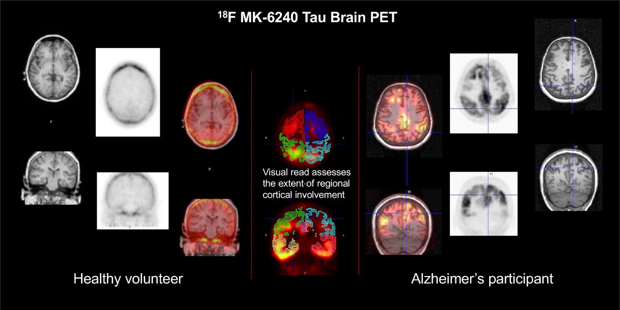
  • FIGURE 2.
    • Download figure
    • Open in new tab
    • Download powerpoint
    FIGURE 2.

    18F-MK-6240 PET in CN volunteer, AD patient, and patient with non-AD tauopathy. “Positive, atypical AD” was added in refined algorithm. Non-AD tauopathy patterns with only subcortical (cluster 3) and no cortical uptake (clusters 1 and 2), as would be expected in progressive supranuclear palsy (PSP), were not encountered in this primarily MCI/AD dataset.

  • FIGURE 3.
    • Download figure
    • Open in new tab
    • Download powerpoint
    FIGURE 3.

    Difficult case showing bilateral anterior mesial temporal uptake, which can be confused with off-target uptake in meninges and floor of calvarium. Fused axial image of MK-6240 with the participant’s T1-weighted MR image (top left), fused coronal image of MK-6240 with the participant’s T1-weighted MR image (bottom left); axial (top right) and coronal (bottom right) views of MRI only.

  • FIGURE 4.
    • Download figure
    • Open in new tab
    • Download powerpoint
    FIGURE 4.

    SUVr for different regional VOIs parsed by visual read of negative or positive. Jack VOI = VOI regions of Jack et al. (15).

Tables

  • Figures
  • Additional Files
    • View popup
    TABLE 1.

    Regions for Visual Assessment

    Brain areaIncluded regionsVisual ratingNo. regionsRationale
    Cluster 1, temporal lobesHippocampus; mesial temporal; inferior temporal; lateral temporalNo uptake (0%); uptake 1%–25%; extension 26%–75%; >75% extension8 regions: 4 each in left and right hemispheresEarliest cortical regions involved in AD per Braak staging
    Cluster 2, extratemporal neocortexOccipital; posterior cingulate; parietal; frontalNo uptake (0%); uptake 1%–25%; extension 26%–75%; >75% extension8 regions: 4 each in left and right hemispheresNext regions involved in AD
    Cluster 3, subcortical areaStriatum-globus; thalamus; dentate nucleus; pons; midbrainPresence or absence5 regionsMay be positive in non-AD tauopathies
    • View popup
    TABLE 2.

    Demographic Data

    Development datasetTest dataset
    Sex (n)Sex (n)
    GroupnAge (y)MFnAge (y)MF
    AD2972.4 (9.9)2092470.4 (10.7)1212
    MCI1771.2 (7.1)1162169.9 (8.0)129
    CN5266.4 (12.1)25274568.6 (7.5)1827
    Other463.3 (4.6)041265.9 (9.4)57
    • Age is mean followed by SD in parentheses.

    • View popup
    TABLE 3.

    Reader Agreement on Visual Assessment as Positive or Negative for Regional Tau

    Regional binary agreement on positive or negativeComplete agreement on spatial extent scoring
    AgreementRegionFleiss κAgreementRegionFleiss κ
    Almost perfectRight parietal0.945SubstantialRight lateral temporal0.748
    Right frontal0.929Left lateral temporal0.716
    Right lateral temporal0.907Right posterior cingulate0.702
    Left parietal0.906Left posterior cingulate0.678
    Left posterior cingulate0.904Right parietal0.655
    Left lateral temporal0.888Left parietal0.621
    Right posterior cingulate0.87Right frontal0.618
    SubstantialLeft frontal0.801Left frontal0.602
    Left occipital0.801ModerateRight inferior temporal0.597
    Left hippocampus0.783Left inferior temporal0.576
    Right inferior temporal0.782Right hippocampus0.552
    Right occipital0.759Right mesial temporal0.552
    Right hippocampus0.756Left hippocampus0.551
    Left inferior temporal0.753Left mesial temporal0.545
    Right mesial temporal0.742Right occipital0.537
    Left mesial temporal0.726Left occipital0.503
    • Overall binary assessment, κ = 0.912.

    • View popup
    TABLE 4.

    Sensitivity and Specificity of MK-6240 Visual Reads and SUVr Analyses Using Clinical Diagnosis as Standard of Truth

    MethodSensitivitySpecificityYouden index
    Visual read
     Reader 00.790.930.72
     Reader 10.750.930.68
     Reader 20.790.910.70
     Consensus0.810.930.74
    Visual VRES (cutoff, 1.5)0.7420.8900.63
    SUVr
     Braak 1–2 (cutoff, 1.4)0.6490.8880.54
     Braak 3–4 (cutoff, 1.3)0.6450.9630.61
     Braak 5–6 (cutoff, 1.2)0.6110.9600.57
     Jack VOI (cutoff, 1.4)0.6500.8800.53
    • Jack VOI = VOI regions of Jack et al. (15).

    • View popup
    TABLE 5.

    Initial and Revised Algorithms for Interpreting MK-6240 PET

    Scan assessmentOriginal algorithmRefined algorithmReason for adjustment
    NegativeNo more than one region positive in cortexAll clusters negativeAllowing one region to be positive was to prevent interpretation of scans as positive when meningeal uptake near inferolateral temporal lobes could be misread as positive region; this was dropped with improved methods or instructions to identify this confounder
    Positive, AD patternEvidence of increased uptake in two cortical regions with at least one region in temporal lobesAt least one cluster 1 region positive and no cluster 3 regions positiveRevision was made after observation of multiple cases with isolated cluster 1 abnormality in just one region
    Positive, atypical AD patternNot assessedIncreased uptake in one or more regions in cluster 2 but not in clusters 1 or 3This rare pattern was noted from review of other MK-6240 datasets and was expected pattern based on published literature on other tau PET tracers
    Positive, non-AD patternAny positive scan not fitting AD criteriaIncreased uptake in one or more regions in clusters 1–3, with at least one region in cluster 3Adjustment was made after formalized uniform assessment for regions involved in non-AD tauopathies

Additional Files

  • Figures
  • Tables
  • Supplemental Data

    Files in this Data Supplement:

    • Supplemental Data
PreviousNext
Back to top

In this issue

Journal of Nuclear Medicine: 64 (3)
Journal of Nuclear Medicine
Vol. 64, Issue 3
March 1, 2023
  • Table of Contents
  • Table of Contents (PDF)
  • About the Cover
  • Index by author
  • Complete Issue (PDF)
Print
Download PDF
Article Alerts
Sign In to Email Alerts with your Email Address
Email Article

Thank you for your interest in spreading the word on Journal of Nuclear Medicine.

NOTE: We only request your email address so that the person you are recommending the page to knows that you wanted them to see it, and that it is not junk mail. We do not capture any email address.

Enter multiple addresses on separate lines or separate them with commas.
A Visual Interpretation Algorithm for Assessing Brain Tauopathy with 18F-MK-6240 PET
(Your Name) has sent you a message from Journal of Nuclear Medicine
(Your Name) thought you would like to see the Journal of Nuclear Medicine web site.
Citation Tools
A Visual Interpretation Algorithm for Assessing Brain Tauopathy with 18F-MK-6240 PET
John P. Seibyl, Jonathan M. DuBois, Annie Racine, Jessica Collins, Qi Guo, Dustin Wooten, Eddie Stage, David Cheng, Roger N. Gunn, Lilly Porat, Alex Whittington, Phillip H. Kuo, Masanori Ichise, Robert Comley, Laurent Martarello, Cristian Salinas
Journal of Nuclear Medicine Mar 2023, 64 (3) 444-451; DOI: 10.2967/jnumed.122.264371

Citation Manager Formats

  • BibTeX
  • Bookends
  • EasyBib
  • EndNote (tagged)
  • EndNote 8 (xml)
  • Medlars
  • Mendeley
  • Papers
  • RefWorks Tagged
  • Ref Manager
  • RIS
  • Zotero
Share
A Visual Interpretation Algorithm for Assessing Brain Tauopathy with 18F-MK-6240 PET
John P. Seibyl, Jonathan M. DuBois, Annie Racine, Jessica Collins, Qi Guo, Dustin Wooten, Eddie Stage, David Cheng, Roger N. Gunn, Lilly Porat, Alex Whittington, Phillip H. Kuo, Masanori Ichise, Robert Comley, Laurent Martarello, Cristian Salinas
Journal of Nuclear Medicine Mar 2023, 64 (3) 444-451; DOI: 10.2967/jnumed.122.264371
Twitter logo Facebook logo LinkedIn logo Mendeley logo
  • Tweet Widget
  • Facebook Like
  • Google Plus One
Bookmark this article

Jump to section

  • Article
    • Visual Abstract
    • Abstract
    • MATERIALS AND METHODS
    • RESULTS
    • DISCUSSION
    • CONCLUSION
    • DISCLOSURE
    • ACKNOWLEDGMENT
    • Footnotes
    • REFERENCES
  • Figures & Data
  • Supplemental
  • Info & Metrics
  • PDF

Related Articles

  • PubMed
  • Google Scholar

Cited By...

  • Performance of blood biomarkers in internal jugular vein for Alzheimer disease pathologies: the Delta Study
  • Updated Appropriate Use Criteria for Amyloid and Tau PET: A Report from the Alzheimers Association and Society for Nuclear Medicine and Molecular Imaging Workgroup
  • Performance of a [18F]Flortaucipir PET Visual Read Method Across the Alzheimer Disease Continuum and in Dementia With Lewy Bodies
  • Tau PET Visual Reads: Research and Clinical Applications and Future Directions
  • Google Scholar

More in this TOC Section

  • 177Lu-PSMA-617 Consolidation Therapy After Docetaxel in Patients with Synchronous High-Volume Metastatic Hormone-Sensitive Prostate Cancer: A Randomized, Phase 2 Trial
  • Transarterial Radioembolization in the TACOME Trial: Dosimetric Analysis and Clinical Features in Predicting Response and Overall Survival
  • Retreatment of Metastatic Castration-Resistant Prostate Cancer Patients with 223Ra Therapy in Daily Practice
Show more Clinical Investigation

Similar Articles

Keywords

  • Neurology
  • PET
  • Alzheimer disease
  • MK-6240
  • tau proteinopathies
SNMMI

© 2025 SNMMI

Powered by HighWire