
Main menu

  • Home
  • Content
    • Current
    • Ahead of print
    • Past Issues
    • JNM Supplement
    • SNMMI Annual Meeting Abstracts
    • Continuing Education
    • JNM Podcasts
  • Subscriptions
    • Subscribers
    • Institutional and Non-member
    • Rates
    • Journal Claims
    • Corporate & Special Sales
  • Authors
    • Submit to JNM
    • Information for Authors
    • Assignment of Copyright
    • AQARA requirements
  • Info
    • Reviewers
    • Permissions
    • Advertisers
  • About
    • About Us
    • Editorial Board
    • Contact Information
  • More
    • Alerts
    • Feedback
    • Help
    • SNMMI Journals
  • SNMMI
    • JNM
    • JNMT
    • SNMMI Journals
    • SNMMI

User menu

  • Subscribe
  • My alerts
  • Log in
  • My Cart

Search

  • Advanced search
Journal of Nuclear Medicine
  • SNMMI
    • JNM
    • JNMT
    • SNMMI Journals
    • SNMMI
  • Subscribe
  • My alerts
  • Log in
  • My Cart
Journal of Nuclear Medicine

Advanced Search

  • Home
  • Content
    • Current
    • Ahead of print
    • Past Issues
    • JNM Supplement
    • SNMMI Annual Meeting Abstracts
    • Continuing Education
    • JNM Podcasts
  • Subscriptions
    • Subscribers
    • Institutional and Non-member
    • Rates
    • Journal Claims
    • Corporate & Special Sales
  • Authors
    • Submit to JNM
    • Information for Authors
    • Assignment of Copyright
    • AQARA requirements
  • Info
    • Reviewers
    • Permissions
    • Advertisers
  • About
    • About Us
    • Editorial Board
    • Contact Information
  • More
    • Alerts
    • Feedback
    • Help
    • SNMMI Journals
  • View or Listen to JNM Podcast
  • Visit JNM on Facebook
  • Join JNM on LinkedIn
  • Follow JNM on Twitter
  • Subscribe to our RSS feeds
Research ArticleClinical Investigation

Predicting Outcomes of Indeterminate Bone Lesions on 18F-DCFPyL PSMA PET/CT Scans in the Setting of High-Risk Primary or Recurrent Prostate Cancer

Tim E. Phelps, Stephanie A. Harmon, Esther Mena, Liza Lindenberg, Joanna H. Shih, Deborah E. Citrin, Peter A. Pinto, Bradford J. Wood, William L. Dahut, James L. Gulley, Ravi A. Madan, Peter L. Choyke and Baris Turkbey
Journal of Nuclear Medicine March 2023, 64 (3) 395-401; DOI: https://doi.org/10.2967/jnumed.122.264334
Tim E. Phelps
1Molecular Imaging Branch, National Cancer Institute, National Institutes of Health, Bethesda, Maryland;
  • Find this author on Google Scholar
  • Find this author on PubMed
  • Search for this author on this site
Stephanie A. Harmon
1Molecular Imaging Branch, National Cancer Institute, National Institutes of Health, Bethesda, Maryland;
  • Find this author on Google Scholar
  • Find this author on PubMed
  • Search for this author on this site
Esther Mena
1Molecular Imaging Branch, National Cancer Institute, National Institutes of Health, Bethesda, Maryland;
  • Find this author on Google Scholar
  • Find this author on PubMed
  • Search for this author on this site
Liza Lindenberg
1Molecular Imaging Branch, National Cancer Institute, National Institutes of Health, Bethesda, Maryland;
  • Find this author on Google Scholar
  • Find this author on PubMed
  • Search for this author on this site
Joanna H. Shih
2Biometric Research Program, National Cancer Institute, National Institutes of Health, Rockville, Maryland;
  • Find this author on Google Scholar
  • Find this author on PubMed
  • Search for this author on this site
Deborah E. Citrin
3Radiation Oncology Branch, National Cancer Institute, National Institutes of Health, Bethesda, Maryland;
  • Find this author on Google Scholar
  • Find this author on PubMed
  • Search for this author on this site
Peter A. Pinto
4Urologic Oncology Branch, National Cancer Institute, National Institutes of Health, Bethesda, Maryland;
  • Find this author on Google Scholar
  • Find this author on PubMed
  • Search for this author on this site
Bradford J. Wood
5Radiology and Imaging Sciences, Clinical Center, National Institutes of Health, Bethesda, Maryland;
6Center for Interventional Oncology, National Cancer Institute, National Institutes of Health, Bethesda, Maryland; and
  • Find this author on Google Scholar
  • Find this author on PubMed
  • Search for this author on this site
William L. Dahut
7Genitourinary Malignancies Branch, National Cancer Institute, National Institutes of Health, Bethesda, Maryland
  • Find this author on Google Scholar
  • Find this author on PubMed
  • Search for this author on this site
James L. Gulley
7Genitourinary Malignancies Branch, National Cancer Institute, National Institutes of Health, Bethesda, Maryland
  • Find this author on Google Scholar
  • Find this author on PubMed
  • Search for this author on this site
Ravi A. Madan
7Genitourinary Malignancies Branch, National Cancer Institute, National Institutes of Health, Bethesda, Maryland
  • Find this author on Google Scholar
  • Find this author on PubMed
  • Search for this author on this site
Peter L. Choyke
1Molecular Imaging Branch, National Cancer Institute, National Institutes of Health, Bethesda, Maryland;
  • Find this author on Google Scholar
  • Find this author on PubMed
  • Search for this author on this site
Baris Turkbey
1Molecular Imaging Branch, National Cancer Institute, National Institutes of Health, Bethesda, Maryland;
  • Find this author on Google Scholar
  • Find this author on PubMed
  • Search for this author on this site
  • Article
  • Figures & Data
  • Supplemental
  • Info & Metrics
  • PDF
Loading

Article Figures & Data

Figures

  • Tables
  • Additional Files
  • Figure
    • Download figure
    • Open in new tab
    • Download powerpoint
  • FIGURE 1.
    • Download figure
    • Open in new tab
    • Download powerpoint
    FIGURE 1.

    Flowchart of patient selection criteria and lesion-level follow-up for patients with at least 1 IBL. Interpretations (benign, malignant, or equivocal) are shown for IBLs with no follow-up (prior imaging) and follow-up (imaging or biopsy).

  • FIGURE 2.
    • Download figure
    • Open in new tab
    • Download powerpoint
    FIGURE 2.

    A 63-y-old patient with BCR PCa and PSA of 0.46 ng/mL. Axial 18F-DCFPyL PET (left), 18F-DCFPyL PET/CT (middle), and CT (right) images show a single area of subtle PSMA-avid uptake with SUVmax of 2.4 in the right fifth rib and no CT correlate (arrows). This IBL was determined to be benign based on negative 17-mo follow-up bone scintigraphy findings.

  • FIGURE 3.
    • Download figure
    • Open in new tab
    • Download powerpoint
    FIGURE 3.

    A 60-y-old patient with BCR PCa and PSA of 3.9 ng/mL. Axial 18F-DCFPyL PET (left), 18F-DCFPyL PET/CT (middle), and CT (right) images show a single area of subtle PSMA-avid uptake with SUVmax of 3.9 in left iliac bone and mixed sclerotic and lytic CT features (arrows). This IBL was stable for 4 mo and negative on 4 other staging modalities; thus, the IBL was interpreted as benign.

  • FIGURE 4.
    • Download figure
    • Open in new tab
    • Download powerpoint
    FIGURE 4.

    A 73-y-old patient with BCR PCa and PSA of 4.9 ng/mL. Axial 18F-DCFPyL PET (A), 18F-DCFPyL PET/CT (B), CT (C), pre-PSMA MRI (D), and follow-up MRI (E) images show a single area of subtle PSMA-avid uptake with SUVmax of 3.3 in left T8 lamina and subtle sclerotic CT features (arrows). This IBL became more prominent and enhanced over a 3-y period between retrospective MRI (D) and follow-up MRI (E) and was determined to be malignant.

  • FIGURE 5.
    • Download figure
    • Open in new tab
    • Download powerpoint
    FIGURE 5.

    Model assessing the likelihood that an IBL is benign or malignant based on 18F-DCFPyL PET/CT imaging variables that predicted for benignancy or malignancy (n = 79).

  • FIGURE 6.
    • Download figure
    • Open in new tab
    • Download powerpoint
    FIGURE 6.

    Sankey diagram showing the relationship of multiple predictive 18F-DCFPyL features (n = 98). Specifically, lesion location, lesion SUVmax, and type of bone findings on a PSMA PET/CT scan are more likely associated with a particular lesion interpretation (right). Pathways with 1 lesion have been removed for clarity.

Tables

  • Figures
  • Additional Files
    • View popup
    TABLE 1.

    Clinical Characteristics of Patients with IBLs

    CharacteristicData
    Patients with IBL48
    Age (y)
     Median66
     Range53–79
    PSA (ng/mL)
     Median4.00
     Range0.44–203.8
    Disease phase
     BCR PCa35
     High-risk PCa13
    TNM stage
     Not available5
     T110
     T213
     T315
     T2 N11
     T3 N13
     T4 N11
    Gleason grade group
     12
     214
     310
     47
     515
    • Data are number of events unless otherwise indicated.

    • View popup
    TABLE 2.

    Histopathologic Assessment of IBL Biopsy Specimens

    Location of biopsied IBLSUVmaxCT morphologyHistopathologic result
    FindingFeatures
    Fifth rib4.1ScleroticBenignFibrous replacement of bone marrow
    Third rib3.6ScleroticBenignTrabecular bone with trilineage hematopoiesis
    Sixth rib2.9ScleroticBenignFragments of bone marrow fibrosis
    Ischium bone17.4Mixed sclerotic/lyticMalignantMetastatic prostate adenocarcinoma involving bone and bone marrow
    Iliac bone1.3NegativeMalignantMetastatic moderately differentiated prostate adenocarcinoma
    Clavicle bone3.1NegativeBenignBone marrow with trilineage hematopoiesis
    Seventh rib2.8ScleroticBenignBone with hematopoietic bone marrow
    Fifth rib15.2Mixed sclerotic/lyticMalignantMetastatic poorly differentiated prostate adenocarcinoma
    • View popup
    TABLE 3.

    18F-DCFPyL PSMA PET/CT-Based Variables That Predict IBL Interpretation (n = 98)

    Predictive variableIncidence (n = 98)IBL interpretationP*
    M (n = 42)B (n = 37)E (n = 19)
    Location0.0201
     Spine28 (28.6%)16120
     Pelvis21 (21.4%)1272
     Rib39 (39.8%)81417
     Other10 (10.2%)640
    SUVmax0.0230
     <564 (65.3%)123616
     ≥534 (34.7%)3013
    • ↵* For 3-way assessment (M vs. B vs. E) statistical significance is evaluated using χ2 test for clustered data.

    • M = malignant; B = benign; E = equivocal.

    • View popup
    TABLE 4.

    Logistic Regression Analysis Showing OR at 95% CI for Clinically Relevant 18F-DCFPyL PET/CT-Based Features That Predict IBLs as Benign vs. Malignant (n = 79)

    18F-DCFPyL PET/CT featureUnivariable analysisMultivariable analysis
    ORPORP
    IBL located in pelvis1.68 (0.48–5.96)0.516——
    IBL located in spine1.42 (0.55–3.65)0.613——
    IBL located in ribs0.38 (0.14–1.03)0.170——
    IBL SUVmax9.29 (3.49–24.75)0.001613.87 (1.91–100.9)0.0089
    Other bone metastases9.87 (2.00–48.82)0.011211.35 (3.05–42.25)0.0030
    Other lymph node metastases2.26 (0.62–8.26)0.315——
    • Data in parentheses are 95% CI. Features significantly associated with malignant interpretation were included in multivariable analysis.

    • View popup
    TABLE 5.

    Model Predicting the Likelihood That IBLs Identified on 18F-DCFPyL PET/CT at 2 Hour of Uptake Are Benign or Malignant (n = 79)
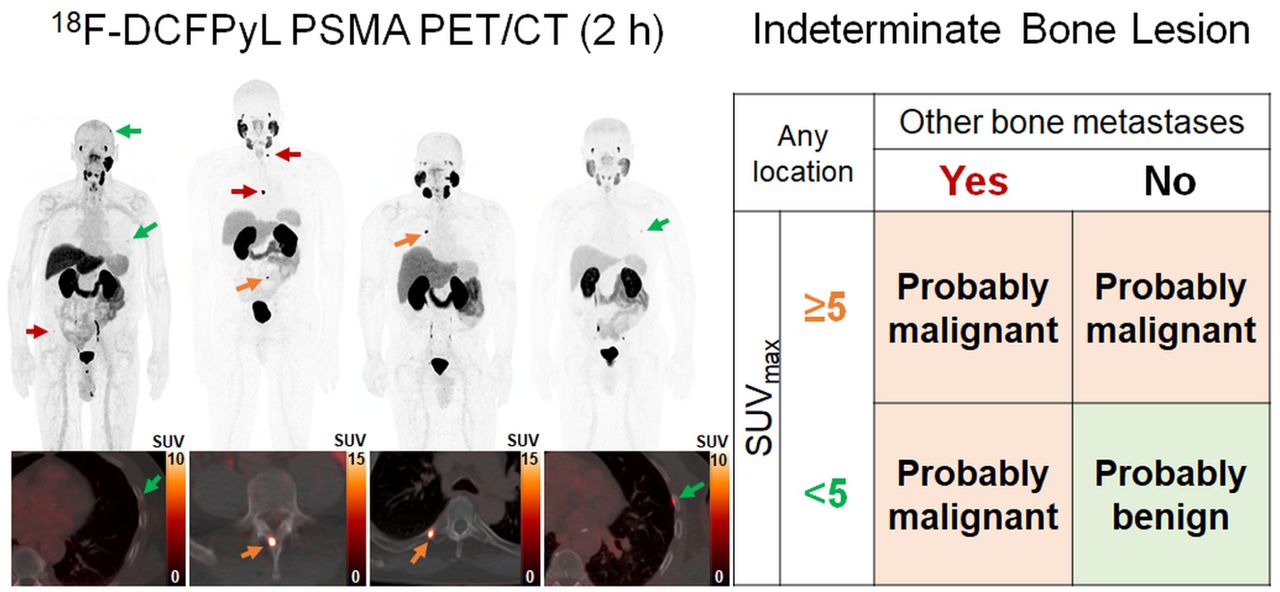
    Other PSMA-avid bone findingsIBL locationIBL SUVmaxLikelihood of interpretationInterpretation accuracy
    No bone metastasesTypical<581.0% benignTrue positive (n = 17); false negative (n = 4)
    No bone metastasesTypical≥594.7% malignantTrue positive (n = 18); false positive (n = 1)
    No bone metastasesAtypical<594.4% benignTrue negative (n = 17); false negative (n = 1)
    No bone metastasesAtypical≥5100% malignantTrue positive (n = 8)
    Bone metastasesAny<577.8% malignantTrue positive (n = 7); false positive (n = 2)
    Bone metastasesAny≥5100% malignantTrue positive (n = 4)

Additional Files

  • Figures
  • Tables
  • Supplemental Data

    Files in this Data Supplement:

    • Supplemental Data
PreviousNext
Back to top

In this issue

Journal of Nuclear Medicine: 64 (3)
Journal of Nuclear Medicine
Vol. 64, Issue 3
March 1, 2023
  • Table of Contents
  • Table of Contents (PDF)
  • About the Cover
  • Index by author
  • Complete Issue (PDF)
Print
Download PDF
Article Alerts
Sign In to Email Alerts with your Email Address
Email Article

Thank you for your interest in spreading the word on Journal of Nuclear Medicine.

NOTE: We only request your email address so that the person you are recommending the page to knows that you wanted them to see it, and that it is not junk mail. We do not capture any email address.

Enter multiple addresses on separate lines or separate them with commas.
Predicting Outcomes of Indeterminate Bone Lesions on 18F-DCFPyL PSMA PET/CT Scans in the Setting of High-Risk Primary or Recurrent Prostate Cancer
(Your Name) has sent you a message from Journal of Nuclear Medicine
(Your Name) thought you would like to see the Journal of Nuclear Medicine web site.
Citation Tools
Predicting Outcomes of Indeterminate Bone Lesions on 18F-DCFPyL PSMA PET/CT Scans in the Setting of High-Risk Primary or Recurrent Prostate Cancer
Tim E. Phelps, Stephanie A. Harmon, Esther Mena, Liza Lindenberg, Joanna H. Shih, Deborah E. Citrin, Peter A. Pinto, Bradford J. Wood, William L. Dahut, James L. Gulley, Ravi A. Madan, Peter L. Choyke, Baris Turkbey
Journal of Nuclear Medicine Mar 2023, 64 (3) 395-401; DOI: 10.2967/jnumed.122.264334

Citation Manager Formats

  • BibTeX
  • Bookends
  • EasyBib
  • EndNote (tagged)
  • EndNote 8 (xml)
  • Medlars
  • Mendeley
  • Papers
  • RefWorks Tagged
  • Ref Manager
  • RIS
  • Zotero
Share
Predicting Outcomes of Indeterminate Bone Lesions on 18F-DCFPyL PSMA PET/CT Scans in the Setting of High-Risk Primary or Recurrent Prostate Cancer
Tim E. Phelps, Stephanie A. Harmon, Esther Mena, Liza Lindenberg, Joanna H. Shih, Deborah E. Citrin, Peter A. Pinto, Bradford J. Wood, William L. Dahut, James L. Gulley, Ravi A. Madan, Peter L. Choyke, Baris Turkbey
Journal of Nuclear Medicine Mar 2023, 64 (3) 395-401; DOI: 10.2967/jnumed.122.264334
Twitter logo Facebook logo LinkedIn logo Mendeley logo
  • Tweet Widget
  • Facebook Like
  • Google Plus One
Bookmark this article

Jump to section

  • Article
    • Visual Abstract
    • Abstract
    • MATERIALS AND METHODS
    • RESULTS
    • DISCUSSION
    • CONCLUSION
    • DISCLOSURE
    • Footnotes
    • REFERENCES
  • Figures & Data
  • Supplemental
  • Info & Metrics
  • PDF

Related Articles

  • PubMed
  • Google Scholar

Cited By...

  • No citing articles found.
  • Google Scholar

More in this TOC Section

  • First-in-Human Study of 18F-Labeled PET Tracer for Glutamate AMPA Receptor [18F]K-40: A Derivative of [11C]K-2
  • Detection of HER2-Low Lesions Using HER2-Targeted PET Imaging in Patients with Metastatic Breast Cancer: A Paired HER2 PET and Tumor Biopsy Analysis
  • [11C]Carfentanil PET Whole-Body Imaging of μ-Opioid Receptors: A First in-Human Study
Show more Clinical Investigation

Similar Articles

Keywords

  • prostate cancer
  • PET/CT
  • PSMA
  • bone metastases
  • indeterminate bone lesions
SNMMI

© 2025 SNMMI

Powered by HighWire