


Abstract
3280
Introduction: As part of a clinical imaging study with alpha-particle emitter Ra-224, the energy spectra recorded with SPECT have been investigated. There are several imageable emissions resulting from the Ra-224 decay chain, with the largest contributions from daughter Pb-212. An emission from daughter Tl-208 of 2.6 MeV was suspected to add significant scatter to energy windows at 240 keV and 80 keV. Hence, Monte Carlo simulations were used to examine the contributions of key emissions to the energy windows.
Methods: Experimental energy spectra were recorded on a Siemens Symbia Intevo Bold SPECT/CT with 3/8” crystals with a high energy (HE) collimator, a medium energy low penetration (ME) collimator, and no collimator. The source was a NEMA IEC Body Phantom with approximately 27.8 kBq/ml (1.3 MBq) Ra-224 in the spheres and non-radioactive water in the background. Secondly, Monte Carlo simulations were performed with Geant4 Application for Tomographic emission (GATE) (1, 2). The SPECT detectors were modelled with collimators, crystals, shielding, and back compartments. The back compartments were described in detail, with PMTs, lightguides, and electronics, to accurately model the backscatter of high energy photons (3, 4). The bed and phantom were modelled with STL-files. The source was simulated using a UserSpectrum including all photon emissions from the Ra-224 decay chain with emission probabilities larger than 0.1% per Ra-224 decay. The electromagnetic standard option 4 physics list was used. By calibrating the simulated spectra to experimental results, the energy blurring was determined and modelled with a linear law with 13% resolution at 80 keV and slope -0.09 1/MeV. The obtained spectra were validated against experimental spectra, with ME, HE, and no collimator. After validation, the simulations were repeated including only selected emissions from the Ra-224 decay chain. First, simulations were run with only emitted x-rays with energies of 70-90 keV included as the source. After, only the gamma emissions of 238.6 keV and 241 keV were simulated. Lastly, only the 2.6 MeV gamma emission from Tl-208 was simulated.
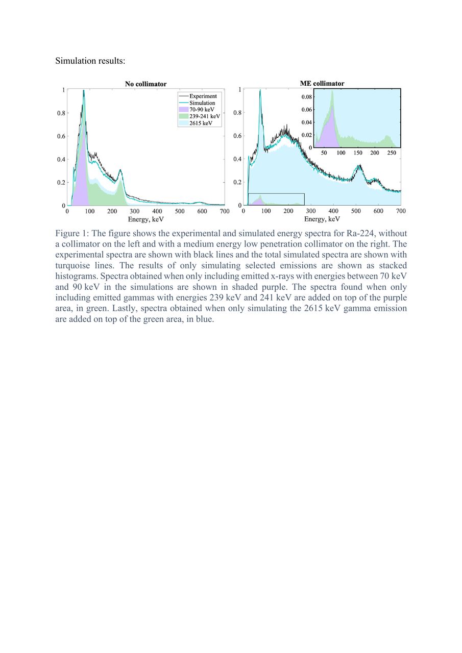
Results: The simulated spectra matched the experimental spectra with reasonable visual agreement, with ME, HE, and no collimator. Without a collimator, the 2.6 MeV emission barely made a contribution and the 70-90 keV emissions made up the majority of the 80 keV window and the 238.6 keV and 241 keV emissions made up most of the 240 keV window. The results were massively different with a collimator, though similar between the collimators, as the 2.6 MeV emission made up the majority of the energy spectrum and the chosen energy windows. Primary photons made up less than 10% of the counts in both energy windows with both collimators. As an example, triple energy window scatter correction with a 20% window on 240 keV with 5% scatter windows gave a scatter fraction above 90%, matching the true scatter well. However, a 40% window on 80 keV with 20% scatter windows gave a scatter fraction below 70%, overestimating primary photons recorded.
Conclusions: The scatter, pair production, and characteristic lead x-rays produced by the 2.6 MeV emission from Tl-208 make up the majority of the Ra-224 energy spectra seen on a SPECT when using a collimator. The scatter and characteristic x-rays resulting from this high energy emission contribute significantly to the energy windows investigated for imaging.
1. Jan S, et al. GATE: a simulation toolkit for PET and SPECT. Phys Med Biol. 2004;49(19):4543-61.
2. Sarrut D, et al. A review of the use and potential of the GATE Monte Carlo simulation code for radiation therapy and dosimetry applications. Med Phys. 2014;41(6):064301.
3. Robinson AP, et al. The influence of triple energy window scatter correction on activity quantification for 177Lu molecular radiotherapy. Phys in Med Biol. 2016;61(14):5107-27.
4. Rault E, et al. Accurate Monte Carlo modelling of the back compartments of SPECT cameras. Phys Med Biol. 2011;56(1):87-104.

In this issue


Jump to section
Related Articles
Cited By...
- No citing articles found.