


Abstract
2478
Introduction: Background: Herein, we demonstrate that a subpopulation of TICs could be specifically defined by the voltage-gated calcium channel α2δ1 subunit. The α2δ1+ TICs are refractory to conventional chemotherapy, and own stem cell-like properties such as self-renewal, and the ability to generate heterogeneous tumors, and tumor recurrence.
Purpose: Herein, we generate and labeled the 1B50-1-F(ab)2 with 64Cu, an anti-α2δ1 antibody, and investigate whether positron-emission tomography (PET) were sensitive approaches for detecting and quantitating α2δ1+ liver cancer stem cells in vivo.
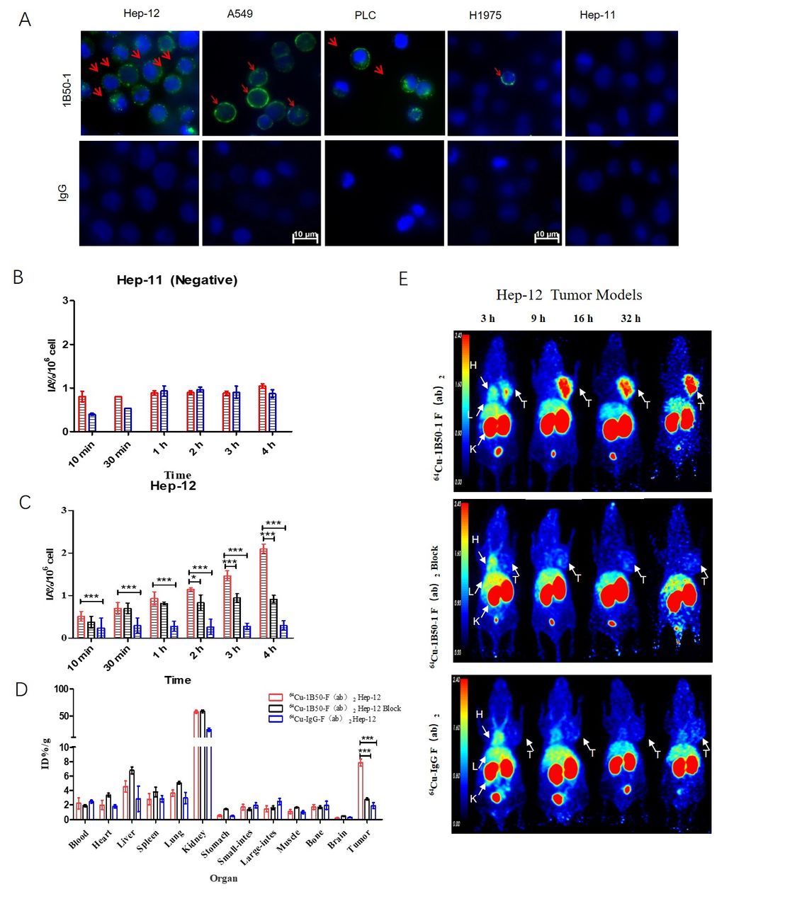
Methods: Methods: 1B50-1-F(ab)2 was labeled with 64Cu (using NOTA-NCS as bi-functional chelator). IgG-F(ab)2 was conjugated with same procedures as a control agent. Properties including stability, immunological competence and binding affinity were evaluated in vitro. The cell uptake experiment was conducted with Hep-12, A549, PLC (α2δ1+) and Hep-11 (α2δ1-) cell lines to determine the α2δ1 specific binding. Micro-PET imaging and bio-distribution studies were performed in Hep-12 and A549 tumor models. After chemotherapy, normal tumor cells would be killed, while α2δ1 positive cells (α2δ1+ TICs) increased relatively. The PLC tumor models was treated with adriamycin for 4 weeks. Then Micro-PET imaging was performed on the treated and untreated PLC tumor models. HE staing, immunofluorescence (IF) and autoradiography of tumor and main organ were carried out to confirm the expression of α2δ1.
Results: Results: 64Cu-NOTA-1B50-1-F(ab)2 were successfully constructed with 80-90% yield over 98% radiochemical purity. Different α2δ1+ TICs proportion in different tumor cell lines: 92-94% in Hep-12 cell lines, 27.2-31.2% in A549 cell lines, 15-20% in PLC cell lines. 64Cu-NOTA-1B50-1-F(ab)2 with a Kd of 5.76 nM. In addition, 64Cu-NOTA-1B50-1-F(ab)2 show the same affinity with the uptake of 2.1 IA%/106 cells in Hep-12 cells. The tumor uptake could be clearly visualized of 64Cu-NOTA-1B50-1-F(ab)2 at 9 h with uptake at 7.85 ± 0.49 ID%/g and remained to be clearly visible up to 32 h post-injection. Low tumor uptake was observed of 64Cu-NOTA-IgG-F(ab)2 with uptake of 1.91 ± 0.41 ID%/g, and 2.8 ± 0.14 ID%/g when blocked at 9 h. In the Micro-PET imaging of PLC tumor models, the uptake of tumor after treatment was significantly higher than that of untreated group.
Conclusions: Conclusion: α2δ1 is a idea target of tumor chemotherapy resistance and recurrence. Targeted molecular imaging allows for real-time, noninvasive, and quantitative detection of α2δ1+ TICs in vivo. It may be possible to early warning of early detection of tumor resistance and recurrence.

In this issue


Jump to section
Related Articles
Cited By...
- No citing articles found.