
Main menu

  • Home
  • Content
    • Current
    • Ahead of print
    • Past Issues
    • JNM Supplement
    • SNMMI Annual Meeting Abstracts
    • Continuing Education
    • JNM Podcasts
  • Subscriptions
    • Subscribers
    • Institutional and Non-member
    • Rates
    • Journal Claims
    • Corporate & Special Sales
  • Authors
    • Submit to JNM
    • Information for Authors
    • Assignment of Copyright
    • AQARA requirements
  • Info
    • Reviewers
    • Permissions
    • Advertisers
  • About
    • About Us
    • Editorial Board
    • Contact Information
  • More
    • Alerts
    • Feedback
    • Help
    • SNMMI Journals
  • SNMMI
    • JNM
    • JNMT
    • SNMMI Journals
    • SNMMI

User menu

  • Subscribe
  • My alerts
  • Log in
  • My Cart

Search

  • Advanced search
Journal of Nuclear Medicine
  • SNMMI
    • JNM
    • JNMT
    • SNMMI Journals
    • SNMMI
  • Subscribe
  • My alerts
  • Log in
  • My Cart
Journal of Nuclear Medicine

Advanced Search

  • Home
  • Content
    • Current
    • Ahead of print
    • Past Issues
    • JNM Supplement
    • SNMMI Annual Meeting Abstracts
    • Continuing Education
    • JNM Podcasts
  • Subscriptions
    • Subscribers
    • Institutional and Non-member
    • Rates
    • Journal Claims
    • Corporate & Special Sales
  • Authors
    • Submit to JNM
    • Information for Authors
    • Assignment of Copyright
    • AQARA requirements
  • Info
    • Reviewers
    • Permissions
    • Advertisers
  • About
    • About Us
    • Editorial Board
    • Contact Information
  • More
    • Alerts
    • Feedback
    • Help
    • SNMMI Journals
  • View or Listen to JNM Podcast
  • Visit JNM on Facebook
  • Join JNM on LinkedIn
  • Follow JNM on Twitter
  • Subscribe to our RSS feeds
Meeting ReportPediatrics

Feasibility of Reduced Count Acquisition of Whole Body 18F-FDG PET in Children and Young Adults Imaged with a Digital PET Scanner

Vinicius Alves, Nadeen Abu Ata, Yinan Li, Susan Sharp, Joseph MacLean, Samuel Brady and Andrew Trout
Journal of Nuclear Medicine August 2022, 63 (supplement 2) 2384;
Vinicius Alves
1Cincinnati Children's Hospital Medical Center
  • Find this author on Google Scholar
  • Find this author on PubMed
  • Search for this author on this site
Nadeen Abu Ata
1Cincinnati Children's Hospital Medical Center
  • Find this author on Google Scholar
  • Find this author on PubMed
  • Search for this author on this site
Yinan Li
1Cincinnati Children's Hospital Medical Center
  • Find this author on Google Scholar
  • Find this author on PubMed
  • Search for this author on this site
Susan Sharp
1Cincinnati Children's Hospital Medical Center
  • Find this author on Google Scholar
  • Find this author on PubMed
  • Search for this author on this site
Joseph MacLean
1Cincinnati Children's Hospital Medical Center
  • Find this author on Google Scholar
  • Find this author on PubMed
  • Search for this author on this site
Samuel Brady
2CCHMC
  • Find this author on Google Scholar
  • Find this author on PubMed
  • Search for this author on this site
Andrew Trout
1Cincinnati Children's Hospital Medical Center
  • Find this author on Google Scholar
  • Find this author on PubMed
  • Search for this author on this site
  • Article
  • Figures & Data
  • Info & Metrics
Loading

Abstract

2384

Introduction: Newer PET scanners with increased z-axis fields of view and with improved detector technology provide greater sensitivity and thus capture more counts for image generation, potentially allowing for reduced administered activities or reduced image acquisition times. These potentials are particularly relevant to the clinical care of children. Our study aims to quantify the impact of simulated shorter acquisition times (and by analogy lower administered activities) for 18F-FDG PET whole-body PET images obtained in children and young adults with a state-of-the-art digital PET/CT system.

Methods: This retrospective, IRB-approved study included 27 (9 male, 16 female) clinically indicated whole-body FDG PET/CT examinations acquired on a 5-ring 25 cm axial field of view GE Discovery MI Gen 2 PET/CT System (GE Healthcare). Included examinations were selected from clinically performed examinations to be equally distributed by body weight from 0-69.9 kg, with uptake times of 60±10 minutes. Standard-of-care clinical images were acquired at 90 seconds per bed position with 21% overlap. PET examinations were reprocessed based on retrospective trimming of list-mode data to simulate reduced scan acquisition times of 60, 55, 50, 45, 40, and 30 seconds per bed. Images were reconstructed using time-of-flight reconstructions with SharpIR.

A total of 189 image volumes (27 examinations, 7 reconstructions) were generated and independently rated in random order by three board-certified pediatric radiologists with experience in pediatric nuclear medicine. Reviewers were blinded to patient history and simulated activity. Each reconstructed PET volume was scored based on: (1) lesion conspicuity, (2) conspicuity of normal structures, and (3) overall image quality using a 5-rank Likert scale; perceived image noise was also scored using a 4-rank Likert scale. Additionally, for each of the reconstructions, 3-dimensional circular regions-of-interest (ROIs) with a diameter of 3 cm were drawn by a fourth independent observer in (1) a homogeneous area of liver parenchyma, and (2) muscle of the right mid-thigh. The most FDG-avid lesion in the image volume was also contoured. Standardized uptake values (SUV) were recorded for each of the measurements, including SUVmean, SUVmax, and SUVstandard deviation. SUVstandard deviation / SUVmean was calculated as a surrogate of image noise.

Likert scores were converted to numerical values for statistical analysis. Analysis of variance (ANOVA) Friedman's test and Dunn's test were used to compare scores across acquisition times.

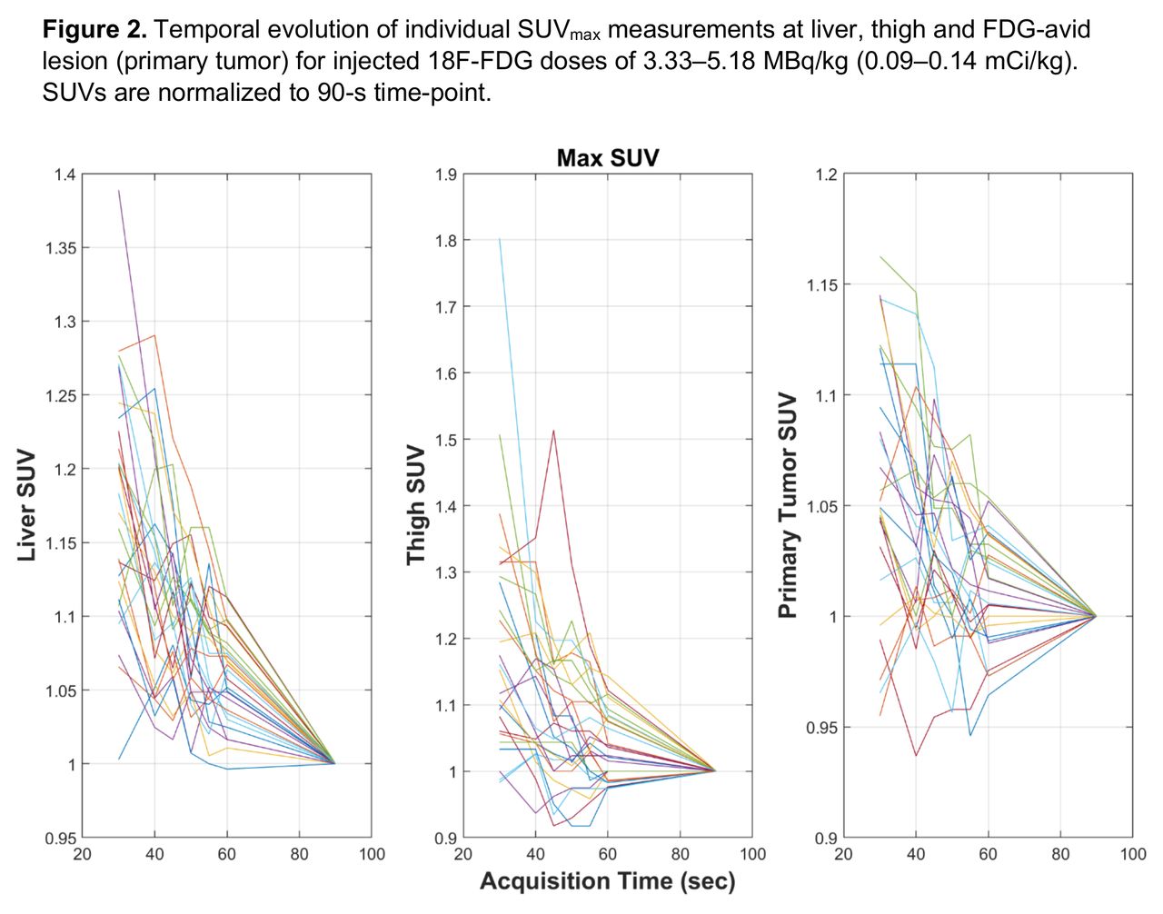
Results: Patients included in this study had a mean age of 10.8 ± 8.3 yr, a weight of 36.3 ± 19.7 kg, and received 0.12 ± 0.01 mCi/kg 18F-FDG. As compared to the 90 sec/bed standard-of-care protocol, a significant difference was observed for lesion conspicuity scores at &le; 40 sec/bed (p=0.007), for conspicuity of normal structures and overall image quality at &le; 45 sec/bed (p=0.038 and p=0.006, respectively), and image noise at &le; 55 sec/bed (p=0.025) (Figure 1). Quantitatively, no statistical difference was observed for SUVmean in liver, thigh, or lesion across all reconstructed acquisition times, although SUVstandard deviation/ SUVmean and SUVmax showed a trend of increase with decreased acquisition times&nbsp;(p<0.0001).&nbsp;Significant differences were present for SUVstandard deviation/SUVmean of liver at &le; 60 sec/bed (p= 0.044); thigh at &le; 55 sec/bed (p = 0.018) and lesions at &le; 50 sec/bed (p = 0.015).&nbsp;Figure 2 demonstrates the decreasing variability in SUVmax with increasing acquisition times.

Conclusions: For children and young adults receiving 0.12 mCi/kg injected activity of 18F-FDG and imaged with a state-of-the-art extended field of view digital PET/CT system, an acquisition time of 60 sec/bed (or 0.8 mCi/kg injected activity imaged at 90 sec/bed) shows no significant impact in image quality or quantitative measures.

Figure
  • Download figure
  • Open in new tab
  • Download powerpoint
Figure
  • Download figure
  • Open in new tab
  • Download powerpoint
Previous
Back to top

In this issue

Journal of Nuclear Medicine
Vol. 63, Issue supplement 2
August 1, 2022
  • Table of Contents
  • Index by author
Article Alerts
Sign In to Email Alerts with your Email Address
Email Article

Thank you for your interest in spreading the word on Journal of Nuclear Medicine.

NOTE: We only request your email address so that the person you are recommending the page to knows that you wanted them to see it, and that it is not junk mail. We do not capture any email address.

Enter multiple addresses on separate lines or separate them with commas.
Feasibility of Reduced Count Acquisition of Whole Body 18F-FDG PET in Children and Young Adults Imaged with a Digital PET Scanner
(Your Name) has sent you a message from Journal of Nuclear Medicine
(Your Name) thought you would like to see the Journal of Nuclear Medicine web site.
Citation Tools
Feasibility of Reduced Count Acquisition of Whole Body 18F-FDG PET in Children and Young Adults Imaged with a Digital PET Scanner
Vinicius Alves, Nadeen Abu Ata, Yinan Li, Susan Sharp, Joseph MacLean, Samuel Brady, Andrew Trout
Journal of Nuclear Medicine Aug 2022, 63 (supplement 2) 2384;

Citation Manager Formats

  • BibTeX
  • Bookends
  • EasyBib
  • EndNote (tagged)
  • EndNote 8 (xml)
  • Medlars
  • Mendeley
  • Papers
  • RefWorks Tagged
  • Ref Manager
  • RIS
  • Zotero
Share
Feasibility of Reduced Count Acquisition of Whole Body 18F-FDG PET in Children and Young Adults Imaged with a Digital PET Scanner
Vinicius Alves, Nadeen Abu Ata, Yinan Li, Susan Sharp, Joseph MacLean, Samuel Brady, Andrew Trout
Journal of Nuclear Medicine Aug 2022, 63 (supplement 2) 2384;
Twitter logo Facebook logo LinkedIn logo Mendeley logo
  • Tweet Widget
  • Facebook Like
  • Google Plus One
Bookmark this article

Jump to section

  • Article
  • Figures & Data
  • Info & Metrics

Related Articles

  • No related articles found.
  • Google Scholar

Cited By...

  • No citing articles found.
  • Google Scholar

More in this TOC Section

  • Effect of phosphorus-32 application combined with pulsed laser on superficial strawberry hemangioma in infants
  • 2-[18F] FDG-PET/CT as a Predictor for Bone Marrow Involvement in Paediatric Patients with Langerhans Cell Histiocytosis
  • Comparison between the diagnostic accuracy of F-18 FDG PET/CT scan and Tc-99m MDP bone scintigraphy for detection of distant metastasis in extra-ocular retinoblastoma - a single centre initial experience
Show more Pediatrics

Similar Articles

SNMMI

© 2025 SNMMI

Powered by HighWire