


Article Figures & Data
Figures
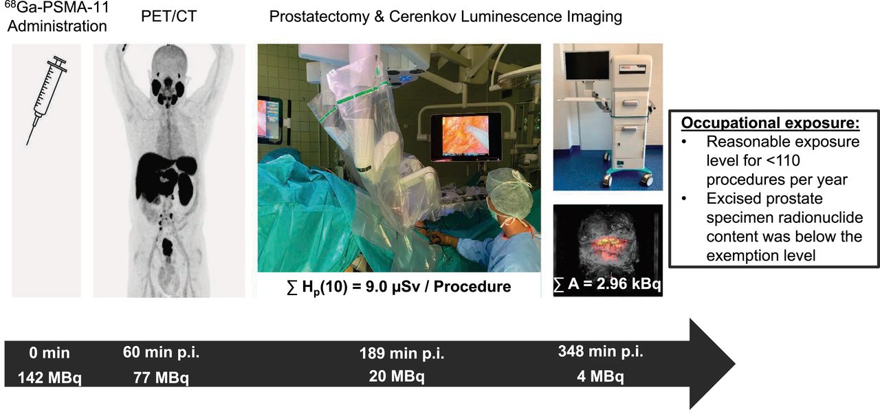
- FIGURE 1.
One-stop-shop protocol, including tracer administration (A), PET/CT imaging (B), and CLI during prostatectomy (C–E), permitted by remainder of 68Ga-PSMA-11 uptake in prostate. Temporal sequence (black arrow) shows median time points after injection (p.i.) and decay-corrected whole-body activity expected for each step of protocol.
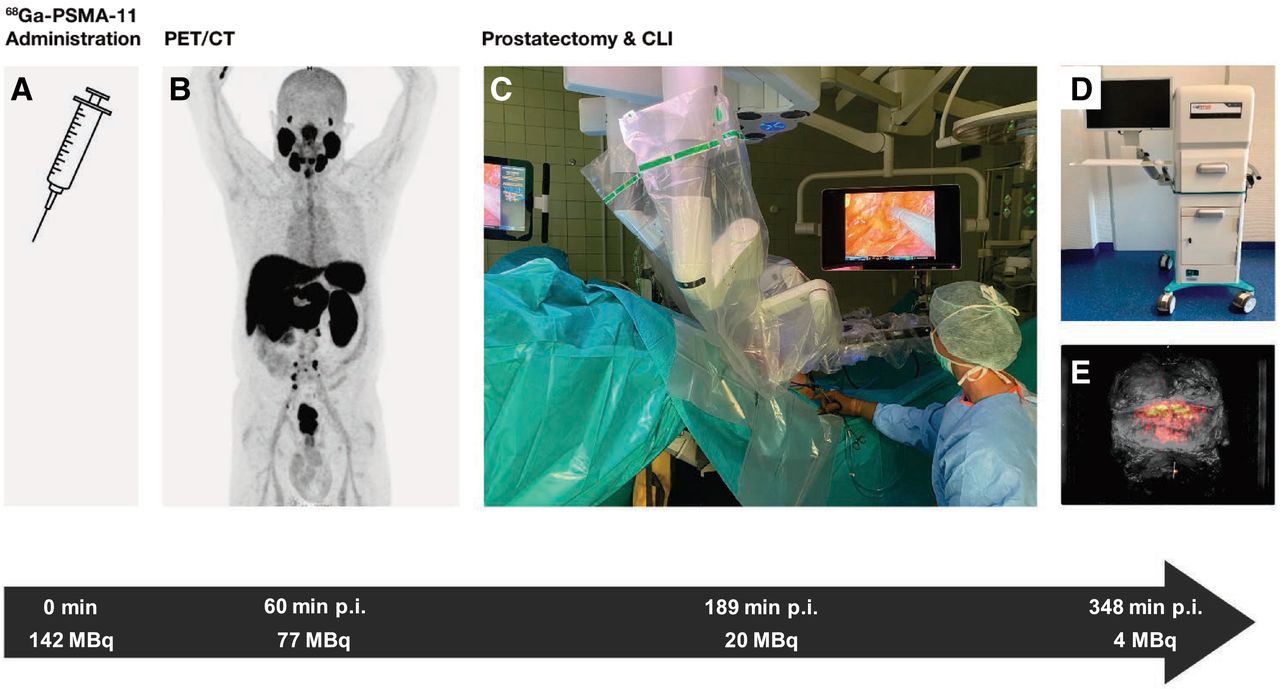
- FIGURE 2.
Setup in operating theater and respective distances used for calculated personal equivalent doses based on patient-specific dose rate constants.
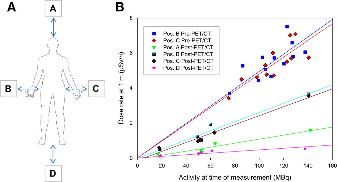
- FIGURE 3.
Dose rate measurements at predefined positions (Pos.) (A) and respective linear regression lines (B). Positions are as follows: A = at head; B = right side; C = left side; D = at feet. Post-PET/CT = dose rate readings immediately before entering operating room; Pre-PET/CT = measurements immediately after tracer injection.
- FIGURE 4.
EPD measurements (black bars) alongside computed values based on point source (PS) model (medium gray bars) at measured distance r. Variations of minus −10 cm (dark gray bars) and +10 cm (light gray bars) from distance r using PS assumption are shown.
- FIGURE 5.
Activity quantification of excised prostate specimens decay corrected to time of excision. Black bars represent PET-based quantification (median, 3.83 kBq; IQR, 2.83–8.50 kBq). Gray bars represent HPGe measurement (GeDet) (median, 2.96 kBq; IQR, 2.23–7.65 kBq).


Tables
- TABLE 1.
Summary of Dose Rate Measurements, Activity at Time of Measurement, and Respective Patient-Specific Dose Rate Constants (Dose Rate/Activity)
Position Activity, MBq Dose Rate/Activity, μSv ⋅ h−1 ⋅ MBq−1 r2 Average SD Average SD Point source (1 m) 69.96 2.3 0.145 0.002 Patient before PET (1 m) 112.2 17.7 B 0.047 0.004 0.70 C 0.047 0.005 0.66 Patient after PET (1 m) 64.5 41.7 A 0.011 0.003 0.92 B 0.026 0.004 0.96 C 0.024 0.005 0.97 D 0.003 0.004 0.84 Characteristic Value Patient and imaging* Age, in y 63 (56.5–69) BMI, in kg/m2 30.37 (24.25–34.68) Injected activity, in MBq 122 (99.25–185) Activity derived from HPGe, in kBq/mL, corrected to time of excision 2.96 (2.24–7.65) Activity derived from PET/CT in prostate, in kBq/mL, corrected to time of excision 3.83 (2.83–8.50) Duration from tracer injection to CLI, in min 328.5 (298.75–371.75) Duration from skin incision to CLI, in min 130.2 (125.4–145.2) Surgical and oncologic† Organ-confined PC 5 (50) Locally advanced PC 5 (50) Initial PSA, in ng/mL* 12.5 (8.3–15.25) Risk stratification according to D’Amico (34) Intermediate-risk PC 5 (50) High-risk PC 5 (50) ISUP-GGG 2 4 (40) 3 4 (40) 4 0 5 2 (20) Prostate specimen weight, in g* 43.5 (41.25–55)


