


SNMMI announced on June 13 at its Annual Meeting in Vancouver, Canada, the 2022 Henry N. Wagner, Jr., Image of the Year, chosen from more than 1,280 submitted scientific abstracts and voted on by reviewers and society leadership. As part of the presentation “Predicting remodeling and outcome from molecular imaging of fibroblast activation in patients after acute myocardial infarction,” the image from Diekmann et al. from the Hannover Medical School (Germany) contrasted 68Ga–fibroblast-activation protein–46 (68Ga-FAP-46) PET/CT, 99mTc-tetrofosmin perfusion SPECT, and cardiac MR images acquired in patients within 11 days after acute myocardial infarction.
“Molecular PET imaging of the fibroblast activation protein has recently been evaluated in patients after acute myocardial infarction,” stated Johanna Diekmann, MD, lead author of the presentation. “In our study, we sought to obtain further insights by correlating FAP-targeted PET imaging with tissue characteristics from cardiac MRI, as well as functional outcome.”
The study included 35 patients, in whom infarct size was determined from SPECT, cardiac FAP volume was calculated from 68Ga-FAP-46 PET/CT uptake values and polar mapping analyses, and MRI provided information on functional parameters, area of injury, and tissue characteristics. Cardiac function in a subgroup of 14 patients was followed up by echocardiography or cardiac MRI at a median of 139.5 days.
In all patients the area of FAP upregulation was significantly larger than that in either SPECT perfusion defect size or infarct area as defined by late gadolinium-enhanced MRI. FAP volume on PET/CT was significantly correlated at baseline with multiple cardiac parameters, including infarct area, left ventricular (LV) mass, and end-systolic and -diastolic volumes. PET/CT segmental analysis showed FAP upregulation in 308 of 496 (62%) myocardial segments. MRI late gadolinium enhancement was found in only 56% of FAP-positive segments, elevated T1 in 74%, and elevated T2 in 68%. Fourteen percent (44/308) of FAP-positive segments showed neither prolonged T1 or T2 nor significant late gadolinium enhancement, with a weak correlation between myocardial FAP volume and LV ejection fraction (LVEF) at baseline. Early myocardial PET values showed a stronger correlation with LVEF at follow-up, suggesting a relationship between the extent of fibroblast activation and more severe adverse ventricular remodeling. The authors concluded that because a higher extent of myocardial FAP upregulation was predictive of subsequent LV dysfunction and exceeded infarct area as defined by other modalities, fibroblast activation in noninfarcted myocardial areas may contribute to adverse outcomes. FAP imaging with PET/CT may be a complementary biomarker with applications in establishing treatment strategies to mitigate profibrotic activity outside of the primary infarct region to prevent adverse remodeling.
“Myocardial infarction is an important contributor to the development of heart failure, but the early molecular processes involved in the transition from initial injury to heart failure are undertreated,” said Diekmann. “New antifibrotic therapies (such as CAR-T cell therapies) could significantly change future therapy of heart failure. Using FAP PET to select patients suitable for therapy would open a new major application for PET in fibrosis and cardiac diseases.”
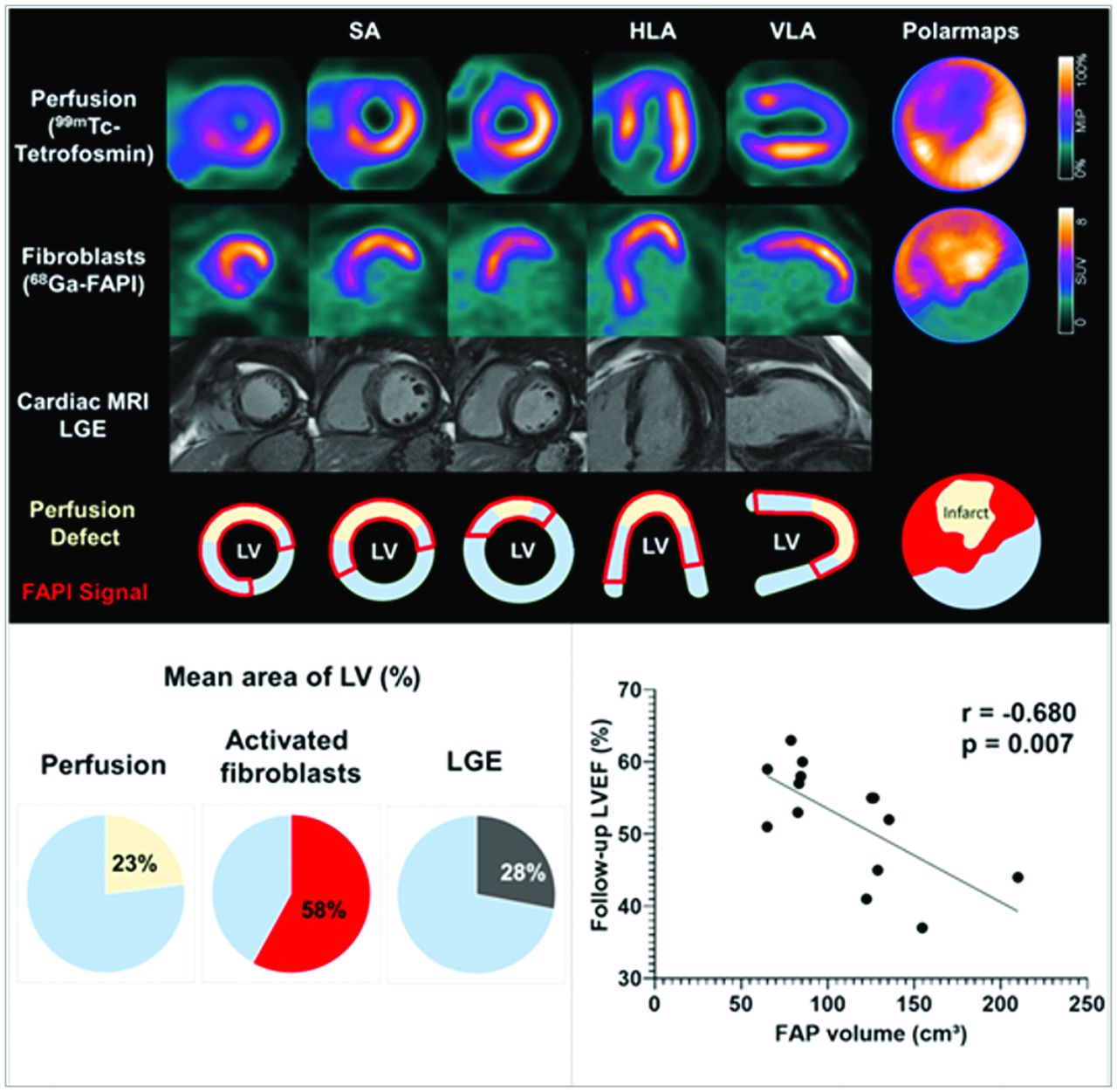
SNMMI 2022 Image of the Year. Top: Representative case with acute anterior wall myocardial infarction. 99mTc-tetrofosmin perfusion SPECT, 68Ga–fibroblast-activation protein–46 (68Ga-FAP-46) PET/CT, and late gadolinium-enhanced (LGE) cardiac MR images were compared in patients within 11 days after acute myocardial infarction. Bottom: Areas of myocardial FAP upregulation on PET/CT were significantly greater than those with either SPECT or MRI and were predictive of subsequent left ventricular (LV) dysfunction. Global myocardial FAP volume early after acute myocardial infarction inversely correlated with left ventricular ejection fraction (LVEF) at follow-up in the chronic stage. The findings suggest promise for 68Ga-FAP-46 PET/CT as a complementary biomarker in predicting adverse cardiac remodeling.
- © 2022 by the Society of Nuclear Medicine and Molecular Imaging.
In this issue


Jump to section
Related Articles
Cited By...
- No citing articles found.