


Article Figures & Data
Figures
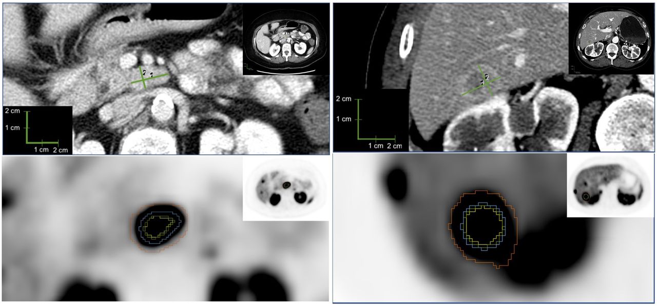
- FIGURE 1.
Example lesion 1. (A) CT evaluation of lymph node, with longest diameter of 2.2 cm (1) and longest perpendicular diameter of 1.3 cm (2). (B) PET evaluation of lymph node, with yellow representing 50% threshold segmentation; green, 42% threshold segmentation; blue, manual segmentation; and red, BSL segmentation.
- FIGURE 2.
Example lesion 2. (A) CT evaluation of hepatic metastasis, with longest diameter of 2.3 cm (1) and longest perpendicular diameter of 1.8 cm (2). (B) PET evaluation of hepatic metastasis, with yellow representing 50% threshold segmentation; green, 42% threshold segmentation; blue, manual segmentation; and red, BSL segmentation.
- FIGURE 3.
Correlation charts of FTV calculations to morphologic measurements. (A) Manual volume from PET imaging (VOLNM). (B) BSL (VOLBSL). (C) Threshold of 50% relative to SUVmax (VOL50). (D) Threshold of 42% relative to SUVmax (VOL42).
- FIGURE 4.
Bland–Altman scatterplots showing relative difference between FTV method as labeled and morphologic volume on y-axis and mean volume of FTV method as labeled and morphologic volume on x-axis. Dashed lines represent upper limits of agreement, lower limits of agreement, and bias (or mean difference). Log transformation was used to correct skewness in distribution of volumes. (A) Manual volume from PET imaging (VOLNM). (B) BSL (VOLBSL). (C) Threshold of 50% relative to SUVmax (VOL50). (D) Threshold of 42% relative to SUVmax (VOL42).


Tables
Characteristic Data Patients 20 (100%) Sex Female 13 (65%) Male 7 (35%) Age at 68Ga-DOTATATE PET (y) Mean ± SD 56 ± 12 Range 28–78 NET primary tumor subtype (%) Pancreatic 11 (55%) Small intestine 5 (25%) Other 3 (15%) Unknown 1 (5%) Gastroenteropancreatic NET grade G1 (Ki-67 < 3%) 5 (29%) G2 (Ki-67 = 3%–20%) 9 (53%) G3 (Ki-67 > 20%) 3 (18%) Local recurrence 1 (5%) Metastases No 2 (10%) Yes 18 (90%) Metastatic sites Liver 18 Nodes 8 Bone 3 Adrenal 2 Mesenteric 2 Cardiac 1 Splenic 1 Clinical syndrome Nonfunctioning tumor 12 (60%) Functioning tumor 8 (40%) Data are number, except for age.
Treatment n Resection of primary tumor 9 (45%) Additional treatments Liver-directed therapy 7 (35%) Chemotherapy 4 (20%) Radiotherapy 1 (5%) Peptide radionuclide receptor therapy 0 Treatment with cold SSA at time of 68Ga-DOTATATE PET/CT 8 (40%) Parameter Data SUVmax Mean ± SD 36.9 ± 27.0 Range 1.3–188.3 Lesions analyzed 94 Site of lesions Liver 69 (73.4%) Node 10 (10.6%) Pancreas 5 (5.3%) Bone 5 (5.3%) Bowel 2 (2.1%) Perihepatic implant 2 (2.1%) Mesenteric node 1 (1.1%) Data are number, except for SUVmax.


