


Article Figures & Data
Figures
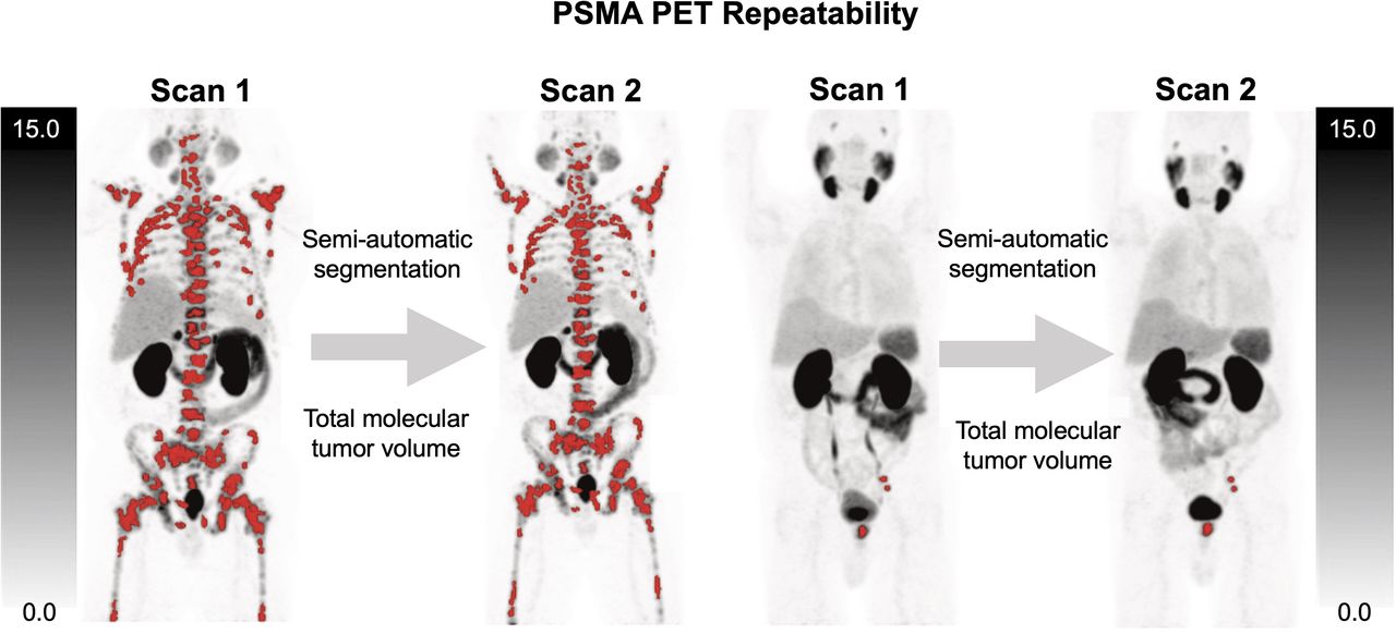
- FIGURE 1.
Semiautomatic total tumor segmentations with red overlay designating sites of segmented lesions in scans 1 and 2 for patient with disease limited to prostate and left pelvic lymph nodes (A) and patient with extensive skeletal metastases (B). Interval between scans was 2 d for both patients.
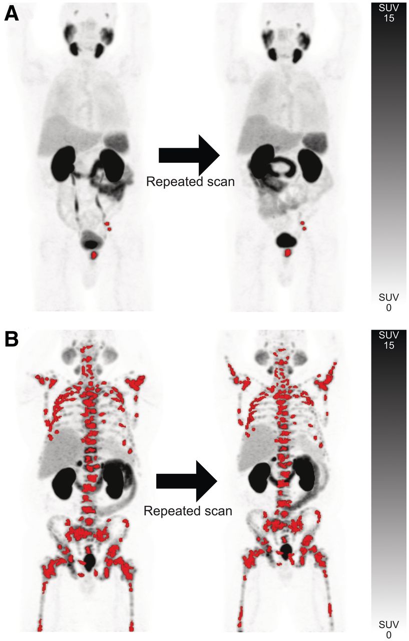
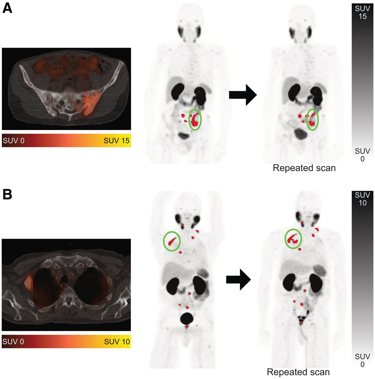
- FIGURE 2.
Examples of segmentation challenges on 68Ga-PSMA-HBED-CC PET/CT. Segmented tumor metastases are shown in red. (A) Metastasis in os ilium was segmented as single lesion on first scan but as 3 separate lesions in second scan (encircled). (B) Metastasis in rib was segmented accurately on first scan but inaccurately on second scan, with isocontour including portion of lung (encircled). Error was resolved manually.
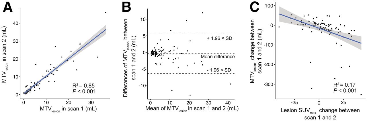
- FIGURE 3.
Analysis of individual manually segmented 68Ga-PSMA-HBED-CC–avid lesions. Linear regression and Bland–Altman plots (A and B) of MTVlesion show correlation between scans. (C) Association is noted between MTVlesion and SUVmax changes between scans 1 and 2.
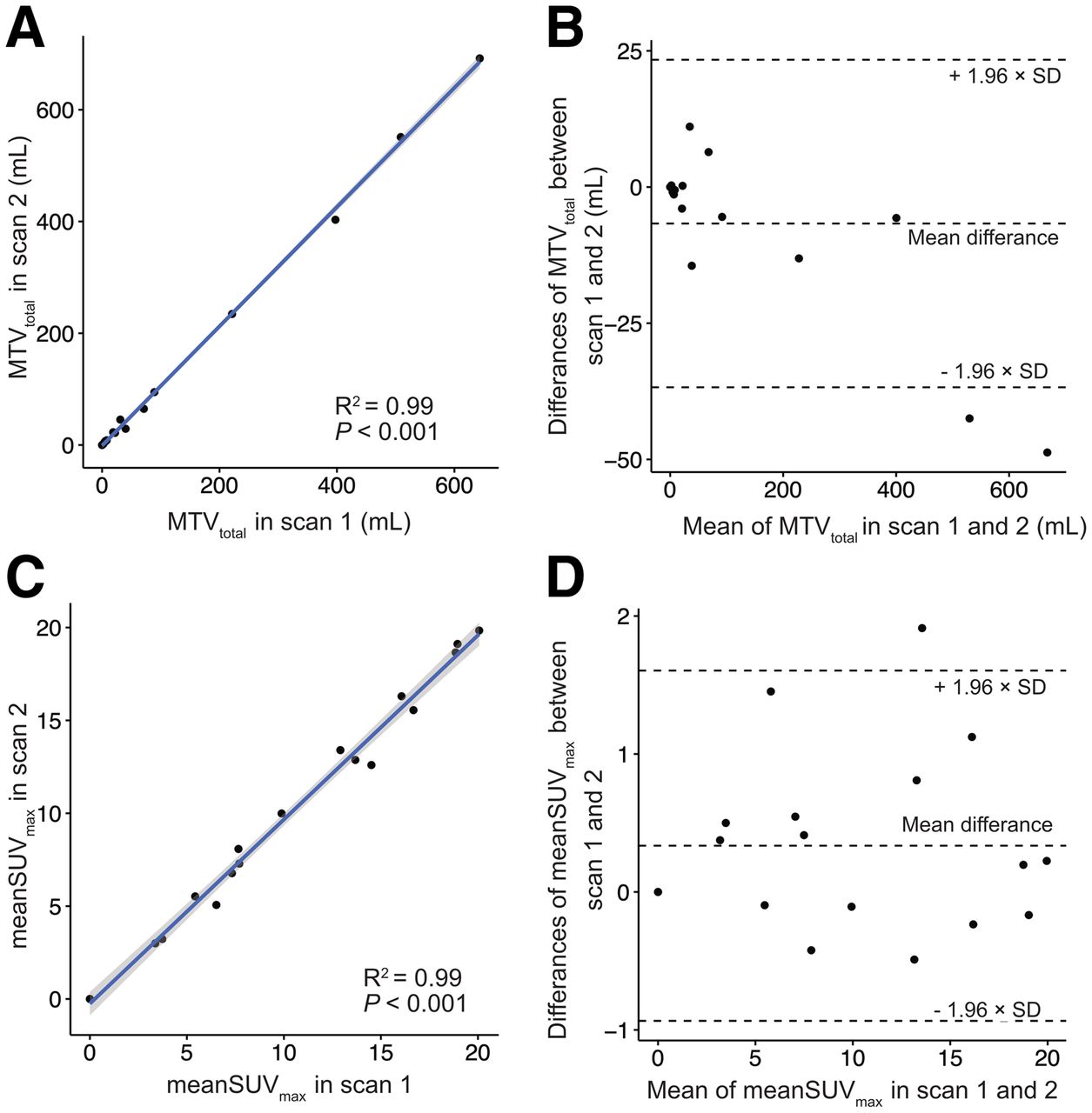
- FIGURE 4.
Analysis of semiautomatic whole-body segmentation of 68Ga-PSMA-HBED-CC–avid lesions. Linear regression (A and C) and Bland–Altman plots (B and D) of MTVtotal and mean SUVmax show excellent correlation between scans and suggest no association between total tumor volume or lesion intensity and test–retest differences. Results for readers 1 and 2 were averaged for purposes of these graphs. (A and C) MTVtotal and mean SUVmax for scan 1 are plotted separately against same metric for scan 2. (B and D) Mean of MTVtotal or mean SUVmax between scans 1 and 2 was plotted against absolute difference in metric between 2 scans.
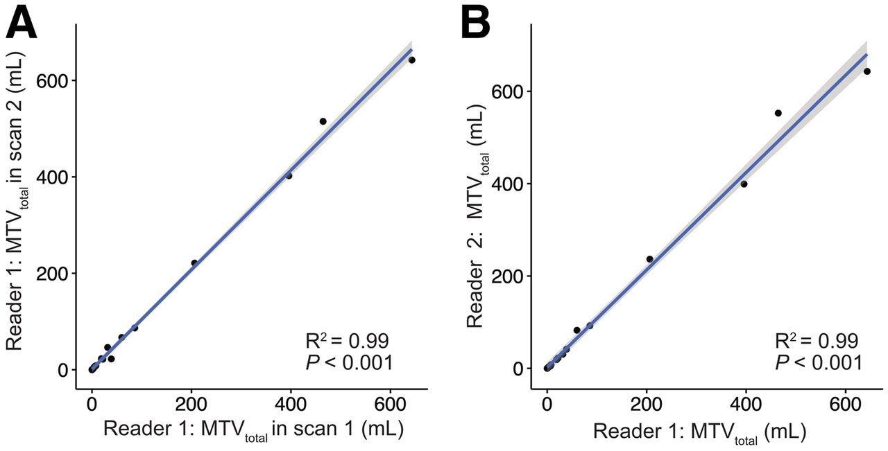
- FIGURE 5.
Graphical analysis of intra- and interreader agreement in reporting MTVtotal, showing high correlation in measures between scans 1 and 2 for same reader (reader 1) (A) and showing high correlation in measures between 2 independent readers for same scan (scan 1) (B).
- FIGURE 6.
Graphical analysis of prostate-specific antigen vs. MTVtotal, with log–log plot showing moderate correlation.


Tables
MTVtotal (mL) Patient no. PSA within ≤90 d (ng/mL) Gleason score at diagnosis R1, scan 1 R2, scan 1 R1, scan 2 R2, scan 2 1 0.15 7 (4 + 5) 0 0 0 0 2 4.35 6 (3 + 3) 4.81 5.88 4.81 5.88 3 104.5 9 (4 + 5) 395.7 404.02 399.18 402.22 4 0.14 9 (4 + 5) 59.91 62.59 82.42 66.9 5 0.66 9 (5 + 4) 6.42 6.77 5.18 7.56 6 0.22 9 (5 + 4) 3.78 4.67 3.78 4.67 7 56.3 Presumptive diagnosis 38.89 35.59 41.36 22.49 8 95.5 7 (4 + 3) 206.38 247.85 236.35 221.08 9 276.3 9 (4 + 5) 643.19 741.4 643.19 642.43 10 0.04 Presumptive diagnosis 0 0 0 0 11 0.64 9 (4 + 5) 7.78 8.33 7.78 8.33 12 2.8 Lymph node biopsy 31.49 44.68 30.53 46.24 13 40.1 10 (5 + 5) 464.53 587.13 552.7 515.05 14 19.7 7 (3 + 4) 18.87 22.83 18.87 22.83 15 2.5 Bone biopsy 2.26 1.96 2.26 1.96 16 54.1 9 (5 + 4) 85.89 102.6 92.3 86.56 17 2.5 9 (5 + 4) 21.78 21.81 22.29 21.81 18 2.5 9 (5 + 4) 6.52 6.31 5.53 7.34 PSA = prostate-specific antigen; R1 = reader 1; R2 = reader 2.
Metric wCV (%) RC (%) 95% CI of RC (%) MTVlesion 27.7 76.9 62.9–95.9 PSMA-TLlesion 23.3 64.7 53.4–80.67 PSMA-TLQlesion 34.5 95.7 81.5–114.5 Lesion SUVmax 12.4 34.4 29.6–41.2 Lesion SUVpeak 9.9 27.3 23.3–32.8 Lesion SUVmean 11.8 32.7 27.5–40.2 Metric wCV (%) RC (%) 95% CI of RC MTVsubgroup 12.0 33.1 24.2–46.2 Subgroup MTVmean 12.0 33.1 24.8–47.7 PSMA-TLsubgroup 7.4 20.6 16.0–26.9 PSMA-TLQsubgroup 18.4 51.0 36.5–78.0 Subgroup mean SUVmax 12.3 34.0 20.0–59.4 Subgroup mean SUVpeak 6.6 18.3 13.3–24.5 Subgroup mean SUVmean 9.1 25.2 17.5–35.7 Metric R1 wCV (%) R2 wCV (%) Mean wCV (%) R1 RC (%) R2 RC (%) Mean RC (%) 95% CI of mean RC MTVtotal 13.4 11.9 12.7 37.0 33.0 35.0 24.9–49.7 Total MTVmean 13.4 11.9 12.7 37.1 33.0 35.0 25.0–48.8 PSMA-TLtotal 8.4 12.1 10.3 23.3 33.5 28.4 20.7–41.9 PSMA-TLQtotal 19.4 17.3 18.4 53.9 48.0 50.9 32.7–84.7 Total mean SUVmax 8.4 8.6 8.5 23.3 23.9 23.6 17.0–32.4 Total mean SUVmean 8.1 8.0 8.1 22.6 22.2 22.4 16.4–30.7 R1 = reader 1; R2 = reader 2.
Metric R1, R2 RC (%) R2, R1 RC (%) Mean RC (%) 95% CI of mean RC MTVtotal 29.9 44.7 37.3 27.9–49.3 Total MTVmean 29.9 44.7 37.3 29.9–44.7 PSMA-TLtotal 24.9 37.2 31.0 24.5–39.5 PSMA-TLQtotal 52.5 58.4 55.5 38.1–83.6 Total mean SUVmax 28.3 20.7 24.5 17.5–33.5 Total mean SUVmean 27.4 18.7 23.1 17.2–31.1 R1, R2 = first scan read by reader 1, second scan read by reader 2; R2, R1 = first scan read by reader 2, second scan read by reader 1.
Supplemental Data
Files in this Data Supplement:


