


Article Figures & Data
Figures
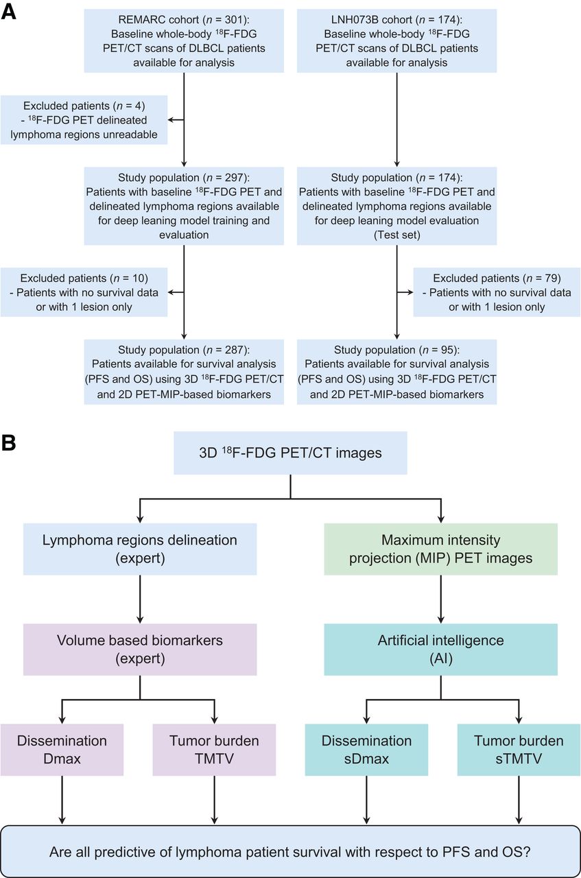
- FIGURE 1.
(A) Study flowchart. (B) Study design.
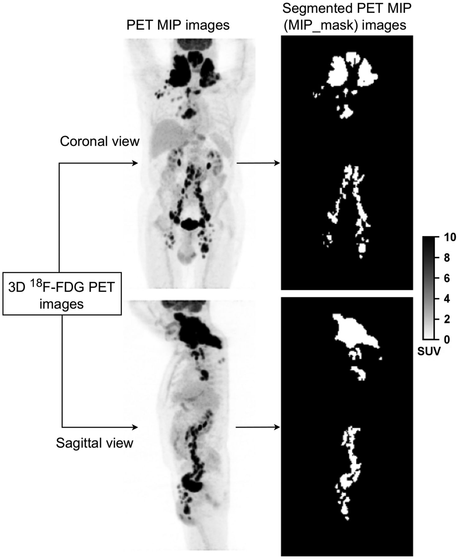
- FIGURE 2.
Example of 18F-FDG PET MIP images (left) and associated lymphoma regions (right) based on expert delineation of the 3D 18F-FDG PET images.
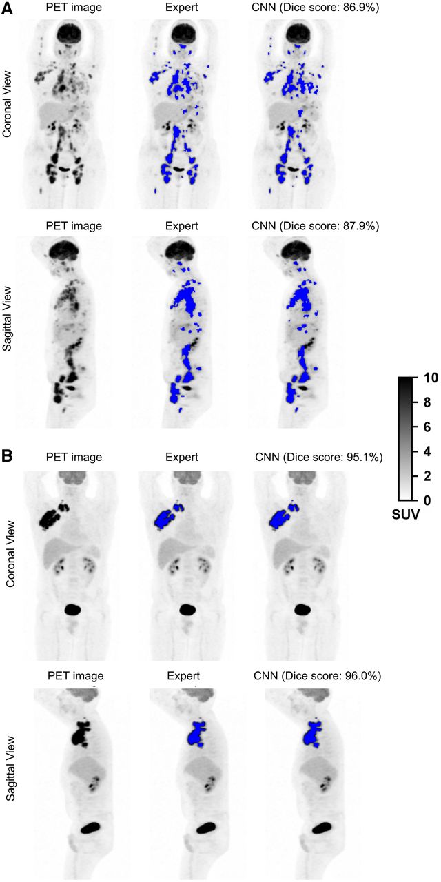
- FIGURE 3.
18F-FDG PET MIP images and segmentation results (blue color overlapped over PET MIP images) by experts (MIP_masks) and by CNN for 4 patients: from REMARC cohort (A) and from LNH073B cohort (B).
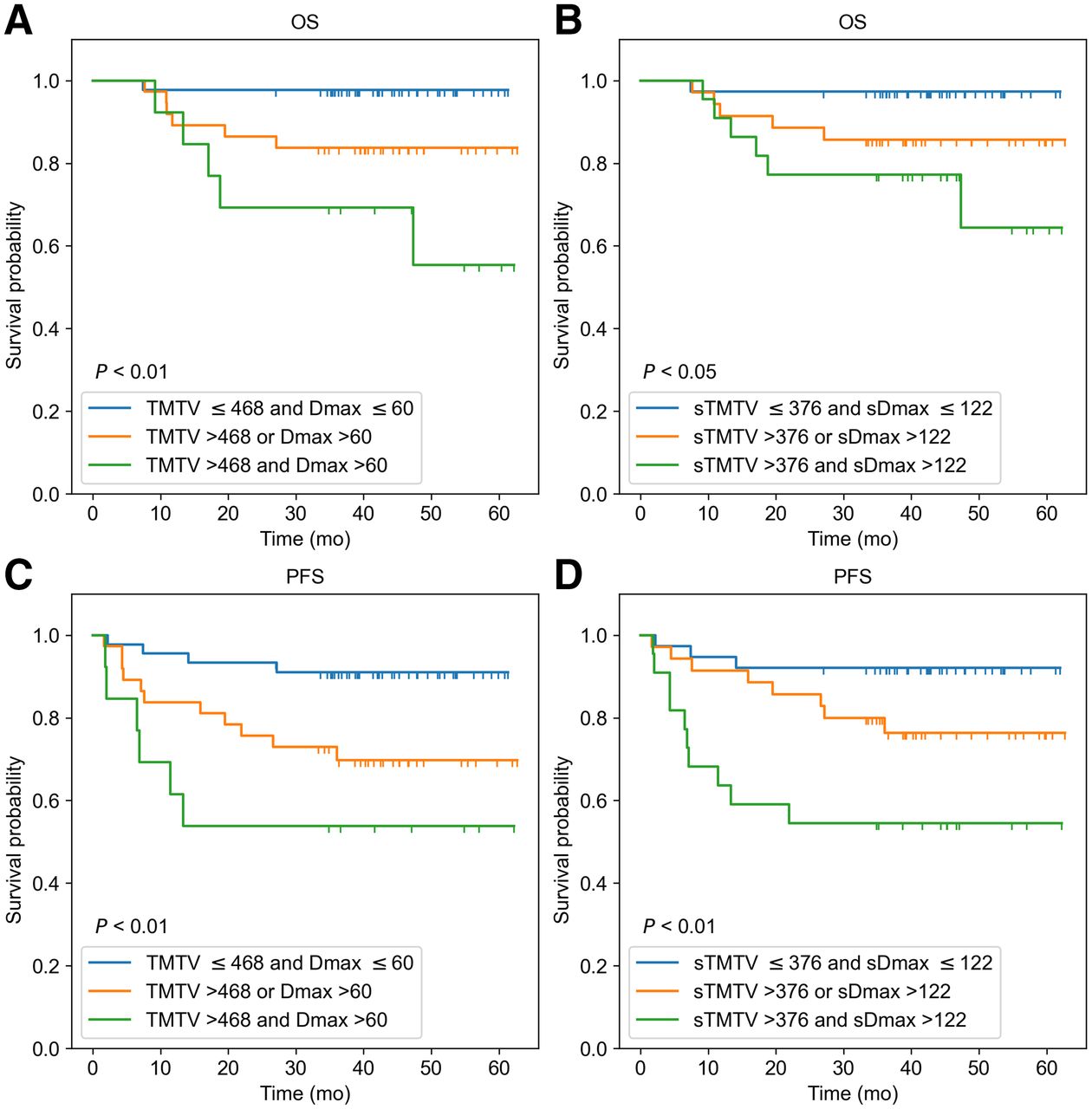
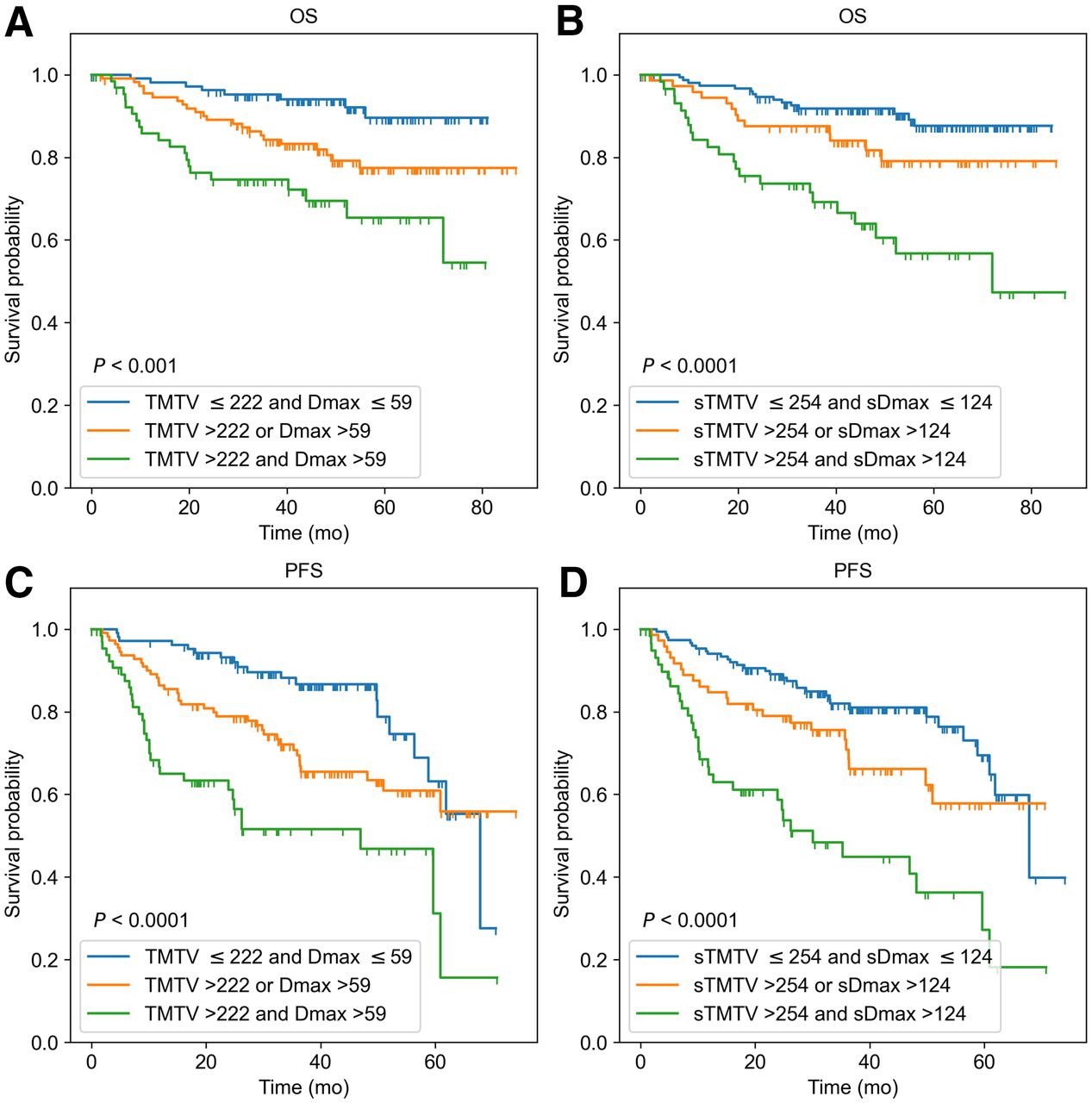
- FIGURE 4.
Kaplan–Meier estimates of OS and PFS from REMARC cohort according to 3D 18F-FDG PET/CT image–based features TMTV (cm3) and Dmax (cm) (A and C), and according to PET MIP image–based features sTMTV (cm2) and sDmax (cm) estimated from AI (B and D).
- FIGURE 5.
Kaplan–Meier estimates of OS and PFS from LNH073B cohort according to 3D 18F-FDG PET/CT image–based features TMTV (cm3) and Dmax (cm) (A and C), and according to PET MIP image–based features sTMTV (cm2) and sDmax (cm) estimated from AI (B and D).


Tables
Characteristic REMARC LNH073B No. of patients 287 95 Sex No. of men 165 (57.5%) 42 (44%) No. of women 122 (42.5%) 53 (56%) Median age (y) 68 (IQR, 64.0–73.0) 46 (IQR, 33.25–55.0) Median weight (kg) 72 (IQR, 63.0–84.2) 68 (IQR, 58.0–80.0) Median height (cm) 167.5 (IQR, 160.0–175.0) (1 case missed) 173 (IQR, 140.0–193.0) Ann Arbor stage <I 1 (0.4%) 0 (0%) ≥II 286 (99.6%) 95 (100%) Performance status 0 115 (40%) 0 (0%) 1 121 (42%) 27 (28.4%) 2 42 (14.6%) 43 (45.3%) 3 2 (0.7%) 20 (21.1%) 4 2 (0.7%) 5 (5.3%) Missing 5 (1.7%) NA IQR = interquartile range (quartile 1 to quartile 3); NA = not applicable.
Cohort sTMTV/sDmax Mean SD Minimum Q1 (25%) Median Q3 (75%) Maximum REMARC sTMTV (cm2) 252.27 245.75 0.48 77.04 174.24 350.56 1339.36 sDmax (cm) 100.16 49.89 0.40 66.20 98.0 135.0 225.20 LNH073B sTMTV (cm2) 388.12 249.91 63.68 224.48 307.2 450.08 1186.24 sDmax (cm) 121.82 41.10 43.20 92.00 116.40 145.60 222.40 Q1 = first quartile (25% percentile); Q3 = third quartile (75% percentile).
- TABLE 3.
Results of the Univariate Analyses for PFS and OS Using Time-Dependent AUC Analysis and Cox Models (HR)
3D 18F-FDG PET/CT estimates 2D PET MIP estimates Data PFS/OS Metrics TMTV Dmax sTMTV sDmax REMARC PFS AUC 0.67 (0.60–0.73) 0.65 (0.58–0.72) 0.65 (0.58–0.72) 0.68 (0.62–0.75) HR 11.24 (2.10–46.20) 9.0 (2.53–23.63) 11.81 (3.29–31.77) 12.49 (3.42–34.50) OS AUC 0.67 (0.58–0.76) 0.62 (0.53–0.71) 0.67 (0.58–0.76) 0.68 (0.59–0.76) HR 16.43 (2.42–77.29) 8.60 (1.47–28.33) 22.14 (4.73–69.06) 22.79 (3.80–79.21) LNH073B PFS AUC 0.62 (0.49–0.75) 0.56 (0.39–0.72) 0.66 (0.53–0.80) 0.58 (0.41–0.74) HR 13.79 (0.45–86.80) 32.83 (0.4–220.8) 9.24 (0.95–37.94) 16.79 (0.69–86.41) OS AUC 0.65 (0.46–0.82) 0.51 (0.31–0.72) 0.64 (0.45–0.82) 0.50 (0.29–0.72) HR 64.30 (0.74–384.80) 49.21 (0.07–258.3) 14.17 (0.59–67.02) 20.39 (0.08–93.66)
Supplemental Data
Files in this Data Supplement:
In this issue


Jump to section
Related Articles
Cited By...
- The Use of Maximum-Intensity Projections and Deep Learning Adds Value to the Fully Automatic Segmentation of Lesions Avid for [18F]FDG and [68Ga]Ga-PSMA in PET/CT
- Integration of clinical, pathological, radiological, and transcriptomic data improves the prediction of first-line immunotherapy outcome in metastatic non-small cell lung cancer
- Promising Candidate Prognostic Biomarkers in [18F]FDG PET Images: Evaluation in Independent Cohorts of Non-Small Cell Lung Cancer Patients
- Tumor Location Relative to the Spleen Is a Prognostic Factor in Lymphoma Patients: A Demonstration from the REMARC Trial