


Article Figures & Data
Figures
- FIGURE 1.
OI calculation. OI was designed to identify voxels with stable high activity over time using 2 consecutive tau PET scans.
- FIGURE 2.
Examples of low OI and high OI. Three consecutive 3-dimensional scatterplots are displayed in each box for 4 different examples, representing tau PET SUVR of each voxel in each scan from individual subject. (A) Low-OI cases. (B) High-OI cases. Below each rendering, median SUVR represents median value for all voxels in each region. Color bar indicates intensity of each voxel. Font color of median SUVR is red when >1.29 and blue when <1.29. Arrows in B indicate regions showing spatial consistency. Various anatomic regions are plotted and labeled in each panel.
- FIGURE 3.
Relationship between OI and baseline SUVR in single ROI. Bilateral ROIs were included in calculations. Small panel inside figure illustrates enlarged view of lower SUVR range (from 0.9 to 1.5).
- FIGURE 4.
Relationship between meta-ROI OI and meta-ROI SUVR. (A) Scatterplot (left) of baseline SUVR and OI for meta-ROI. Histograms are displayed along SUVR and OI axis, respectively. Low SUVR range (<1.5) was magnified in separate scatterplot (right) with linear regression (solid black line) and 95% confidence band (dotted black lines). (B) Scatterplot of meta-ROI OI and ΔSUVR with regression. (C) Comparison of ΔSUVR between SUVR-based subgroups. (D) SUVR-based subgroups in C were further separated into low-OI and high-OI categories. *P < 0.05, post hoc Dunn tests. **P < 0.05, post hoc Dunn tests. ***P < 0.005, post hoc Dunn tests.
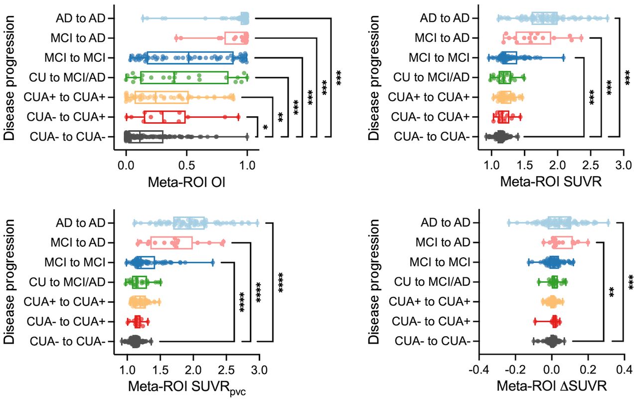
- FIGURE 5.
Association of OI with disease progression. (A) Tau PET variables in different clinical groups. OI, baseline SUVR, baseline SUVR with partial-volume correction (SUVRpvc), and ΔSUVR from meta-ROI of CUA− to CUA− were compared with those of other groups. *P < 0.05, post hoc Dunn tests. **P < 0.05, post hoc Dunn tests. ***P < 0.005, post hoc Dunn tests.
- FIGURE 6.
Result for ADNI cohort. (A) Scatterplot of baseline SUVR and OI for meta-ROI. (B) Comparison of ΔSUVR between SUVR-based subgroups. (C) SUVR-based subgroups in B were further separated into low-OI and high-OI categories. (D) OI, baseline SUVR, and ΔSUVR from meta-ROI of CUA− to CUA− were compared with those of other groups. *P < 0.05, post hoc Dunn tests. **P < 0.05, post hoc Dunn tests. ***P < 0.005, post hoc Dunn tests.


Additional Files
Supplemental Data
Files in this Data Supplement:


