


Article Figures & Data
Figures
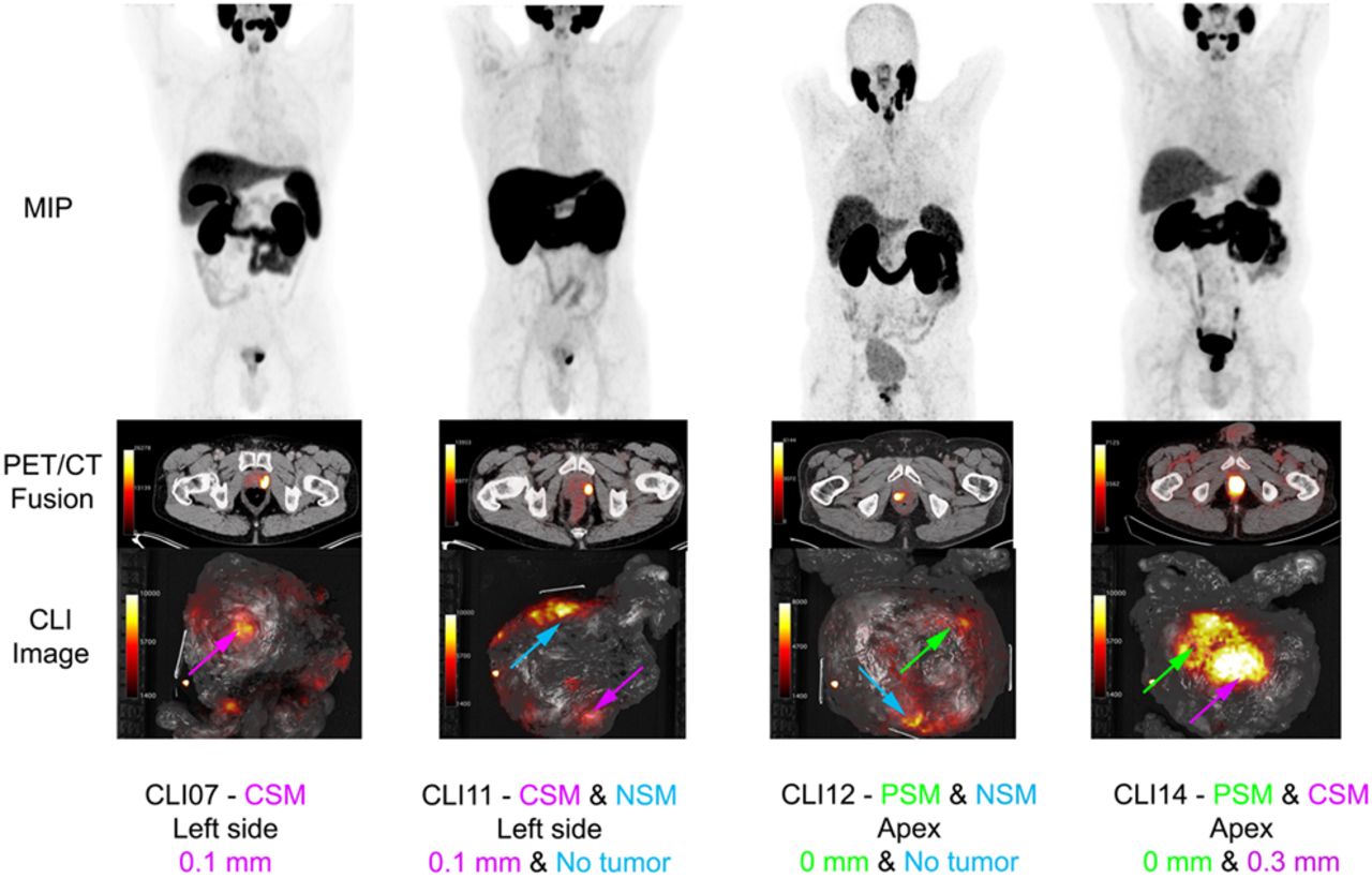
- FIGURE 1.
Examples of PET/CT and CLI images from 4 patients: maximum-intensity projections (MIPs) of preoperative PSMA PET, transversal PET/CT images at height of primary tumor, and CLI images of excised prostate specimens without optical filter. Arrows locate hot-spot areas; green, PSM; blue, NSM; and pink, CSM with tumor ≤ 1 mm from margin, according to pathologist. Corresponding tumor–to–specimen edge distances on histopathology are noted below images.
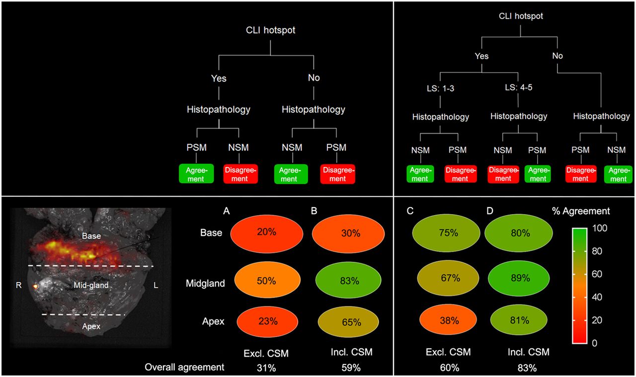
- FIGURE 2.
Agreement between CLI and histopathology in all patients divided into 3 regions of prostate. (A) Agreement between CLI hot spot (yes or no) and histopathology (PSM or NSM), excluding CSM. (B) Agreement including CSM (≤1 mm). (C) Agreement when adding LS to CLI hot spot. (D) Agreement with LS, including CSM. Overall agreement is noted below circles. Excl. = excluding; incl. = including.
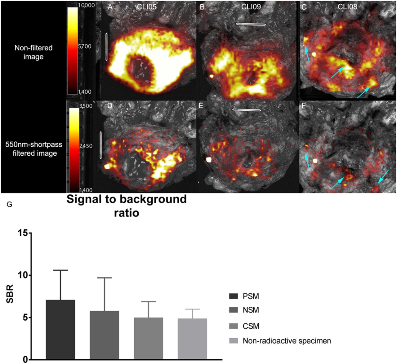
- FIGURE 3.
CLI images from prostate base in 68Ga-PSMA-11 patients and bar chart showing SBR of CLI and chemiluminescence. (A and B) Unfiltered images of chemiluminescence at base. (D and E) Corresponding 550-nm short-pass filtered images. (C and F) Base of patient 8, who had multiple PSMs at base (arrows). These images show that visual distinction between chemiluminescence and actual PSMs is difficult. (G) Bar chart displaying SBR of chemiluminescence in nonradioactive specimens and that of PSM, NSM, and CSM in patient nonfiltered and filtered images. Average SBR is derived from all patient data, in which lesions on all sides of prostate were included. Note difference in scaling.
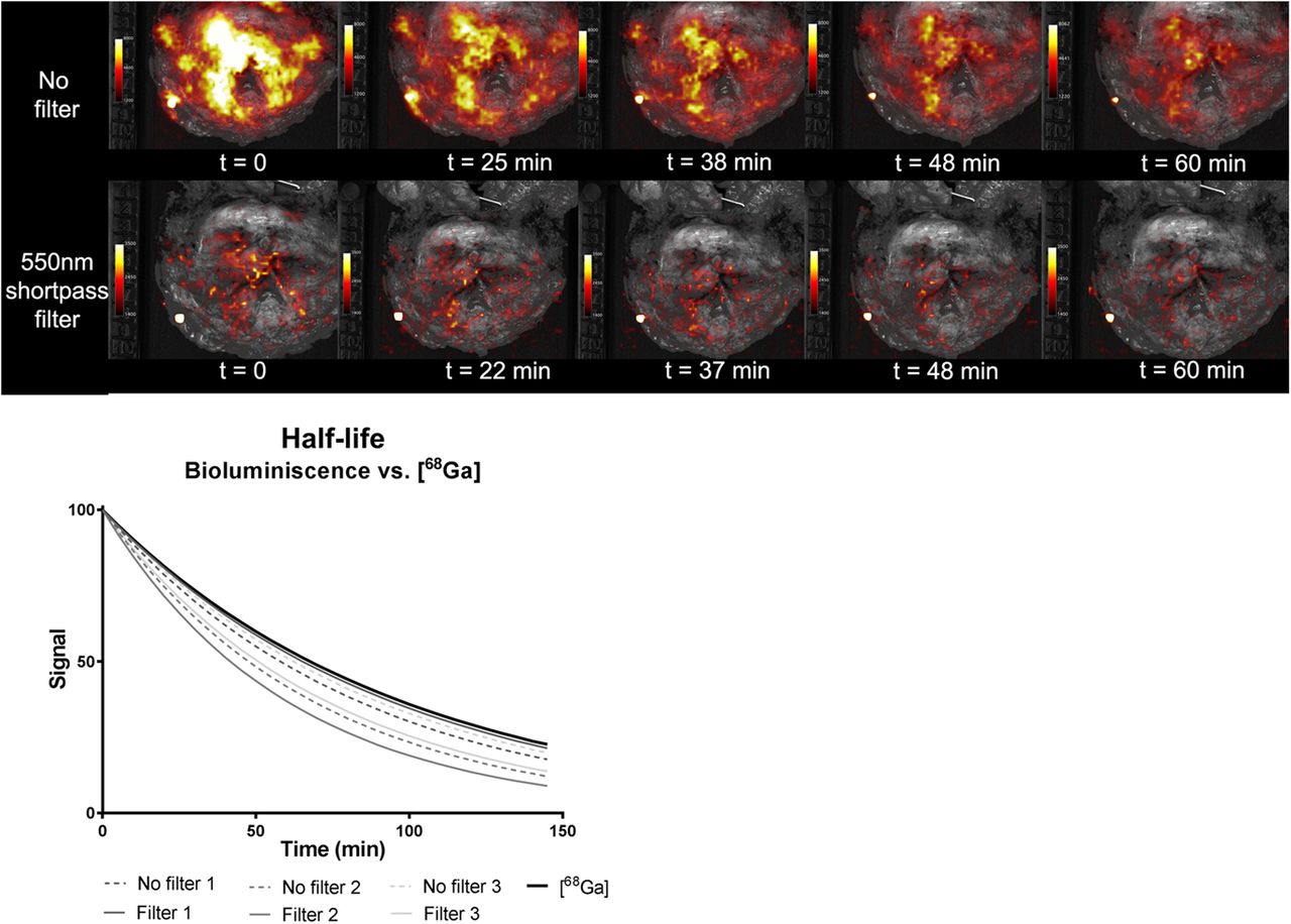
- FIGURE 4.
Sequential imaging of nonradioactive prostate specimens to determine effect of time on intensity of chemiluminescence signal. Same scaling is used in all images. Graph displays half-life of chemiluminescence in 3 nonradioactive specimens on filtered and nonfiltered images and of 68Ga.
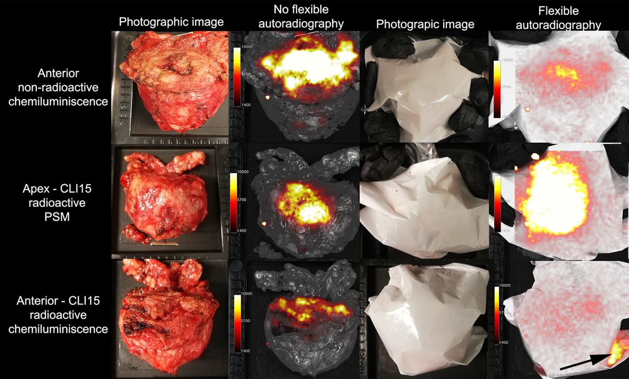
- FIGURE 5.
FAR images of 68Ga-PSMA-11–containing prostate specimens and nonradioactive prostate specimens to investigate effect of FAR on tumor and chemiluminescent signal levels. FAR reduced chemiluminescence in both nonradioactive (60%) and radioactive specimen (70%) (top and bottom rows). FAR amplified signal originating from tumor (center row). Arrow shows contamination of scintillator. Same scaling is used for all images.


Tables
- TABLE 1
Patient Demographics, TNM Classification, Tumor Characteristics, TBR on Preoperative PET/CT Scan, and Information Regarding CLI Imaging
Patient no. Age (y) Preoperative TNM Postoperative TNM PSA (µg/L) Gleason score, preoperative biopsy TBR on PSMA PET Intraoperative injected activity (MBq) Time between injection and CLI imaging (h:min) Estimated* activity in prostate at start of CLI imaging (kBq) 1 67 cT3bN0M0 pT3aN0R1 29.9 4 + 4 = 8 4.9 118.0 01:35 111 2 71 cT2bN0M0 pT3aN0R1 4.4 4 + 4 = 8 8.1 68.4 01:04 93 3 58 cT1cN0M0 pT2cN0R0 5.3 4 + 4 = 8 1.8 88.1 01:25 26 4 73 cT3bN1M0 pT3bN1R0 8.3 4 + 5 = 9 8.9 75.6 00:59 92 5 66 cT3bN0M0 pT3bN0R0 2.7 4 + 4 = 8 2.1 96.7 01:00 52 6 63 cT2aN0M0 pT2N0R0 6.4 4 + 5 = 9 3.8 64.6 01:02 37 7 55 cT2cN0M0 pT3aN1R0 9.3 4 + 4 = 8 3.7 62.1 01:14 61 8 48 cT3bN1M0 pT3bN1R1 4.4 4 + 5 = 9 4.4 23.4 01:05 48 9 73 cT2cN0M0 PT3aN0R0 5.6 4 + 3 = 7 2.7 44.7 00:44 26 10 69 cT3aN0M0 pT3aN0R1 65 4 + 5 = 9 2.4 47.9 00:56 151 11 72 cT2bN0M0 pT3bN1R0 8.7 4 + 4 = 8 2.8 47.6 01:05 42 12 76 cT1cN0M0 pT2N0R1 9.2 3 + 4 = 7 2.5 65.4 01:02 69 13 65 cT2cN0M0 pT3aN0R1 5.1 4 + 3 = 7 1.7 121.1 01:26 8 14 67 cT2bN0M0 pT3bN0R1 18.6 4 + 5 = 9 3.7 37.4 01:14 14 15 74 cT1cN0M0 pT2cN0R0 7.9 4 + 4 = 8 3.5 70.2 01:45 33 ↵* Based on uptake on preoperative PET imaging (uptake in prostate as percentage injected activity).
PSA = prostate-specific antigen; TBR = tumor-to-background ratio.
Patient no. Location of PSM on histopathology LS SBR on CLI Gleason score at PSM Extent of PSM on histopathology (mm) Extent of PSM on CLI (mm) 1 Apex right 5 13.2 8 3 6.9 2 Apex left 5 5.9 8 4 4.3 8 Base central 3 7.1 10 14 14 Base left 3 6.1 10 14 12 Apex right 5 8.7 10 15 12 Base right 3 6.1 9 10 9.6 10 Base central 3 5.1 8 9.0 6.7 12 Apex posterior/mid prostate 5 2.8 3 + 4 = 7 6.0 6.5 13 Apex left 5 2.9 8 1.0 1.4 14 Apex posterior/mid prostate 5 12.3 10 2.0 12.0
Supplemental Data
Files in this Data Supplement:


