


Article Figures & Data
Figures
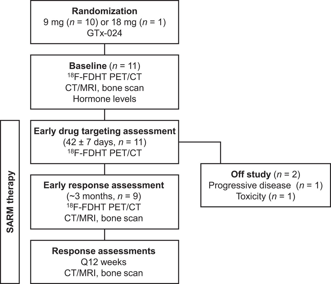
- FIGURE 1.
Study schema.
- FIGURE 2.
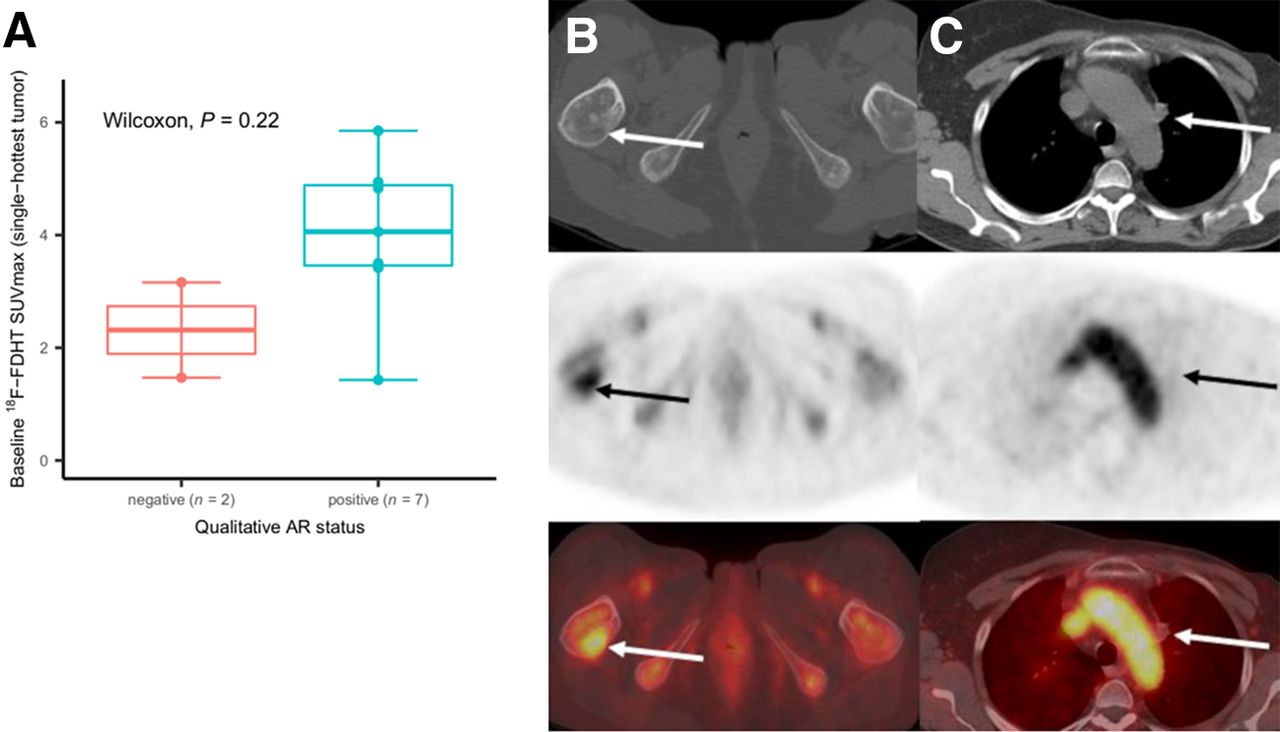
Baseline 18F-FDHT uptake and qualitative AR status. (A) For 9 participants with archival tissue, median baseline 18F-FDHT SUVmax was 4.1 (range, 1.4–5.9) for 7 participants with AR+ tumors and 2.3 (range, 1.5–3.2) for 2 with AR− tumors (P = 0.22). Individual dots on box plot represent individual-participant data. (B, top row: axial CT; middle row: axial PET; bottom row: axial fused PET/CT) Participant 6, with AR+ tumor and 18F-FDHT uptake in right femur metastasis (arrows, SUVmax of 4.9). (C, top row: axial CT; middle row: axial PET; bottom row: axial fused PET/CT) Participant 8, with AR− tumor and no 18F-FDHT uptake in prevascular lymph node (arrows, SUVmax of 1.5).
- FIGURE 3.
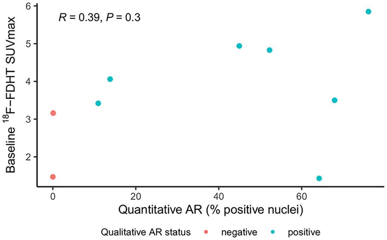
Baseline 18F-FDHT uptake and quantitative AR status. Weak, but not statistically significant, correlation was observed between quantitative AR expression levels and baseline 18F-FDHT uptake (Pearson ρ = 0.39, P = 0.30).
- FIGURE 4.
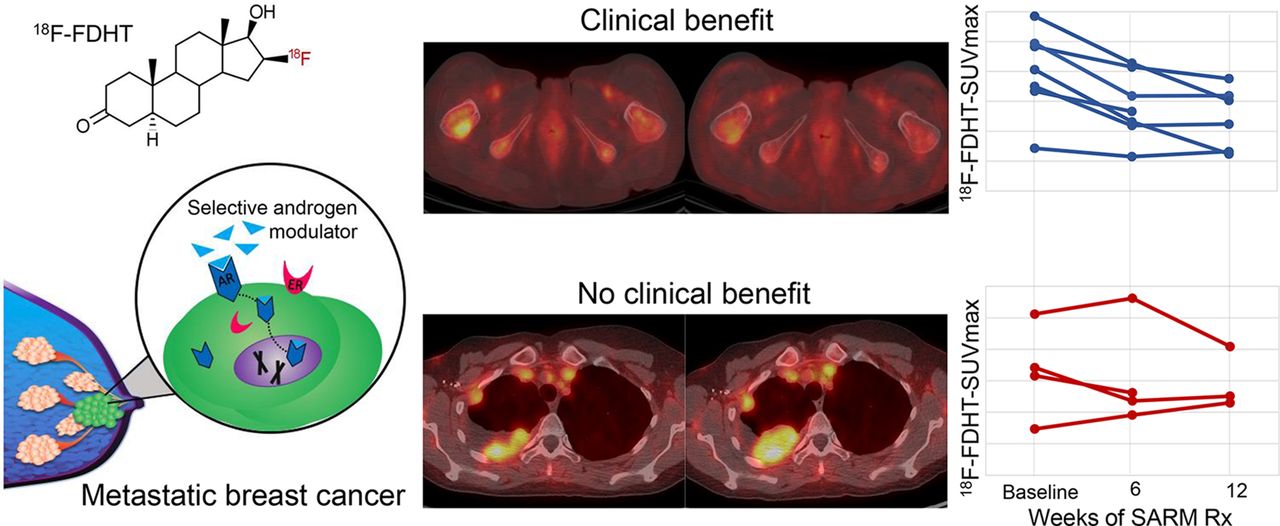
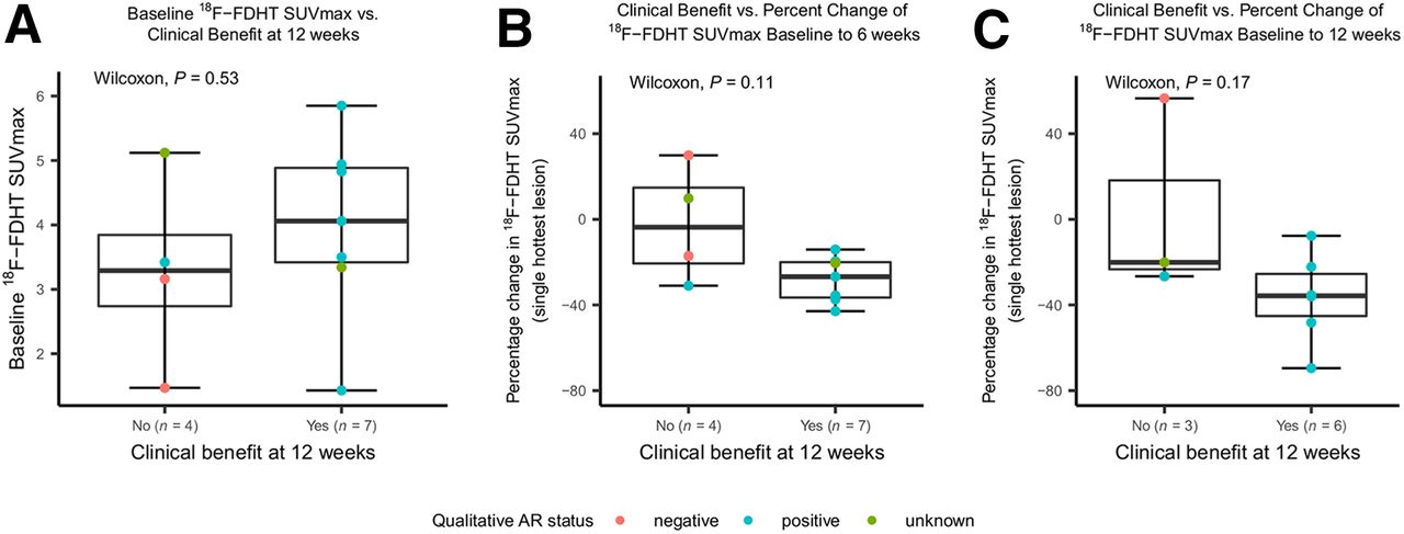
CB at 12 weeks after starting therapy vs. baseline and change in 18F-FDHT uptake. (A) For 7 participants with CB, median baseline 18F-FDHT SUVmax was 4.1 (range, 1.4–5.9), compared with 3.3 (range, 1.5–5.1) for 4 participants with disease progression (P = 0.53). Individual dots on scatterplot represent individual-participant data. (B and C) Participants with CB at 12 wk tended to have larger declines in 18F-FDHT uptake at 6 wk after starting GTx-024 (median decline, 26.8%; range, −42.9% to −14.1%) than did those with disease progression (median decline, 3.7%; range, −31% to +29%; P = 0.11) (B) and tended to have larger declines in 18F-FDHT uptake at 12 wk after starting GTx-024 (median decline, 35.7%; range, −69.5% to −7.7%) than did those with disease progression (median decline, 20.1%; range, −26.6% to +56.5%; P = 0.17) (C).
- FIGURE 5.
(A, left-most panel: maximum-intensity-projection PET; 2nd panel: axial CT; 3rd panel: axial PET; 4th panel: axial fused PET/CT) 18F-FDHT–avid AR+ tumor at baseline (top row, arrow [SUVmax, 4.9]) and decline in 18F-FDHT uptake 6 wk after starting GTx-024 (bottom row, arrow). Best overall response was stable disease 12 wk after starting therapy. (B, left-most panel: maximum-intensity-projection PET; 2nd panel: axial CT; 3rd panel: axial PET; 4th panel: axial fused PET/CT) 18F-FDHT–avid AR+ tumor at baseline (top row, lower arrow [SUVmax, 5.1]) and no decline in 18F-FDHT uptake and increased tumor size 6 wk after starting GTx-024 (bottom row, arrows). Best overall response was PD 12 wk after starting therapy.


Tables
Characteristic Data Age (y) Median 59 Range 49–73 Histology (n) Invasive ductal carcinoma 9 Invasive lobular carcinoma 2 Receptor status (n) ER+/PR+/HER2− 6 ER+/PR−/HER2− 5 Metastases at diagnosis (n) Yes 4 No 7 Disease-free interval* (y) Metastases at diagnosis (n = 4) Not applicable No metastases at diagnosis (n = 7) 7 (range, 3–19) Median lines of treatment before enrollment (n) Adjuvant chemotherapy Metastases at diagnosis (n = 4) Not applicable No metastases at diagnosis (n = 7) 1 (0–1) Adjuvant endocrine therapy Metastases at diagnosis (n = 4) Not applicable No metastases at diagnosis (n = 7) 1 (0–2) Chemotherapy for metastatic disease Metastases at diagnosis (n = 4) 1 (0–1)† No metastases at diagnosis (n = 7) 0 (0–1) Endocrine therapy for metastatic disease Metastases at diagnosis (n = 4) 2 (1–4) No metastases at diagnosis (n = 7) 2 (1–6) CDK4/6 inhibitor Metastases at diagnosis (n = 4) 2 No metastases at diagnosis (n = 7) 6 mTOR inhibitor Metastases at diagnosis (n = 4) 1 No metastases at diagnosis (n = 7) 2 Dual PI3 kinase and mTOR inhibitor Metastases at diagnosis (n = 4) 0 No metastases at diagnosis (n = 7) 1 Radiation therapy to metastatic disease Metastases at diagnosis (n = 4) 1 (bone) No metastases at diagnosis (n = 7) 2 (bone) Median metastatic sites at enrollment (n) 2 (range, 1–4) Location of metastatic sites at enrollment (n) Bone 8 (bone only, 5) Viscera (liver, vaginal cuff) 4 Pleura 5 Serosa/peritoneum 2 Lymph node 2 *Time from start of adjuvant therapy to first diagnosis of recurrence or metastatic disease.
↵†n = 1 with high-dose chemotherapy and stem cell transplantation.
PR = progesterone receptor; HER2 = human epidermal growth factor receptor 2; CDK = cyclin-dependent kinase; mTOR = mammalian target of rapamycin; PI3 = phosphoinositide 3.
Participant no. Lesions (n) AR status Archival tissue location 18F-FDHT SUVmax of hottest lesion at baseline Change in 18F-FDHT SUVmax from baseline to… Outcome Week 6 Week 12 Best overall response Week of best overall response Week 24 response 1* 3 Positive Primary 4.1 −43% −70% NonCR/nonPD 12 CB 2 2 Positive Metastasis 3.5 −37% −36% NonCR/nonPD 12 CB 3 2 Positive Metastasis 1.4 −20% −8% NonCR/nonPD 12 No CB 4 1 Not assessed 3.3 −20% Off study† NonCR/nonPD 6 No CB 5 8 Positive Metastasis 4.8 −14% −22% PR 12 No CB 6 4 Positive Metastasis 4.9 −36% −35% SD 12 No CB 7 5 Positive Primary 5.9 −27% −48% SD 12 No CB 8 1 Negative Metastasis 1.5 +30% +56% PD 12 No CB 9 5 Not assessed 5.1 +10% −20% PD 12 No CB 10 4 Negative Metastasis 3.2 −17% Off study† PD 7 No CB 11 5 Positive Metastasis 3.4 −31% −27% PD 12 No CB *Received 18 mg of GTx-024; all others received 9 mg.
↵†Baseline and week 6 scan only; patient 4 off study week 6 because of toxicity, patient 10 off study week 7 because of progression.
NonCR/nonPD = incomplete response but no PD for participants with nonmeasurable disease by RECIST 1.1; CR = complete response; PR = partial response; SD = stable disease.
Supplemental Data
Files in this Data Supplement:
In this issue


Jump to section
Related Articles
Cited By...
- Report on the PET/CT Image-Based Radiation Dosimetry of [18F]FDHT in Women, a Validated Imaging Agent with New Applications for Evaluation of Androgen Receptor Status in Women with Metastatic Breast Cancer
- Report on the PET/CT Image-Based Radiation Dosimetry of [18F]FDHT in Women, a Validated Imaging Agent with New Applications for Evaluation of Androgen Receptor Status in Women with Metastatic Breast Cancer