
Main menu

  • Home
  • Content
    • Current
    • Ahead of print
    • Past Issues
    • JNM Supplement
    • SNMMI Annual Meeting Abstracts
    • Continuing Education
    • JNM Podcasts
  • Subscriptions
    • Subscribers
    • Institutional and Non-member
    • Rates
    • Journal Claims
    • Corporate & Special Sales
  • Authors
    • Submit to JNM
    • Information for Authors
    • Assignment of Copyright
    • AQARA requirements
  • Info
    • Reviewers
    • Permissions
    • Advertisers
  • About
    • About Us
    • Editorial Board
    • Contact Information
  • More
    • Alerts
    • Feedback
    • Help
    • SNMMI Journals
  • SNMMI
    • JNM
    • JNMT
    • SNMMI Journals
    • SNMMI

User menu

  • Subscribe
  • My alerts
  • Log in
  • My Cart

Search

  • Advanced search
Journal of Nuclear Medicine
  • SNMMI
    • JNM
    • JNMT
    • SNMMI Journals
    • SNMMI
  • Subscribe
  • My alerts
  • Log in
  • My Cart
Journal of Nuclear Medicine

Advanced Search

  • Home
  • Content
    • Current
    • Ahead of print
    • Past Issues
    • JNM Supplement
    • SNMMI Annual Meeting Abstracts
    • Continuing Education
    • JNM Podcasts
  • Subscriptions
    • Subscribers
    • Institutional and Non-member
    • Rates
    • Journal Claims
    • Corporate & Special Sales
  • Authors
    • Submit to JNM
    • Information for Authors
    • Assignment of Copyright
    • AQARA requirements
  • Info
    • Reviewers
    • Permissions
    • Advertisers
  • About
    • About Us
    • Editorial Board
    • Contact Information
  • More
    • Alerts
    • Feedback
    • Help
    • SNMMI Journals
  • View or Listen to JNM Podcast
  • Visit JNM on Facebook
  • Join JNM on LinkedIn
  • Follow JNM on Twitter
  • Subscribe to our RSS feeds
Meeting ReportPoster - PhysicianPharm

Non-invasive input function extraction from dynamic PET using machine learning along with an iterative approach

Hamed Moradi, Viktor Vegh and David Reutens
Journal of Nuclear Medicine May 2021, 62 (supplement 1) 1416;
Hamed Moradi
1ARC Centre for Innovation in Biomedical Imaging Technology, The University of Queensland Brisbane Australia
2Centre for Advanced Imaging, The University of Queensland Brisbane Australia
  • Find this author on Google Scholar
  • Find this author on PubMed
  • Search for this author on this site
Viktor Vegh
2Centre for Advanced Imaging, The University of Queensland Brisbane Australia
1ARC Centre for Innovation in Biomedical Imaging Technology, The University of Queensland Brisbane Australia
  • Find this author on Google Scholar
  • Find this author on PubMed
  • Search for this author on this site
David Reutens
2Centre for Advanced Imaging, The University of Queensland Brisbane Australia
1ARC Centre for Innovation in Biomedical Imaging Technology, The University of Queensland Brisbane Australia
  • Find this author on Google Scholar
  • Find this author on PubMed
  • Search for this author on this site
  • Article
  • Figures & Data
  • Info & Metrics
Loading

Abstract

1416

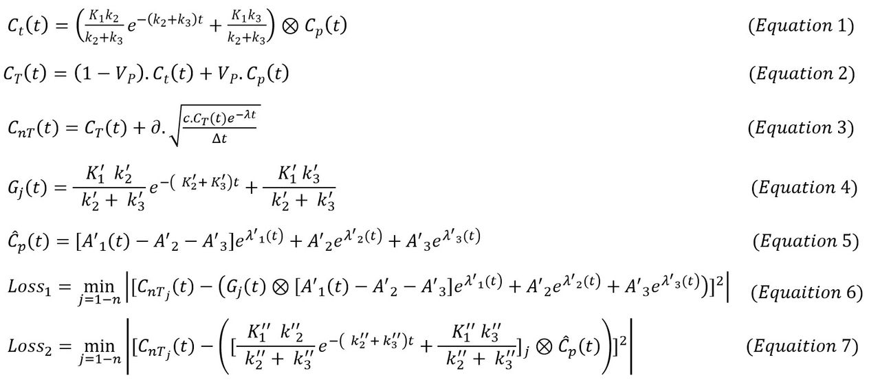
Introduction: Dynamic Positron Emission Tomography (PET) with 18F-FDG is widely used to evaluate neurological disease. The irreversible 2 Tissue Compartmental Model (2TCM) originally developed by Sokoloff et al. is used to extract kinetic parameters (influx rate: K1, ml.g-1.min-1; rate constants: k2,k3, min-1; blood fraction: Vb ) from dynamic FDG-PET data (Equations 1 and 2). The model requires an Arterial Input Function (AIF) obtained via invasive arterial blood sampling which is challenging to use clinically. Here, a non-invasive method of estimating the AIF is proposed. We consider the combination of Machine Learning (ML) along with an iterative approach for AIF estimation using multiple Time Activity Curves (TACs) from one subject.

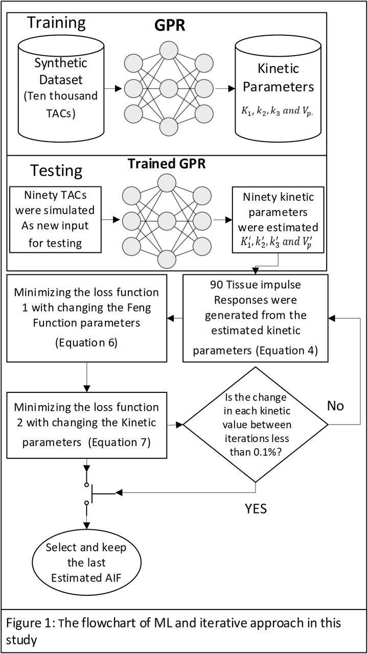
Methods: Ten thousand synthetic TACs were generated based on the 2TCM with a total sampling time of 60 min, including 4×30s, 8×60s, 10×120s and 6×300s frames. The formulation of the input function introduced by Feng et al. was used in this study (Equation 5). Gaussian random noise was added to each noise-free TAC refer to Equation 3. Three noise levels were considered (c = 0.1, 1.0, 2.8). Gaussian Process Regression (GPR) was employed to build the ML kinetic modelling framework. Individual TACs were used as feature vectors, and corresponding kinetic parameters, K1, k2, k3 and Vb were the labelled data. The GPR was trained using the 10,000 noisy synthetic TACs. For testing, two individual subjects including ninety TACs were simulated using two fixed input functions. Each AIF was estimated iteratively as follows. Each of the 90 TACs was fed into the trained GPR network, leading to an initial estimate of the kinetic parameters (K'1, k'2, k'3 and V'b). Ninety tissue impulse responses, Gj(t) where j is TAC number, were generated using the estimated kinetic parameters, refer to Equation 4. Two specific steps were required to extract the AIF. First, the Feng function AIF parameters, A'1, A'2, A'3, λ'1, λ'2, λ'3 were estimated by minimising loss function 1 stated in Equation 6. Second, kinetic parameters were recalculated (K''1, k''2, k''3 and V''b) using the estimated AIF and by minimising loss function 2 stated in Equation 7. Then the recalculated kinetic parameters were employed to refine the estimated AIF parameters in the first loss function. Figure 1 represents the flowchart of ML and iterative approach. The optimisation was performed using the Levenberg-Marquardt algorithm in MATLAB®. This process was repeated resulting in iterative refinement of kinetic parameters until each value had a change less than 0.1% for the iteration.

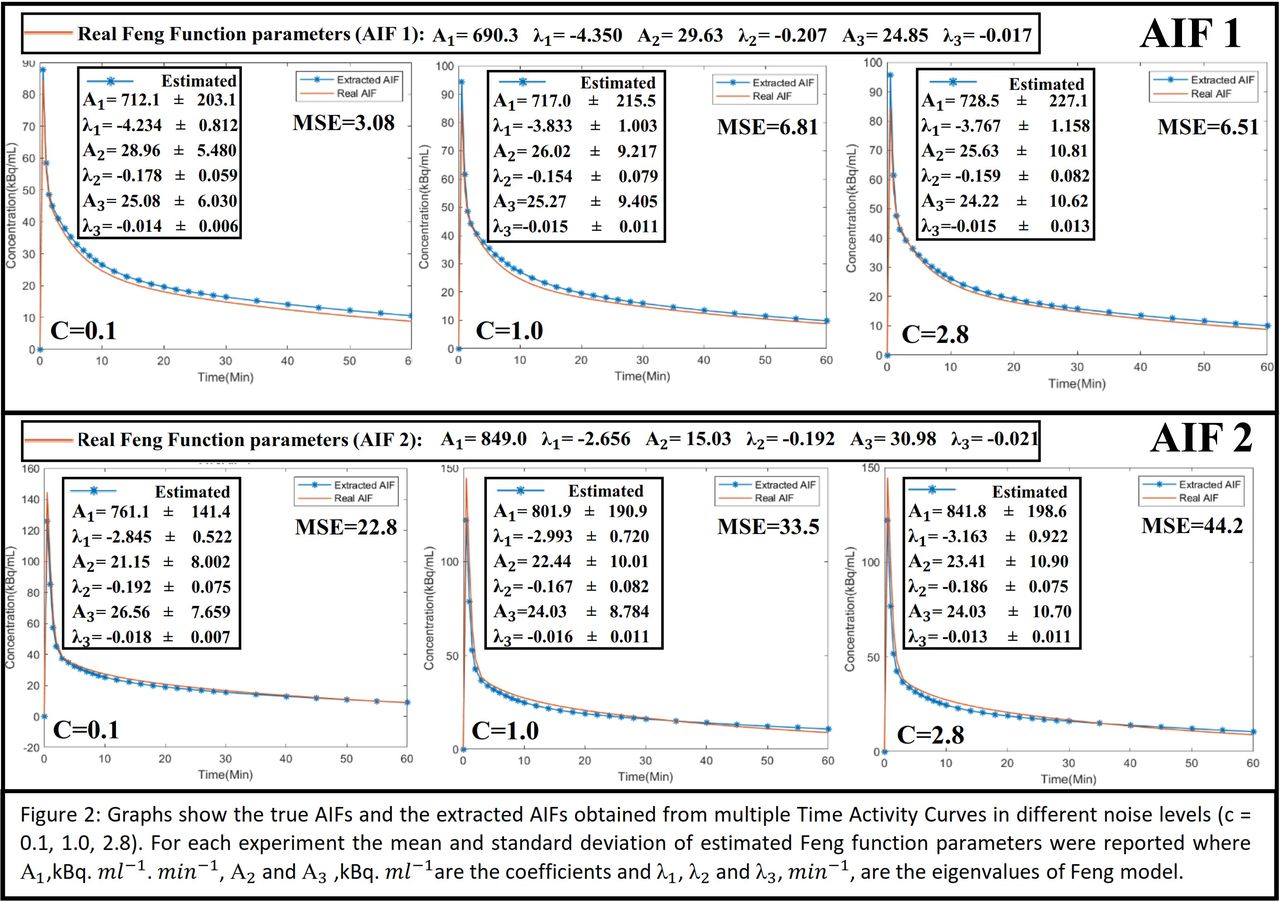
Results: Figure 2 provides the comparison between extracted and real AIF shapes and, the Mean Squared Error (MSE) is stated as a measure of accuracy. It also shows the mean and standard deviation of Feng function parameters based on two separate experiments considering 90 TACs each. It took six and five iterations to generate the first and second AIFs, respectively. The results show that an excellent agreement between the extracted AIF and the real AIF was obtained in both cases.

Conclusions: Our goal was to extract the AIF from multiple pixel location time activity curves. Our findings show that the AIF can be extracted accurately using the outlined ML approach through iterative updates of the AIF parameters. Future work should investigate how the number of time activity curves and anatomical regions considered in AIF estimation impact accuracy. Acknowledgments: This research was conducted at the Australian Research Council Training Centre for Innovation in Biomedical Imaging Technology (IC170100035) and funded by the Australian Government. The authors also acknowledge the facilities and scientific and technical assistance of the National Imaging Facility, a National Collaborative Research Infrastructure Strategy (NCRIS) capacity, at the Centre for Advance Imaging, the University of Queensland.

Figure
  • Download figure
  • Open in new tab
  • Download powerpoint
Figure
  • Download figure
  • Open in new tab
  • Download powerpoint
Figure
  • Download figure
  • Open in new tab
  • Download powerpoint
Previous
Back to top

In this issue

Journal of Nuclear Medicine
Vol. 62, Issue supplement 1
May 1, 2021
  • Table of Contents
  • Index by author
Article Alerts
Sign In to Email Alerts with your Email Address
Email Article

Thank you for your interest in spreading the word on Journal of Nuclear Medicine.

NOTE: We only request your email address so that the person you are recommending the page to knows that you wanted them to see it, and that it is not junk mail. We do not capture any email address.

Enter multiple addresses on separate lines or separate them with commas.
Non-invasive input function extraction from dynamic PET using machine learning along with an iterative approach
(Your Name) has sent you a message from Journal of Nuclear Medicine
(Your Name) thought you would like to see the Journal of Nuclear Medicine web site.
Citation Tools
Non-invasive input function extraction from dynamic PET using machine learning along with an iterative approach
Hamed Moradi, Viktor Vegh, David Reutens
Journal of Nuclear Medicine May 2021, 62 (supplement 1) 1416;

Citation Manager Formats

  • BibTeX
  • Bookends
  • EasyBib
  • EndNote (tagged)
  • EndNote 8 (xml)
  • Medlars
  • Mendeley
  • Papers
  • RefWorks Tagged
  • Ref Manager
  • RIS
  • Zotero
Share
Non-invasive input function extraction from dynamic PET using machine learning along with an iterative approach
Hamed Moradi, Viktor Vegh, David Reutens
Journal of Nuclear Medicine May 2021, 62 (supplement 1) 1416;
Twitter logo Facebook logo LinkedIn logo Mendeley logo
  • Tweet Widget
  • Facebook Like
  • Google Plus One
Bookmark this article

Jump to section

  • Article
  • Figures & Data
  • Info & Metrics

Related Articles

  • No related articles found.
  • Google Scholar

Cited By...

  • No citing articles found.
  • Google Scholar

More in this TOC Section

Poster - PhysicianPharm

  • Preliminary result of Texture Analysis on prediction of overall outcome of neuroendocrine tumors based on pre-therapy heterogeneity of somatostatin receptors on 68Ga Dotatate PET/CT scans.
  • The Effect of BMI on High versus Low Administered Activity Protocol MPI Scan Time Using a CZT Camera in Clinical Practice
  • The Role of 18F-FDG-PET/CT Imaging in Predicting Outcome of Patients with Newly Diagnosed Multiple Myeloma
Show more Poster - PhysicianPharm

PIDS - Data Analysis and Management

  • An empirical update of left ventricular 3D segmentation algorithm in myocardial perfusion SPECT imaging
  • The value of differentiation malignant solitary pulmonary nodules in a digital PET/CT: Impact of image reconstructions on diagnostic performance
  • Restoration of full dynamic data using only early and late dynamic data from amyloid PET image
Show more PIDS - Data Analysis and Management

Similar Articles

SNMMI

© 2025 SNMMI

Powered by HighWire