


Abstract
1245
Introduction: The evaluation of newly developed radiopharmaceuticals always requires animal experiments. The HET-CAM (hen’s egg test- chorioallantoic membrane) model is a promising alternative to reduce the number of necessary animal experiments by preceding studies in chicken eggs in accordance with the 3Rs principles. Our aim was to validate the model, particularly in terms of quantifiability and reproducibility of data using the PSMA-specific tracer 18F-siPSMA-14.
Methods: Tumor xenografts of a PSMA-positive (LNCaP C4-2) and a PSMA-negative cell line (PC-3) were established on the CAM. MRI and PET measurements were performed between embryo development days 12 and 16. The MRI (BioSpec 117/16, Bruker) included an overview scan (Flash3D) of the whole egg and a high-resolution scan (RARE) of the tumor region. After i.v. application of 18F-siPSMA-14 a dynamic PET (Focus 120, Siemens Medical Solutions, Inc.) was performed for 60 min. PET data were evaluated using 3DSlicer [1] and Pmod ver. 4.105 (PMOD Technologies Ltd., Zurich, Switzerland). Subsequently, tumor xenografts were excised and the corresponding radioactivity was determined using a γ-counter (Cobra II, PerkinElmer). To detect the PSMA protein, tumors were lysed and Western blots (WB) were generated.
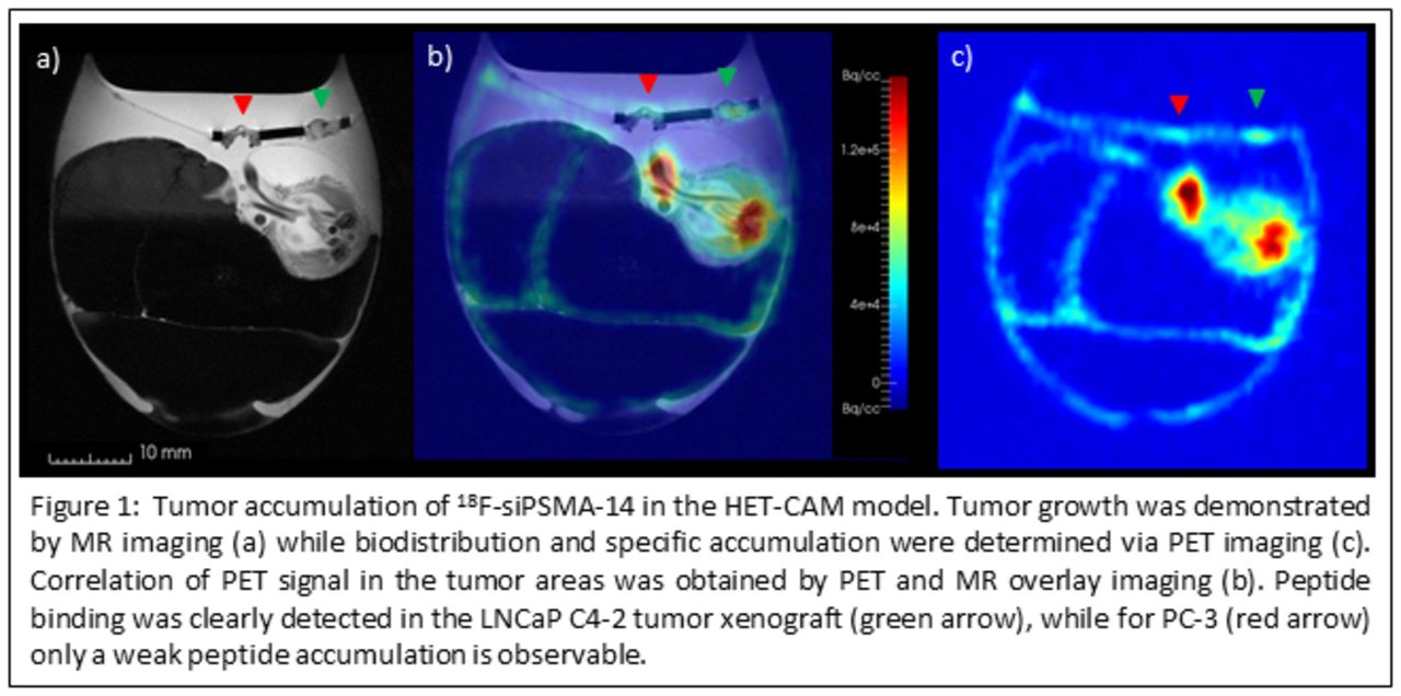
Results: In the WB analysis PSMA was detected only in the LNCaP C4-2 cells. Tumor volumes were determined based on MRI data LNCaP C4-2 (35.4 ± 7.8) mm³ / PC-3 (29.6 ± 9.7) mm³ (n=4). A high accumulated activity could be detected in the LNCaP C4-2 tumor xenografts after superposition of PET and MRI data: LNCaP C4-2 (1.1 ± 1.3) %IA/ml; PC-3 (0.7 ± 1.1) %IA/ml, resulting in a ratio of C4-2/PC-3 = 1.8 ± 1.4. By γ-counter measurements similar results were obtained: LNCaP C4-2 (8.7 ± 1.5) %IA/ml; PC-3 (5.5 ± 1.6) %IA/ml; C4-2/PC-3 = 1.6 ± 0.3.
Conclusions: In both, PET and y-counter the specific accumulation of 18F-siPSMA-14 in PSMA-positive tumors was successfully demonstrated using the HET-CAM model. This platform allows the exact determination of tumor volume and quantification of peptide accumulation. Thus, the HET-CAM model has great potential to assess radiopharmaceutical accumulation and can contribute to reduce the number of animal experiments. References: [1] Fedorov A., Beichel R., Kalpathy-Cramer J., Finet J., Fillion-Robin J-C., Pujol S., Bauer C., Jennings D., Fennessy F., Sonka M., Buatti J., Aylward S.R., Miller J.V., Pieper S., Kikinis R. 3D Slicer as an Image Computing Platform for the Quantitative Imaging Network. Magnetic Resonance Imaging. 2012 Nov;30(9):1323-41.

In this issue


Jump to section
Related Articles
Cited By...
- No citing articles found.