


Abstract
1159
Introduction: Despite low back pain (LBP) being the leading cause of years lived with disability globally, the pathophysiological mechanism behind the condition is still unclear. In positron emission tomography (PET) scans, 18F-fluorodeoxyglucose (FDG) and 18F-sodium fluoride (NaF) can be used as tracers, assuming that the uptake of these, respectively, is associated with inflammation and microcalcification. In this study, PET was used to compare FDG and NaF uptake in different regions of the lumbar spine in individuals with ongoing LBP and pain free controls.
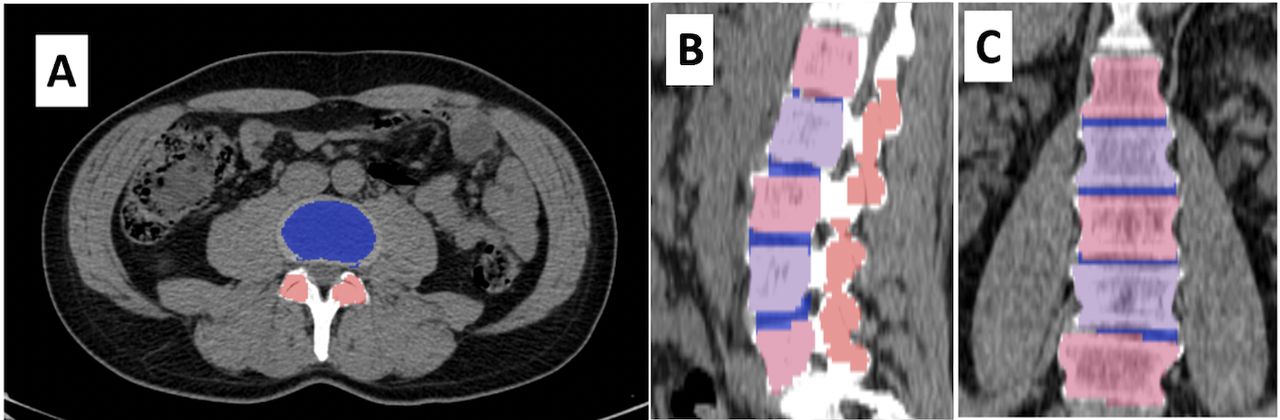
Methods: This case-control study was conducted among 24 individuals who were already enrolled in a prospective cohort study named CAMONA. Our cohort consisted of two sex- and age-matched groups: a case group of 12 individuals with ongoing LBP and a control group of 12 pain free individuals. Using a semi-automatic method, the FDG- and NaF-PET/CT scans were segmented into 3 distinct regions of interest (ROIs): the lumbar bodies, facet joints, and intervertebral discs. The automated part of the image segmentation and analysis was conducted using an artificial intelligence (AI) model. These segmentations were all examined and, if necessary, manually adjusted in order to secure accurate and similar segmentations. The maximum, mean, and total standardized uptake values (SUVmax, SUVmean, and SUVtotal) for both FDG and NaF in the 3 ROIs were measured and compared. Examples of the three ROI segmentations are shown in Figure 1.
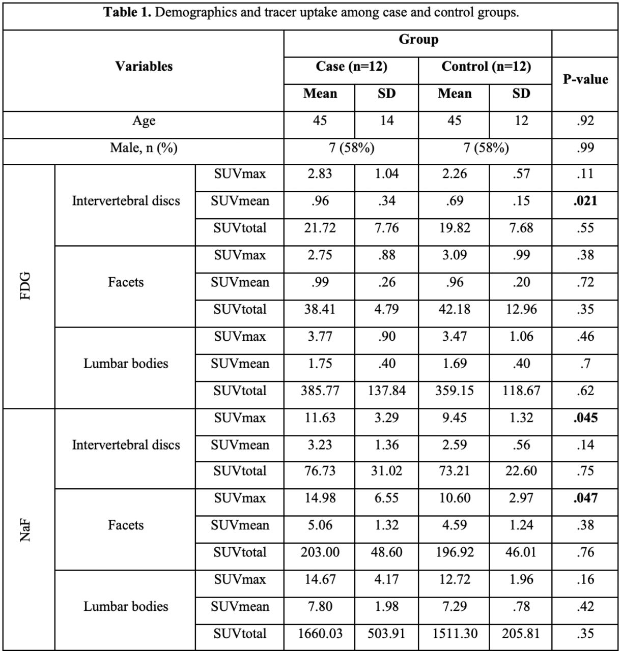
Results: Demographics and tracer uptake in the two groups are listed in Table 1. A statistically significant difference in mean uptake (± standard deviation) between cases and controls was observed for FDG SUVmean in the intervertebral discs (0.96±0.34 vs. 0.69±0.15, p=0.021). Furthermore, statistically significant differences between cases and controls were found with regard to NaF SUVmax in the intervertebral discs (11.63±3.29 vs. 9.45±1.32, p=0.045), and in the facet joints (14.98±6.55 vs. 10.60±2.97, p=0.047). Conclusion: According to the tracer uptake pattern and compared to the control group, individuals with ongoing LBP have inflammation in the intervertebral discs and microcalcification in the intervertebral discs and facet joints indicating previous inflammation in the facet joints. Supporting data: Figure 1. A sample of the semi-automatic segmentation shown in the CT images in transaxial (A), sagittal (B), and coronal (C) planes. Vertebral bodies are shown with alternating light blue and light red colors, facet joints with a dark red color, and intervertebral discs with dark blue.


In this issue


Jump to section
Related Articles
Cited By...
- No citing articles found.