
Main menu

  • Home
  • Content
    • Current
    • Ahead of print
    • Past Issues
    • JNM Supplement
    • SNMMI Annual Meeting Abstracts
    • Continuing Education
    • JNM Podcasts
  • Subscriptions
    • Subscribers
    • Institutional and Non-member
    • Rates
    • Journal Claims
    • Corporate & Special Sales
  • Authors
    • Submit to JNM
    • Information for Authors
    • Assignment of Copyright
    • AQARA requirements
  • Info
    • Reviewers
    • Permissions
    • Advertisers
  • About
    • About Us
    • Editorial Board
    • Contact Information
  • More
    • Alerts
    • Feedback
    • Help
    • SNMMI Journals
  • SNMMI
    • JNM
    • JNMT
    • SNMMI Journals
    • SNMMI

User menu

  • Subscribe
  • My alerts
  • Log in
  • My Cart

Search

  • Advanced search
Journal of Nuclear Medicine
  • SNMMI
    • JNM
    • JNMT
    • SNMMI Journals
    • SNMMI
  • Subscribe
  • My alerts
  • Log in
  • My Cart
Journal of Nuclear Medicine

Advanced Search

  • Home
  • Content
    • Current
    • Ahead of print
    • Past Issues
    • JNM Supplement
    • SNMMI Annual Meeting Abstracts
    • Continuing Education
    • JNM Podcasts
  • Subscriptions
    • Subscribers
    • Institutional and Non-member
    • Rates
    • Journal Claims
    • Corporate & Special Sales
  • Authors
    • Submit to JNM
    • Information for Authors
    • Assignment of Copyright
    • AQARA requirements
  • Info
    • Reviewers
    • Permissions
    • Advertisers
  • About
    • About Us
    • Editorial Board
    • Contact Information
  • More
    • Alerts
    • Feedback
    • Help
    • SNMMI Journals
  • View or Listen to JNM Podcast
  • Visit JNM on Facebook
  • Join JNM on LinkedIn
  • Follow JNM on Twitter
  • Subscribe to our RSS feeds
Meeting ReportOral - PhysicianPharm

Development of the Lymphatic System in the 4D XCAT Phantom for Improved Multimodality Imaging Research

Roberto Fedrigo, Paul Segars, Patrick Martineau, Kerry Savage, Carlos Uribe and Arman Rahmim
Journal of Nuclear Medicine May 2021, 62 (supplement 1) 113;
Roberto Fedrigo
2University of British Columbia Vancouver BC Canada
1BC Cancer Research Institute Vancouver BC Canada
  • Find this author on Google Scholar
  • Find this author on PubMed
  • Search for this author on this site
Paul Segars
3Duke University Durham NC United States
  • Find this author on Google Scholar
  • Find this author on PubMed
  • Search for this author on this site
Patrick Martineau
4BC Cancer Vancouver BC Canada
  • Find this author on Google Scholar
  • Find this author on PubMed
  • Search for this author on this site
Kerry Savage
4BC Cancer Vancouver BC Canada
  • Find this author on Google Scholar
  • Find this author on PubMed
  • Search for this author on this site
Carlos Uribe
4BC Cancer Vancouver BC Canada
2University of British Columbia Vancouver BC Canada
  • Find this author on Google Scholar
  • Find this author on PubMed
  • Search for this author on this site
Arman Rahmim
2University of British Columbia Vancouver BC Canada
4BC Cancer Vancouver BC Canada
1BC Cancer Research Institute Vancouver BC Canada
  • Find this author on Google Scholar
  • Find this author on PubMed
  • Search for this author on this site
  • Article
  • Figures & Data
  • Info & Metrics
Loading

Abstract

113

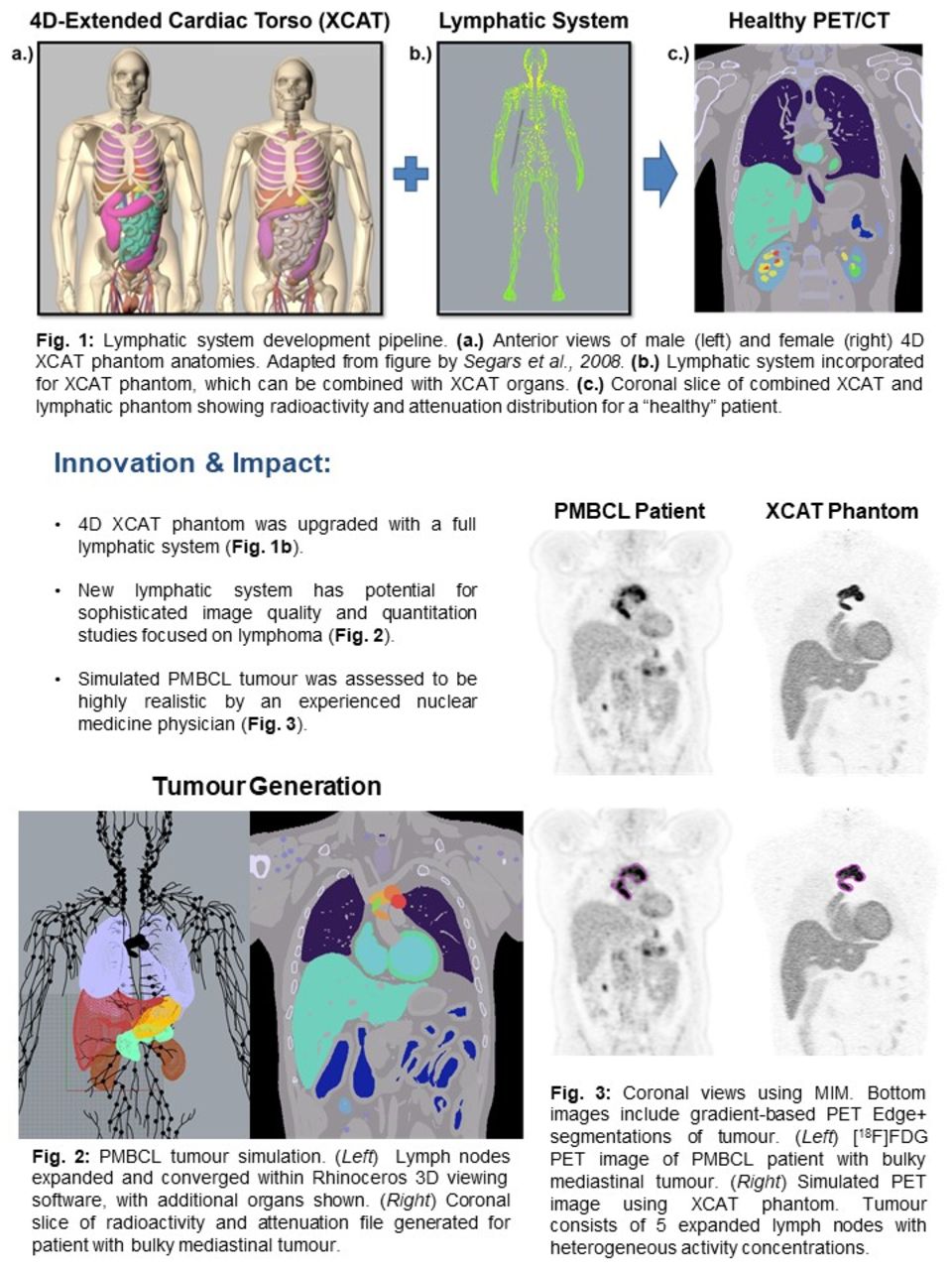
Objectives: Non-Hodgkin’s lymphoma classically presents with lymphadenopathy and bulky lymph node conglomerates. FDG PET/CT scans are used to stage and assess treatment response. Total metabolic tumour volume (TMTV) quantification has shown promising results for predicting therapy response and overall survival of lymphoma patients. However, TMTV accuracy can be impacted by the selected reconstruction parameters and segmentation method. Conventionally, the NEMA Image Quality phantom is used to evaluate image characteristics, but does not represent realistic patient anatomy or tumour properties. Simulated phantoms, such as the 4D extended cardiac-torso (XCAT) phantom used for multimodality imaging research, allows for more realistic patient modelling. The XCAT phantom defines the activity and attenuation for a simulated patient, which includes a complete set of organs, muscle, bone, soft tissue, while also accounting for age, sex, and body mass index (BMI), which allows phantom studies to be performed at a population scale. However, the XCAT phantom does not currently include the lymphatic system, critical for evaluating bulky nodal malignancies in lymphoma. The aim of this study was to incorporate a full lymphatic system into the XCAT phantom, and to generate realistic simulated PET/CT images via guidance from lymphoma patient studies, to enable lymphoma PET/CT optimization studies for improved image quality and quantification.

Methods: A template lymphatic system model based on anatomical data from the Visible Human Project of the National Library of Medicine was used to define 276 lymph nodes and corresponding vessels using non-uniform rational basis spline (NURBS) surfaces. The multichannel large deformation diffeomorphic metric mapping (MC-LDDMM) method was used to propagate from the template phantom to different XCAT anatomies. This allows for the lymphatic system to be investigated on patients with different genders, weight, sizes, age, and other anatomical differences. Lymph node properties were modified using the Rhinoceros 3D viewing software. To determine typical activity concentrations observed in lymphoma, FDG PET/CT images of 5 patients from a cohort of PMBCL positive scans were analyzed using MIM (MIM Software, USA). The XCAT general parameter script was used to input organ concentrations and generate binary files with uptake and attenuation information. The phantom was used as the input to a MATLAB-based PET simulation and reconstruction tool (Ashrafinia et al., 2017) generating simulated PET/CT images for a GE Discovery RX scanner, reconstructed with OSEM (2 iterations, 24 subsets).

Results: The lymphatic system was added to the XCAT phantom with the capability to select male/female anatomy or patient size. Lymph nodes can be scaled, asymmetrically stretched, and translated within the intuitive Rhinoceros interface, to allow for realistic simulation of different lymph node pathologies. Bulky, heterogeneous PMBCL tumours were generated in the mediastinum using expanded lymph nodes. Simulated PET images from the XCAT phantom were optimized to represent FDG PET/CT images of PMBCL patients and was assessed to be realistic by an experienced nuclear medicine clinician.

Conclusions: An upgraded XCAT phantom with a fully-simulated lymphatic system was created. Realistic simulated PET/CT images were generated using the phantom with uptake values measured from real patient scans. Made publicly available, the XCAT phantom with the new lymphatic system has the potential of enabling studies to optimize image quality and quantitation, towards improved assessment of lymphoma including predictive modeling (e.g. improved TMTV and radiomics research).

Figure
  • Download figure
  • Open in new tab
  • Download powerpoint
View this table:
  • View inline
  • View popup

Measured [18F]FDG activity concentrations for PMBCL patients in different regions-of-interest.

Previous
Back to top

In this issue

Journal of Nuclear Medicine
Vol. 62, Issue supplement 1
May 1, 2021
  • Table of Contents
  • Index by author
Article Alerts
Sign In to Email Alerts with your Email Address
Email Article

Thank you for your interest in spreading the word on Journal of Nuclear Medicine.

NOTE: We only request your email address so that the person you are recommending the page to knows that you wanted them to see it, and that it is not junk mail. We do not capture any email address.

Enter multiple addresses on separate lines or separate them with commas.
Development of the Lymphatic System in the 4D XCAT Phantom for Improved Multimodality Imaging Research
(Your Name) has sent you a message from Journal of Nuclear Medicine
(Your Name) thought you would like to see the Journal of Nuclear Medicine web site.
Citation Tools
Development of the Lymphatic System in the 4D XCAT Phantom for Improved Multimodality Imaging Research
Roberto Fedrigo, Paul Segars, Patrick Martineau, Kerry Savage, Carlos Uribe, Arman Rahmim
Journal of Nuclear Medicine May 2021, 62 (supplement 1) 113;

Citation Manager Formats

  • BibTeX
  • Bookends
  • EasyBib
  • EndNote (tagged)
  • EndNote 8 (xml)
  • Medlars
  • Mendeley
  • Papers
  • RefWorks Tagged
  • Ref Manager
  • RIS
  • Zotero
Share
Development of the Lymphatic System in the 4D XCAT Phantom for Improved Multimodality Imaging Research
Roberto Fedrigo, Paul Segars, Patrick Martineau, Kerry Savage, Carlos Uribe, Arman Rahmim
Journal of Nuclear Medicine May 2021, 62 (supplement 1) 113;
Twitter logo Facebook logo LinkedIn logo Mendeley logo
  • Tweet Widget
  • Facebook Like
  • Google Plus One
Bookmark this article

Jump to section

  • Article
  • Figures & Data
  • Info & Metrics

Related Articles

  • No related articles found.
  • Google Scholar

Cited By...

  • No citing articles found.
  • Google Scholar

More in this TOC Section

Oral - PhysicianPharm

  • Therapeutic Efficacy of Pb-214-labeled Trastuzumab in a Preclinical Model of Ovarian Cancer
  • Automatic Lung Lobe Analysis with COVID-19 Support
  • Molecular Imaging of Multiple Myeloma Targeting CD46 Using ImmunoPET
Show more Oral - PhysicianPharm

Advances in Data Sciences and Image Analysis

  • The impact of noise level mismatch between training and testing images for deep learning-based PET denoising
  • Feasibility of imaging synaptic density in the human spinal cord using 11C-UCB-J PET
  • Linking radiomic PET features with metabolic tissue parameters using a hybrid mathematical model of tumor growth
Show more Advances in Data Sciences and Image Analysis

Similar Articles

SNMMI

© 2025 SNMMI

Powered by HighWire