
Main menu

  • Home
  • Content
    • Current
    • Ahead of print
    • Past Issues
    • JNM Supplement
    • SNMMI Annual Meeting Abstracts
  • Subscriptions
    • Subscribers
    • Institutional and Non-member
    • Rates
    • Corporate & Special Sales
    • Journal Claims
  • Authors
    • Submit to JNM
    • Information for Authors
    • Assignment of Copyright
    • AQARA requirements
  • Info
    • Permissions
    • Advertisers
    • Continuing Education
  • About
    • About Us
    • Editorial Board
    • Contact Information
  • More
    • Alerts
    • Feedback
    • Help
    • SNMMI Journals
  • SNMMI
    • JNM
    • JNMT
    • SNMMI Journals
    • SNMMI

User menu

  • Subscribe
  • My alerts
  • Log in
  • Log out
  • My Cart

Search

  • Advanced search
Journal of Nuclear Medicine
  • SNMMI
    • JNM
    • JNMT
    • SNMMI Journals
    • SNMMI
  • Subscribe
  • My alerts
  • Log in
  • Log out
  • My Cart
Journal of Nuclear Medicine

Advanced Search

  • Home
  • Content
    • Current
    • Ahead of print
    • Past Issues
    • JNM Supplement
    • SNMMI Annual Meeting Abstracts
  • Subscriptions
    • Subscribers
    • Institutional and Non-member
    • Rates
    • Corporate & Special Sales
    • Journal Claims
  • Authors
    • Submit to JNM
    • Information for Authors
    • Assignment of Copyright
    • AQARA requirements
  • Info
    • Permissions
    • Advertisers
    • Continuing Education
  • About
    • About Us
    • Editorial Board
    • Contact Information
  • More
    • Alerts
    • Feedback
    • Help
    • SNMMI Journals
  • Follow JNM on Twitter
  • Visit JNM on Facebook
  • Join JNM on LinkedIn
  • Subscribe to our RSS feeds
Research ArticleClinical (Radiobiology/Dosimetry/Safety)

Dosimetry for Radiopharmaceutical Therapy: Current Practices and Commercial Resources

Jacek Capala, Stephen A. Graves, Aaron Scott, George Sgouros, Sara St. James, Pat Zanzonico and Brian E. Zimmerman
Journal of Nuclear Medicine December 2021, 62 (Supplement 3) 3S-11S; DOI: https://doi.org/10.2967/jnumed.121.262749
Jacek Capala
1National Cancer Institute, Bethesda, Maryland;
  • Find this author on Google Scholar
  • Find this author on PubMed
  • Search for this author on this site
Stephen A. Graves
2University of Iowa, Iowa City, Iowa;
  • Find this author on Google Scholar
  • Find this author on PubMed
  • Search for this author on this site
Aaron Scott
3Johns Hopkins University, Baltimore, Maryland;
  • Find this author on Google Scholar
  • Find this author on PubMed
  • Search for this author on this site
George Sgouros
3Johns Hopkins University, Baltimore, Maryland;
  • Find this author on Google Scholar
  • Find this author on PubMed
  • Search for this author on this site
Sara St. James
4University of California, San Francisco, California;
  • Find this author on Google Scholar
  • Find this author on PubMed
  • Search for this author on this site
Pat Zanzonico
5Memorial Sloan Kettering Cancer Center, New York, New York; and
  • Find this author on Google Scholar
  • Find this author on PubMed
  • Search for this author on this site
Brian E. Zimmerman
6National Institute of Standards and Technology, Gaithersburg, Maryland
  • Find this author on Google Scholar
  • Find this author on PubMed
  • Search for this author on this site
  • Article
  • Figures & Data
  • Info & Metrics
  • PDF
Loading

Article Figures & Data

Figures

  • Tables
  • FIGURE 1.
    • Download figure
    • Open in new tab
    • Download powerpoint
    FIGURE 1.

    Hybrid SPECT–planar imaging approach to measurement of radiopharmaceutical kinetics.
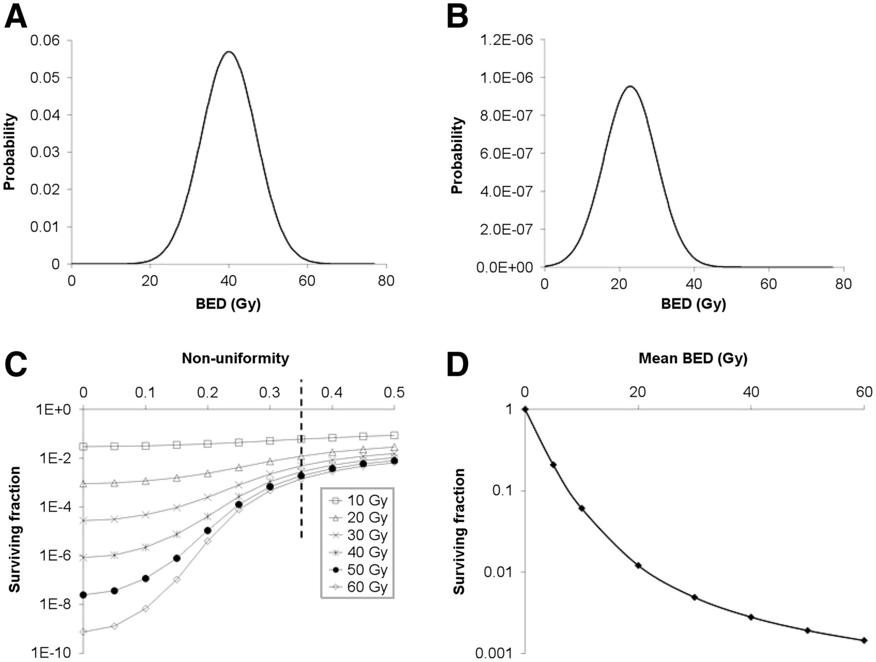
  • FIGURE 2.
    • Download figure
    • Open in new tab
    • Download powerpoint
    FIGURE 2.

    Effect of dose nonuniformity on tumor response. (A) Hypothetical nonuniform dose to tumor cell population represented by normal distribution with average of 40 Gy, SD of 7 Gy, and fractional SD of 7 Gy/40 Gy = 0.175. (B) Overall tumor cell survival fraction as function of dose nonuniformity expressed as fractional SD of average dose from 0 (i.e., uniform dose) to 0.5 and of average tumor dose from 10 Gy (highest curve) to 60 Gy (lowest curve). Tumor cell survival is greater as dose nonuniformity increases. (C) Tumor cell survival probability for dose distribution in A, assuming monoexponential tumor cell survival curve with mean lethal dose Do of 2.85 Gy (i.e., α = 0.35/Gy). Overall tumor cell survival fraction is area under curve. (D) Dose–response for dose nonuniformity (i.e., fractional SD) of 0.35, corresponding to points intersecting dotted vertical line, is concave upward. (Adapted from reference (57).)

Tables

  • Figures
    • View popup
    TABLE 1

    Bioeffects Modeling of RPT: Dose Metrics and Related Parameters (19)

    ParameterDefinition or description
    Lea–Catcheside time factor (46)Radiation delivered at high dose rate is more biologically damaging than same dose delivered at low dose rate as a result of cells’ ability to repair sublethal damage over duration of irradiation. Modifying effect of repair has been modeled with Lea–Catcheside time factor, G(T): Embedded Image where D is total absorbed dose; Embedded Image is dose rate; μ is repair rate, assuming that probability of repair event decreases exponentially as function of time; w is time of first single-strand DNA break; t is time of second break; and T is duration of irradiation. For protracted irradiations such as those encountered in RPT, surviving fraction thus becomes Embedded Image.
    Biologically effective dose (47–49)Variation in biologic response to same absorbed dose delivered at different dose rates or different numbers of fractions has led to concept of biologically effective dose (BED) (or extrapolated tolerance dose, ETD), the absorbed dose required to cause biologic effect if dose were delivered in infinitely small doses per fraction or, equivalently, at very low dose rates: Embedded Image and therefore Embedded Image.
    Equieffective dose (50,51)Equieffective dose (EQDX in Gy) is a quantity that, like BED, is intended to account for differences in fractionation or dose rate; X in this notation refers to reference value of absorbed dose (Gy) per fraction d. It has been recommended that nomenclature for equieffective dose include α/β ratio as well as reference to X: Embedded Image. Newer notation for equieffective dose is thus EQDXα/β, with recommended standard of EQD2α/β, where 2 refers to reference 2-Gy daily fraction. BED is equivalent to EQD0 (52) and is thus particularly relevant to RPT. Low, continuous dose rates delivered by radiopharmaceuticals require modification of this equation to incorporate Lea–Catcheside time factor G(T = ∞) (53–55): Embedded Image
    Equivalent uniform dose (56–58)Tumor therapeutic response and normal-tissue toxicity may not correlate with average absorbed doses even when based on individualized biodistribution and kinetic data because of spatial nonuniformity of dose (Fig. 2 (57)). A quantity has therefore been developed, equivalent uniform dose (EUD), that provides a single value weighted to account for surviving fraction of tumor cells, given spatial distribution of absorbed dose within tumor volume. For any dose distribution, corresponding EUD is absorbed dose (Gy), which, when distributed uniformly across target volume comprised of N voxels, achieves same survival fraction among clonogenic cells: Embedded Image.
    Equivalent uniform biologically effective dose (56–58)EUD has been formulated as equivalent uniform biologic effective dose, EUBED), as first described by O’Donoghue (57). It is often expressed using only linear component of linear-quadratic model: Embedded Image. Solving for EUBED yields Embedded Image
PreviousNext
Back to top

In this issue

Journal of Nuclear Medicine: 62 (Supplement 3)
Journal of Nuclear Medicine
Vol. 62, Issue Supplement 3
December 1, 2021
  • Table of Contents
  • Table of Contents (PDF)
  • About the Cover
  • Index by author
  • Complete Issue (PDF)
Print
Download PDF
Article Alerts
Sign In to Email Alerts with your Email Address
Email Article

Thank you for your interest in spreading the word on Journal of Nuclear Medicine.

NOTE: We only request your email address so that the person you are recommending the page to knows that you wanted them to see it, and that it is not junk mail. We do not capture any email address.

Enter multiple addresses on separate lines or separate them with commas.
Dosimetry for Radiopharmaceutical Therapy: Current Practices and Commercial Resources
(Your Name) has sent you a message from Journal of Nuclear Medicine
(Your Name) thought you would like to see the Journal of Nuclear Medicine web site.
Citation Tools
Dosimetry for Radiopharmaceutical Therapy: Current Practices and Commercial Resources
Jacek Capala, Stephen A. Graves, Aaron Scott, George Sgouros, Sara St. James, Pat Zanzonico, Brian E. Zimmerman
Journal of Nuclear Medicine Dec 2021, 62 (Supplement 3) 3S-11S; DOI: 10.2967/jnumed.121.262749

Citation Manager Formats

  • BibTeX
  • Bookends
  • EasyBib
  • EndNote (tagged)
  • EndNote 8 (xml)
  • Medlars
  • Mendeley
  • Papers
  • RefWorks Tagged
  • Ref Manager
  • RIS
  • Zotero
Share
Dosimetry for Radiopharmaceutical Therapy: Current Practices and Commercial Resources
Jacek Capala, Stephen A. Graves, Aaron Scott, George Sgouros, Sara St. James, Pat Zanzonico, Brian E. Zimmerman
Journal of Nuclear Medicine Dec 2021, 62 (Supplement 3) 3S-11S; DOI: 10.2967/jnumed.121.262749
del.icio.us logo Digg logo Reddit logo Twitter logo CiteULike logo Facebook logo Google logo Mendeley logo
  • Tweet Widget
  • Facebook Like
  • Google Plus One
Bookmark this article

Jump to section

  • Article
    • Abstract
    • DOSE PRESCRIPTION ALGORITHMS
    • PARADIGM FOR PATIENT-SPECIFIC DOSIMETRY
    • MEASUREMENT OF TIME–ACTIVITY DATA
    • CALCULATION OF TIAS
    • DEFINITION OF PATIENT-SPECIFIC ANATOMY
    • CALCULATION OF ABSORBED DOSES
    • BIOEFFECTS MODELING
    • UNCERTAINTIES IN DOSE ESTIMATION
    • COMMERCIAL RESOURCES
    • DISCLOSURE
    • REFERENCES
  • Figures & Data
  • Info & Metrics
  • PDF

Related Articles

  • No related articles found.
  • PubMed
  • Google Scholar

Cited By...

  • No citing articles found.
  • Google Scholar

More in this TOC Section

  • Towards Single Time Point Image-Based Dosimetry of 177Lu-PSMA-617 Therapy
  • Biodistribution, Dosimetry, and Pharmacokinetics of 68Ga-CBP8: A type I collagen targeted PET probe
  • 64Cu Treatment Planning and 67Cu Therapy with Radiolabelled SARTATE ([64Cu/67Cu]MeCOSAR-Octreotate) in Subjects with Unresectable Multifocal Meningioma – Initial Results for Human Imaging, Safety, Biodistribution and Radiation Dosimetry
Show more Clinical (Radiobiology/Dosimetry/Safety)

Similar Articles

Keywords

  • dosimetry
  • radiopharmaceutical therapy
  • SPECT
SNMMI

© 2023 Journal of Nuclear Medicine

Powered by HighWire