
Main menu

  • Home
  • Content
    • Current
    • Ahead of print
    • Past Issues
    • JNM Supplement
    • SNMMI Annual Meeting Abstracts
    • Continuing Education
    • JNM Podcasts
  • Subscriptions
    • Subscribers
    • Institutional and Non-member
    • Rates
    • Journal Claims
    • Corporate & Special Sales
  • Authors
    • Submit to JNM
    • Information for Authors
    • Assignment of Copyright
    • AQARA requirements
  • Info
    • Reviewers
    • Permissions
    • Advertisers
  • About
    • About Us
    • Editorial Board
    • Contact Information
  • More
    • Alerts
    • Feedback
    • Help
    • SNMMI Journals
  • SNMMI
    • JNM
    • JNMT
    • SNMMI Journals
    • SNMMI

User menu

  • Subscribe
  • My alerts
  • Log in
  • My Cart

Search

  • Advanced search
Journal of Nuclear Medicine
  • SNMMI
    • JNM
    • JNMT
    • SNMMI Journals
    • SNMMI
  • Subscribe
  • My alerts
  • Log in
  • My Cart
Journal of Nuclear Medicine

Advanced Search

  • Home
  • Content
    • Current
    • Ahead of print
    • Past Issues
    • JNM Supplement
    • SNMMI Annual Meeting Abstracts
    • Continuing Education
    • JNM Podcasts
  • Subscriptions
    • Subscribers
    • Institutional and Non-member
    • Rates
    • Journal Claims
    • Corporate & Special Sales
  • Authors
    • Submit to JNM
    • Information for Authors
    • Assignment of Copyright
    • AQARA requirements
  • Info
    • Reviewers
    • Permissions
    • Advertisers
  • About
    • About Us
    • Editorial Board
    • Contact Information
  • More
    • Alerts
    • Feedback
    • Help
    • SNMMI Journals
  • View or Listen to JNM Podcast
  • Visit JNM on Facebook
  • Join JNM on LinkedIn
  • Follow JNM on Twitter
  • Subscribe to our RSS feeds
Research ArticleClinical Investigation

Primary Hyperparathyroidism: Defining the Appropriate Preoperative Imaging Algorithm

Elif Hindié, Paul Schwartz, Anca M. Avram, Alessio Imperiale, Frederic Sebag and David Taïeb
Journal of Nuclear Medicine July 2021, 62 (Supplement 2) 3S-12S; DOI: https://doi.org/10.2967/jnumed.120.245993
Elif Hindié
1Department of Nuclear Medicine, University Hospitals of Bordeaux, TRAIL, University of Bordeaux, Bordeaux, France;
  • Find this author on Google Scholar
  • Find this author on PubMed
  • Search for this author on this site
Paul Schwartz
1Department of Nuclear Medicine, University Hospitals of Bordeaux, TRAIL, University of Bordeaux, Bordeaux, France;
  • Find this author on Google Scholar
  • Find this author on PubMed
  • Search for this author on this site
Anca M. Avram
2Division of Nuclear Medicine and Molecular Imaging, Department of Radiology, University of Michigan, Ann Arbor, Michigan;
  • Find this author on Google Scholar
  • Find this author on PubMed
  • Search for this author on this site
Alessio Imperiale
3Nuclear Medicine and Molecular Imaging, ICANS, University Hospitals of Strasbourg, University of Strasbourg, IPHC, Strasbourg, France;
  • Find this author on Google Scholar
  • Find this author on PubMed
  • Search for this author on this site
Frederic Sebag
4Endocrine Surgery, La Timone Hospital, Aix-Marseille University, Marseille, France; and
  • Find this author on Google Scholar
  • Find this author on PubMed
  • Search for this author on this site
David Taïeb
5Nuclear Medicine, La Timone Hospital, CERIMED, Aix-Marseille University, Marseille, France
  • Find this author on Google Scholar
  • Find this author on PubMed
  • Search for this author on this site
  • Article
  • Figures & Data
  • Info & Metrics
  • PDF
Loading

Article Figures & Data

Figures

  • Tables
  • Figure
    • Download figure
    • Open in new tab
    • Download powerpoint
  • FIGURE 1.
    • Download figure
    • Open in new tab
    • Download powerpoint
    FIGURE 1.

    Algorithm for imaging investigations in primary hyperparathyroidism. See also Table 1 for suggested activities and acquisition protocol. *Dual-tracer subtraction scan can be positive in patients with previous negative dual-phase imaging results. †Suggestive lesion seen only on nuclear imaging in area not accessible to US should not be considered discordant or inconclusive. ‡Contrast-enhanced arterial-phase CT acquisition can be added (it can improve PPV of PET/CT in difficult cases). §To be considered only in repeat surgery cases with negative first-line and second-line imaging results.

  • FIGURE 2.
    • Download figure
    • Open in new tab
    • Download powerpoint
    FIGURE 2.

    Simultaneous dual-tracer 99mTc-sestamibi and 123I scan in 39-y-old patient with PHPT. (A) Parallel-hole collimator images with 99Tc-sestamibi, 123I, and subtraction. Parallel-hole subtraction image shows residual focus on right side of neck, barely visible focus on left side (arrow), and no ectopic foci. (B) Pinhole images over thyroid area with 99Tc-sestamibi, 123I, and subtraction. Pinhole subtraction image clearly shows 2 residual foci. Surgery confirmed diagnosis of double adenomas, and calcium level at 6 mo was normal. This example shows better resolution offered by pinhole images and reveals importance of computer subtraction, as visual comparison alone could have missed left-side focus. Computer subtraction should be well balanced, avoiding oversubtraction.
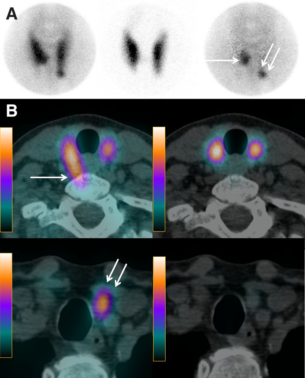
  • FIGURE 3.
    • Download figure
    • Open in new tab
    • Download powerpoint
    FIGURE 3.

    Simultaneous dual-tracer 99mTc-sestamibi and 123I scan in 62-y-old patient with PHPT. (A) Pinhole images of thyroid area with 99mTc-sestamibi, 123I, and subtraction. Pinhole subtraction image clearly identifies 2 residual foci: 1 at inner part of lower third of right thyroid lobe (arrow) and other at lower pole of left thyroid lobe (double arrow). (B) Axial SPECT/CT fusion images with 99mTc-sestamibi (right) and 123I (left), obtained at 2 different levels. On SPECT/CT axial views, right parathyroid lesion is located posteriorly, in paraesophageal position (arrow). This location is typical of prolapsed superior parathyroid gland (minor ectopia). Left inferior parathyroid lesion (double arrow) is located below lower pole of left thyroid lobe. Calcium level 6 mo after resection of 2 adenomas was normal.

  • FIGURE 4.
    • Download figure
    • Open in new tab
    • Download powerpoint
    FIGURE 4.

    84-y-old patient with PHPT and history of total thyroidectomy. Neck ultrasound results were negative. Simultaneous dual-tracer 99mTc-sestamibi and 123I parathyroid scan was performed after patient was instructed to withhold thyroid hormone therapy for 2 wk. (A) Pinhole images of neck show two 99mTc-sestamibi–avid foci (1 on each side of neck), but 123I scan clearly shows that focus on left side corresponds to thyroid remnant (arrow). (B) 99mTc-sestamibi and 123I SPECT/CT axial views without subtraction reveal posteriorly seated right parathyroid lesion and left thyroid remnant. Targeted surgery could be performed with resection of 600-mg right parathyroid adenoma.

  • FIGURE 5.
    • Download figure
    • Open in new tab
    • Download powerpoint
    FIGURE 5.

    18F-fluorocholine PET/CT performed 35 min after intravenous injection of 18F-fluorocholine in 71-y-old patient with PHPT. Results of neck ultrasonography and dual-phase 99mTc-sestamibi parathyroid scan performed at another institution before referral were negative. 18F-fluorocholine PET (A) with maximum-intensity-projection and axial view images show 18F-fluorocholine–avid focus corresponding on axial PET/CT fusion and CT images (B) to small tissue lesion behind right sternoclavicular junction (arrows). Parathyroid adenoma (500 mg) was resected.

  • FIGURE 6.
    • Download figure
    • Open in new tab
    • Download powerpoint
    FIGURE 6.

    18F-fluorocholine PET/CT in 74-y-old patient with PHPT. Neck ultrasonography described doubtful left inferior parathyroid lesion, and 99mTc-sestamibi/123I subtraction scintigraphy results were negative. 18F-fluorocholine PET/CT included conventional nonenhanced plus contrast-enhanced arterial-phase CT acquisitions. (A) Maximum-intensity-projection 18F-fluorocholine PET image reveal faint cervical focus (superior arrow) and multiple mediastinal foci (inferior arrow). (B) PET/CT fusion and arterial-phase contrast-enhanced CT (CECT) and PET axial images show choline-avid lesion with high contrast enhancement contiguous to left posterior side of esophagus, suggestive of ectopic parathyroid lesion (arrows). (C) PET/CT fusion and arterial-phase CECT and PET axial images of superior mediastinum show choline-avid lymph node with low contrast enhancement (arrows). At surgery, retroesophageal parathyroid adenoma was resected.

Tables

  • Figures
    • View popup
    TABLE 1

    Summary of Techniques Used for Parathyroid Scintigraphy and PET/CT

    TechniqueRadiopharmaceutical*Effective dose (mSv)†Protocol of administrationSuggested acquisition protocolTotal acquisition time
    99mTc-sestamibi/123I (subtraction)123I (∼12 MBq)1.5T0: 123I (intravenous or oral)99mTc and 123I simultaneous dual-isotope acquisition starting 3–5 min after 99mTc-sestamibi injection‡<1 h
    99mTc-sestamibi (∼600 MBq)5.4T+2 h: 99mTc-sestamibi (intravenous)Image of thyroid/parathyroid bed with pinhole collimator (15 min)
    Planar image of neck and mediastinum (5 min)§
    Then, dual-isotope SPECT/CT acquisition (∼25 min)
    99mTc-sestamibi (dual-phase)99mTc-sestamibi (740–900 MBq)6.7–8.1T0: 99mTc-sestamibi (intravenous)T+15 min (early phase); planar image of neck and mediastinum (5 min)§∼90 min in 2 sessions
    Image of thyroid/parathyroid bed with pinhole collimator (15 min)
    T+2 h (delayed phase); same protocol as early phase
    SPECT/CT (∼25 min) at early, delayed, or both phases
    18F-fluorocholine PET/CT (± CECT)18F-fluorocholine (100–200 MBq or 2–3 MBq/kg)2.0–4.0T0: 18F-fluorocholine (intravenous)PET acquisition starting ∼45 min (30–60 min) after injection¶20–30 min
    Nonenhanced CT from mandible to carina
    If arterial-phase CECT is added, then acquisition starts ∼30 s (25–40 s) after beginning of contrast medium injection
    • * Values in parentheses are activities.

    • ↵† Not including CT dose (25). For 123I, we considered thyroid uptake of 20%.

    • ↵‡ Example of nonoverlapping windows (29): for 99mTc, 140 keV ± 7% (130–150 keV); for 123I, 159 keV −4% to +10% (153–175 keV).

    • ↵§ Includes submandibular salivary glands and upper part of myocardium.

    • ↵¶ Early phase (∼ 5 min after injection) is additionally obtained by some authors.

    • CECT = contrast-enhanced CT; T = time.

PreviousNext
Back to top

In this issue

Journal of Nuclear Medicine: 62 (Supplement 2)
Journal of Nuclear Medicine
Vol. 62, Issue Supplement 2
July 1, 2021
  • Table of Contents
  • Table of Contents (PDF)
  • About the Cover
  • Index by author
Print
Download PDF
Article Alerts
Sign In to Email Alerts with your Email Address
Email Article

Thank you for your interest in spreading the word on Journal of Nuclear Medicine.

NOTE: We only request your email address so that the person you are recommending the page to knows that you wanted them to see it, and that it is not junk mail. We do not capture any email address.

Enter multiple addresses on separate lines or separate them with commas.
Primary Hyperparathyroidism: Defining the Appropriate Preoperative Imaging Algorithm
(Your Name) has sent you a message from Journal of Nuclear Medicine
(Your Name) thought you would like to see the Journal of Nuclear Medicine web site.
Citation Tools
Primary Hyperparathyroidism: Defining the Appropriate Preoperative Imaging Algorithm
Elif Hindié, Paul Schwartz, Anca M. Avram, Alessio Imperiale, Frederic Sebag, David Taïeb
Journal of Nuclear Medicine Jul 2021, 62 (Supplement 2) 3S-12S; DOI: 10.2967/jnumed.120.245993

Citation Manager Formats

  • BibTeX
  • Bookends
  • EasyBib
  • EndNote (tagged)
  • EndNote 8 (xml)
  • Medlars
  • Mendeley
  • Papers
  • RefWorks Tagged
  • Ref Manager
  • RIS
  • Zotero
Share
Primary Hyperparathyroidism: Defining the Appropriate Preoperative Imaging Algorithm
Elif Hindié, Paul Schwartz, Anca M. Avram, Alessio Imperiale, Frederic Sebag, David Taïeb
Journal of Nuclear Medicine Jul 2021, 62 (Supplement 2) 3S-12S; DOI: 10.2967/jnumed.120.245993
Twitter logo Facebook logo LinkedIn logo Mendeley logo
  • Tweet Widget
  • Facebook Like
  • Google Plus One
Bookmark this article

Jump to section

  • Article
    • Visual Abstract
    • Abstract
    • IMAGING IS RECOMMENDED BEFORE FIRST PARATHYROID SURGERY IRRESPECTIVE OF SURGICAL APPROACH
    • ADVANTAGES AND DISADVANTAGES OF TARGETED SURGERY: IMPLICATIONS REGARDING CHOICE OF IMAGING
    • WHY IS 99mTc-SESTAMIBI/123I SUBTRACTION IMAGING BETTER OPTION THAN DUAL-PHASE SESTAMIBI SCANNING FOR GUIDING TARGETED SURGERY?
    • SECOND-LINE IMAGING STRATEGY WHEN SCINTIGRAPHY AND NECK ULTRASOUND RESULTS ARE NEGATIVE, DISCORDANT, OR INCONCLUSIVE
    • IS IT TOO EARLY TO PROPOSE 18F-FLUOROCHOLINE PET/CT AS FIRST-LINE IMAGING?
    • LOCALIZATION PROCEDURES BEFORE REPEAT SURGERY FOR PERSISTENT OR RECURRENT HYPERPARATHYROIDISM
    • CONCLUSION
    • DISCLOSURE
    • ACKNOWLEDGMENT
    • REFERENCES
  • Figures & Data
  • Info & Metrics
  • PDF

Related Articles

  • No related articles found.
  • PubMed
  • Google Scholar

Cited By...

  • Radiopharmaceutical Dosimetry for Cancer Therapy: From Theory to Practice
  • Google Scholar

More in this TOC Section

  • SNMMI Procedure Standard/EANM Practice Guideline for Brain [18F]FDG PET Imaging, Version 2.0
  • Meeting Upcoming Clinical and Diagnostic Needs in Oncologic Imaging: A Structured Reporting System for Fibroblast-Activation-Protein–Targeted Imaging—FAP-RADS Version 1.0
  • Imaging Efficacy of [18F]CTT1057 PET for the Detection of PSMA-Positive Tumors Using Histopathology as Standard of Truth: Results from the GuideView Phase 2/3 Prospective Multicenter Study
Show more Clinical Investigation

Similar Articles

Keywords

  • primary hyperparathyroidism
  • sestamibi
  • dual-isotope
  • subtraction
  • SPECT/CT
  • 18F-fluorocholine PET/CT
SNMMI

© 2025 SNMMI

Powered by HighWire