
Main menu

  • Home
  • Content
    • Current
    • Ahead of print
    • Past Issues
    • JNM Supplement
    • SNMMI Annual Meeting Abstracts
    • Continuing Education
    • JNM Podcasts
  • Subscriptions
    • Subscribers
    • Institutional and Non-member
    • Rates
    • Journal Claims
    • Corporate & Special Sales
  • Authors
    • Submit to JNM
    • Information for Authors
    • Assignment of Copyright
    • AQARA requirements
  • Info
    • Reviewers
    • Permissions
    • Advertisers
  • About
    • About Us
    • Editorial Board
    • Contact Information
  • More
    • Alerts
    • Feedback
    • Help
    • SNMMI Journals
  • SNMMI
    • JNM
    • JNMT
    • SNMMI Journals
    • SNMMI

User menu

  • Subscribe
  • My alerts
  • Log in
  • My Cart

Search

  • Advanced search
Journal of Nuclear Medicine
  • SNMMI
    • JNM
    • JNMT
    • SNMMI Journals
    • SNMMI
  • Subscribe
  • My alerts
  • Log in
  • My Cart
Journal of Nuclear Medicine

Advanced Search

  • Home
  • Content
    • Current
    • Ahead of print
    • Past Issues
    • JNM Supplement
    • SNMMI Annual Meeting Abstracts
    • Continuing Education
    • JNM Podcasts
  • Subscriptions
    • Subscribers
    • Institutional and Non-member
    • Rates
    • Journal Claims
    • Corporate & Special Sales
  • Authors
    • Submit to JNM
    • Information for Authors
    • Assignment of Copyright
    • AQARA requirements
  • Info
    • Reviewers
    • Permissions
    • Advertisers
  • About
    • About Us
    • Editorial Board
    • Contact Information
  • More
    • Alerts
    • Feedback
    • Help
    • SNMMI Journals
  • View or Listen to JNM Podcast
  • Visit JNM on Facebook
  • Join JNM on LinkedIn
  • Follow JNM on Twitter
  • Subscribe to our RSS feeds
Research ArticleBasic Science Investigation

Comparison of CD38-Targeted α- Versus β-Radionuclide Therapy of Disseminated Multiple Myeloma in an Animal Model

Megan Minnix, Vikram Adhikarla, Enrico Caserta, Erasmus Poku, Russell Rockne, John E. Shively and Flavia Pichiorri
Journal of Nuclear Medicine June 2021, 62 (6) 795-801; DOI: https://doi.org/10.2967/jnumed.120.251983
Megan Minnix
1Department of Molecular Imaging and Therapy, Beckman Research Institute, City of Hope, Duarte, California
2Irell and Manella Graduate School of Biological Sciences, Beckman Research Institute, City of Hope, Duarte, California
  • Find this author on Google Scholar
  • Find this author on PubMed
  • Search for this author on this site
Vikram Adhikarla
3Department of Computational and Quantitative Medicine, Division of Mathematical Oncology, Beckman Research Institute, City of Hope, Duarte, California
  • Find this author on Google Scholar
  • Find this author on PubMed
  • Search for this author on this site
Enrico Caserta
4Briskin Myeloma Center and Department of Hematologic Malignancies Research Institute, City of Hope, Duarte, California; and
  • Find this author on Google Scholar
  • Find this author on PubMed
  • Search for this author on this site
Erasmus Poku
5Radiopharmacy, City of Hope, Duarte, California
  • Find this author on Google Scholar
  • Find this author on PubMed
  • Search for this author on this site
Russell Rockne
3Department of Computational and Quantitative Medicine, Division of Mathematical Oncology, Beckman Research Institute, City of Hope, Duarte, California
  • Find this author on Google Scholar
  • Find this author on PubMed
  • Search for this author on this site
John E. Shively
1Department of Molecular Imaging and Therapy, Beckman Research Institute, City of Hope, Duarte, California
  • Find this author on Google Scholar
  • Find this author on PubMed
  • Search for this author on this site
Flavia Pichiorri
4Briskin Myeloma Center and Department of Hematologic Malignancies Research Institute, City of Hope, Duarte, California; and
  • Find this author on Google Scholar
  • Find this author on PubMed
  • Search for this author on this site
  • Article
  • Figures & Data
  • Supplemental
  • Info & Metrics
  • PDF
Loading

Article Figures & Data

Figures

  • Tables
  • Additional Files
  • Figure1
    • Download figure
    • Open in new tab
    • Download powerpoint
  • FIGURE 1.
    • Download figure
    • Open in new tab
    • Download powerpoint
    FIGURE 1.

    High-dose 177Lu-DOTA-daratumumab (11.1 MBq) for treatment of disseminated MM. (A) Representative bioluminescence images for each group, imaged weekly, with intensity as indicated by color bar. Single mouse survived until day 36 (not shown in A). (B) Myeloma burden as quantified on BLI images, in radiance (daratumumab, P > 0.999; 11.1 MBq of 177Lu-DOTA-daratumumab, P = 0.038) and as quantified on Kaplan–Meier survival plot (daratumumab, P > 0.999; 11.1 MBq of 177Lu-DOTA-daratumumab, P = 0.045). (C) Whole-body toxicity as measured by weight (daratumumab, P = 0.883; 11.1 MBq of 177Lu-DOTA-daratumumab, P = 0.914). n = 4 for all groups. Dara = daratumumab.
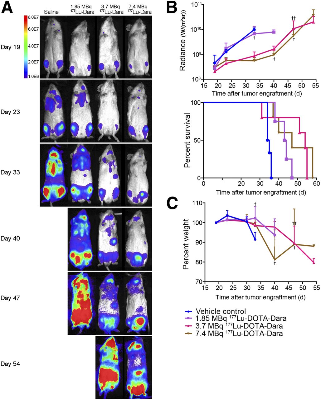
  • FIGURE 2.
    • Download figure
    • Open in new tab
    • Download powerpoint
    FIGURE 2.

    Dose response of 177Lu-DOTA-daratumumab (1.85, 3.7, and 7.4 MBq) for treatment of disseminated MM model. (A) Representative BLI images for each group, with intensity as indicated by color bar. (B) MM burden as quantified on BLI images, in radiance (1.85 MBq of 177Lu-DOTA-daratumumab, P = 0.91; 3.7 MBq of 177Lu-DOTA-daratumumab, P = 0.015; 7.4 MBq of 177Lu-DOTA-daratumumab, P = 0.014) and as quantified on Kaplan–Meier survival plot (1.85-MBq dose, P < 0.01; 3.7-MBq dose, P = 0.0310; 7.4-MBq dose, P < 0.01). Crosses indicate days on which mice were euthanized. (C) Whole-body toxicity as measured by weight (1.85-MBq dose, P = 0.997; 3.7-MBq dose, P = 0.821; 7.4-MBq dose, P = 0.750). n = 6 for saline group, 4 for 1.85-MBq group, and 5 for 3.7- and 7.4-MBq groups. Dara = daratumumab.
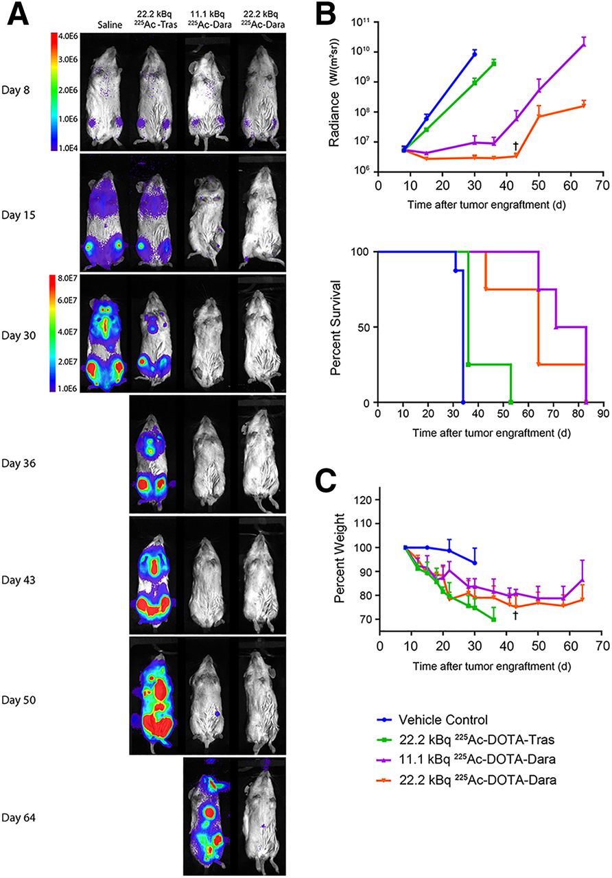
  • FIGURE 3.
    • Download figure
    • Open in new tab
    • Download powerpoint
    FIGURE 3.

    High dose of 225Ac-DOTA-daratumumab (11.1 and 22.2 kBq) for treatment of disseminated MM. (A) Representative BLI for each group, with intensity as indicated by color bar. To visually compare groups, >30-d separate scale was used. (B) MM burden as quantified on BLI, in radiance (22.2 kBq of 225Ac-DOTA-trastuzumab, P = 0.035; 11.1 kBq of 225Ac-DOTA-daratumumab, P = 0.015; 22.2 kBq of 225Ac-DOTA-daratumumab, P = 0.015) and as quantified on Kaplan–Meier survival plot (22.2 kBq of 225Ac-DOTA-trastuzumab, P < 0.01; 11.1 kBq of 225Ac-DOTA-daratumumab, P < 0.01; 22.2 kBq of 225Ac-DOTA-daratumumab, P < 0.01). Crosses indicate days on which mice were euthanized. (C) Whole-body toxicity as measured by weight (22.2 kBq of 225Ac-DOTA-trastuzumab, P = 0.0096; 11.1 kBq of 225Ac-DOTA-daratumumab, P = 0.0306; 22.2 kBq of 225Ac-DOTA-daratumumab, P = 0.0048). n = 8 for saline group and 4 for treated groups. Dara = daratumumab; Tras = trastuzumab.
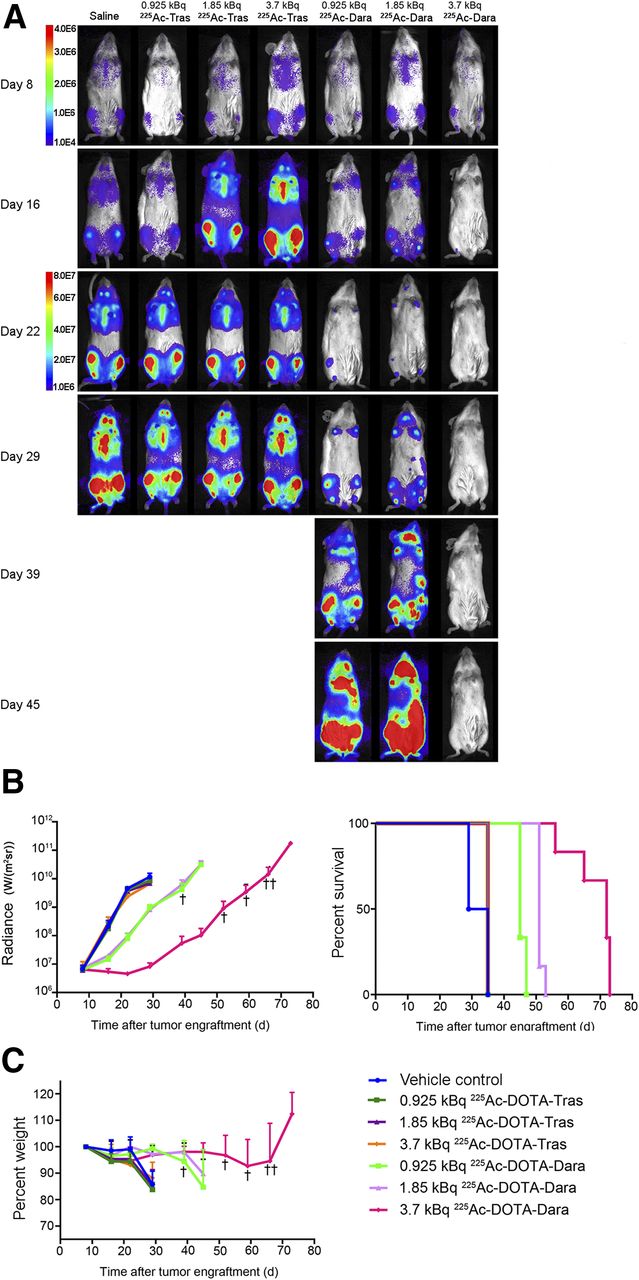
  • FIGURE 4.
    • Download figure
    • Open in new tab
    • Download powerpoint
    FIGURE 4.

    Dose response of 225Ac-DOTA-daratumumab (0.925, 1.85, and 3.7 kBq) for treatment of disseminated MM. (A) Representative BLI for each group, with intensity as indicated by color bar. After day 52, single mouse survived until day 66 (not shown in A). To visually compare groups, >30-d separate scale was used. (B) MM burden as quantified on BLI, in radiance (225Ac-DOTA-trastuzumab groups: 0.925 kBq, P = 0.96; 1.85 kBq, P = 0.67; 3.7 kBq, P = 0.42) (225Ac-DOTA-daratumumab groups: 0.925 kBq, P < 0.01; 1.85 kBq, P < 0.01; 3.7 kBq, P < 0.01) and as quantified on Kaplan–Meier survival plot (225Ac-DOTA-trastuzumab groups: 0.925 kBq, P = 0.048; 1.85 kBq, P = 0.048; 3.7 kBq, P = 0.048) (225Ac-DOTA-daratumumab groups: 0.925 kBq, P < 0.01; 1.85 kBq P < 0.01; 3.7 kBq, P < 0.01). Crosses indicate days on which mice were euthanized. (C) Whole-body toxicity as measured by weight (225Ac-DOTA-trastuzumab groups: 0.925 kBq, P = 0.992; 1.85 kBq, P = 0.999; 3.7 kBq, P = 0.999) (225Ac-DOTA-daratumumab groups: 0.925 kBq, P ≥ 0.999; 1.85 kBq, P ≥ 0.999; 3.7 kBq, P = 0.995). n = 8 for saline group and 6 for therapy groups. Dara = daratumumab; Tras = trastuzumab.
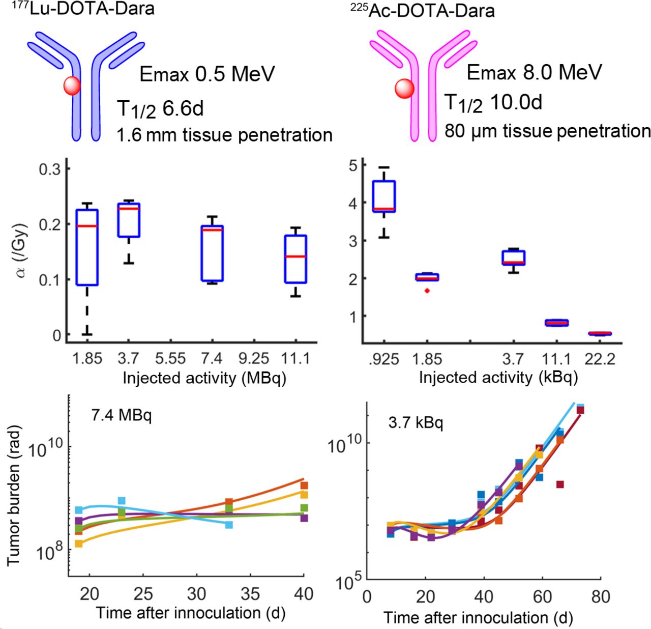
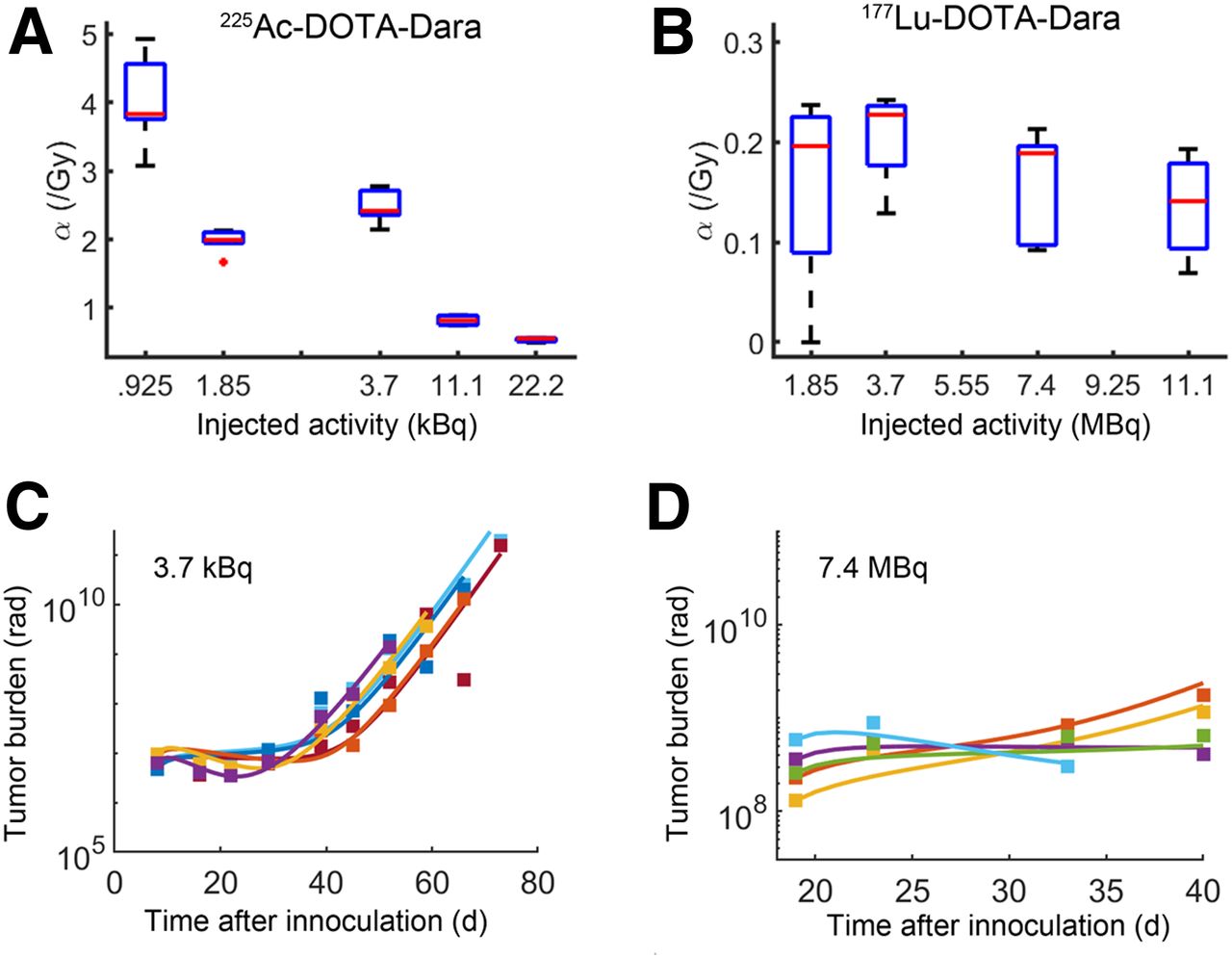
  • FIGURE 5.
    • Download figure
    • Open in new tab
    • Download powerpoint
    FIGURE 5.

    Radiobiologic analysis of 225Ac-DOTA-daratumumab and 177Lu-DOTA-daratumumab therapy. Radiosensitivity parameter α (Gy−1) is calculated for all dose levels of 225Ac and 177Lu DOTA-daratumumab treatments. (A) We observed nonlinear relationship between radiosensitivity and dose for 225Ac. Although 0.925 kBq results in largest value of α, this dose level did not confer survival advantage. Model predicts 3.7 kBq of 225Ac-DOTA-daratumumab to provide largest radiosensitivity and therapeutic benefit relative to 1.85-, 11.1-, and 22.2-kBq doses. (B) Low-LET 177Lu results in 10-fold lower α than does high-LET 225Ac and less pronounced correlation with injected activity. (C and D) Tumor burden measured by bioluminescence over time and mathematic model fits for 3.7 kBq of 225Ac and 7.4 MBq of 177Lu, respectively. Difference in duration of response can be seen between 225Ac and 177Lu, 60–80 d vs. 30–40 d. Dara = daratumumab.

Tables

  • Figures
  • Additional Files
    • View popup
    TABLE 1

    Efficacy, Toxicity, and Whole-Body Absorbed Dose for 177Lu Radioimmunotherapy in MM1-S Disseminated MM

    177Lu-DOTA-daratumumab
    ParameterVehicle control (n ≤ 10)*Daratumumab (n ≤ 4)1.85 MBq (n ≤ 4)3.7 MBq (n ≤ 5)7.4 MBq (n ≤ 5)11.1 MBq (n ≤ 4)
    Duration of tumor growth delay (days after MM1-S injection)000333332
    Start of weight loss (days after MM1-S injection)28†26†33†33‡33‡26‡
    Median survival (days after MM1-S injection)333144544736
    Whole-body absorbed dose (Gy)——0.91.94.16.4
    • ↵* Combined vehicle control groups from all 177Lu experiments.

    • ↵† Weight loss due to MM burden.

    • ↵‡ Weight loss due to radioimmunotherapy toxicity.

    • Mean value of all mice in each condition is given except for survival, which is median.

    • View popup
    TABLE 2

    Efficacy, Toxicity, and Whole-Body Absorbed Dose for 225Ac-Targeted α-Therapy in MM1-S Disseminated MM

    225Ac-DOTA-trastuzumab225Ac-DOTA-daratumumab
    ParameterVehicle control (n ≤ 16)*0.925 kBq (n ≤ 6)1.85 kBq (n ≤ 6)3.7 kBq (n ≤ 6)22.2 kBq (n ≤ 4)0.925 kBq (n ≤ 6)1.85 kBq (n ≤ 6)3.7 kBq (n ≤ 6)11.1 kBq (n ≤ 4)22.2 kBq (n ≤ 4)
    Duration of tumor growth delay (days after MM1-S injection)000001616293643
    Start of weight loss (days after MM1-S injection)22†22†22†22†0‡39†39†730‡0‡
    Median survival (days after MM1-S injection)33353535364551727764
    Whole-body absorbed dose (Gy)—0.20.30.74.20.20.40.82.34.7
    • ↵* Combined vehicle control groups from all 225Ac experiments.

    • ↵† Weight loss due to MM burden.

    • ↵‡ Weight loss due to radioimmunotherapy toxicity.

    • Mean value of all mice in each condition is given except for survival, which is median.

Additional Files

  • Figures
  • Tables
  • Supplemental Data

    Files in this Data Supplement:

    • Supplemental Data
PreviousNext
Back to top

In this issue

Journal of Nuclear Medicine: 62 (6)
Journal of Nuclear Medicine
Vol. 62, Issue 6
June 1, 2021
  • Table of Contents
  • Table of Contents (PDF)
  • About the Cover
  • Index by author
Print
Download PDF
Article Alerts
Sign In to Email Alerts with your Email Address
Email Article

Thank you for your interest in spreading the word on Journal of Nuclear Medicine.

NOTE: We only request your email address so that the person you are recommending the page to knows that you wanted them to see it, and that it is not junk mail. We do not capture any email address.

Enter multiple addresses on separate lines or separate them with commas.
Comparison of CD38-Targeted α- Versus β-Radionuclide Therapy of Disseminated Multiple Myeloma in an Animal Model
(Your Name) has sent you a message from Journal of Nuclear Medicine
(Your Name) thought you would like to see the Journal of Nuclear Medicine web site.
Citation Tools
Comparison of CD38-Targeted α- Versus β-Radionuclide Therapy of Disseminated Multiple Myeloma in an Animal Model
Megan Minnix, Vikram Adhikarla, Enrico Caserta, Erasmus Poku, Russell Rockne, John E. Shively, Flavia Pichiorri
Journal of Nuclear Medicine Jun 2021, 62 (6) 795-801; DOI: 10.2967/jnumed.120.251983

Citation Manager Formats

  • BibTeX
  • Bookends
  • EasyBib
  • EndNote (tagged)
  • EndNote 8 (xml)
  • Medlars
  • Mendeley
  • Papers
  • RefWorks Tagged
  • Ref Manager
  • RIS
  • Zotero
Share
Comparison of CD38-Targeted α- Versus β-Radionuclide Therapy of Disseminated Multiple Myeloma in an Animal Model
Megan Minnix, Vikram Adhikarla, Enrico Caserta, Erasmus Poku, Russell Rockne, John E. Shively, Flavia Pichiorri
Journal of Nuclear Medicine Jun 2021, 62 (6) 795-801; DOI: 10.2967/jnumed.120.251983
Twitter logo Facebook logo LinkedIn logo Mendeley logo
  • Tweet Widget
  • Facebook Like
  • Google Plus One
Bookmark this article

Jump to section

  • Article
    • Visual Abstract
    • Abstract
    • MATERIALS AND METHODS
    • RESULTS
    • DISCUSSION
    • CONCLUSION
    • DISCLOSURE
    • Footnotes
    • REFERENCES
  • Figures & Data
  • Supplemental
  • Info & Metrics
  • PDF

Related Articles

  • This Month in JNM
  • PubMed
  • Google Scholar

Cited By...

  • Targeted Alpha Therapy with [225Ac]Ac-Macropa-Isatuximab for CD38-positive Hematological Malignancies
  • Mathematical Modeling Unveils Optimization Strategies for Targeted Radionuclide Therapy of Blood Cancers
  • Cure of Disseminated Human Lymphoma with [225Ac]Ac-Ofatumumab in a Preclinical Model
  • Improved Tumor Responses with Sequential Targeted {alpha}-Particles Followed by Interleukin 2 Immunocytokine Therapies in Treatment of CEA-Positive Breast and Colon Tumors in CEA Transgenic Mice
  • Google Scholar

More in this TOC Section

  • Establishing In Vitro Dosimetric Models and Dose–Effect Relationships for 177Lu-DOTATATE in Neuroendocrine Tumors
  • Designed Ankyrin Repeat Protein–Mediated Peptide Nucleic Acid–Based Pretargeting: A Proof-of-Principle Study
  • [11C]ZTP-1: An Effective Short-Lived Radioligand for PET of Rat and Monkey Brain Phosphodiesterase Type 4 Subtype B
Show more Basic Science Investigation

Similar Articles

Keywords

  • CD38
  • daratumumab
  • multiple myeloma
  • radioimmunotherapy
  • mathematic modeling
SNMMI

© 2025 SNMMI

Powered by HighWire