


Article Figures & Data
Figures
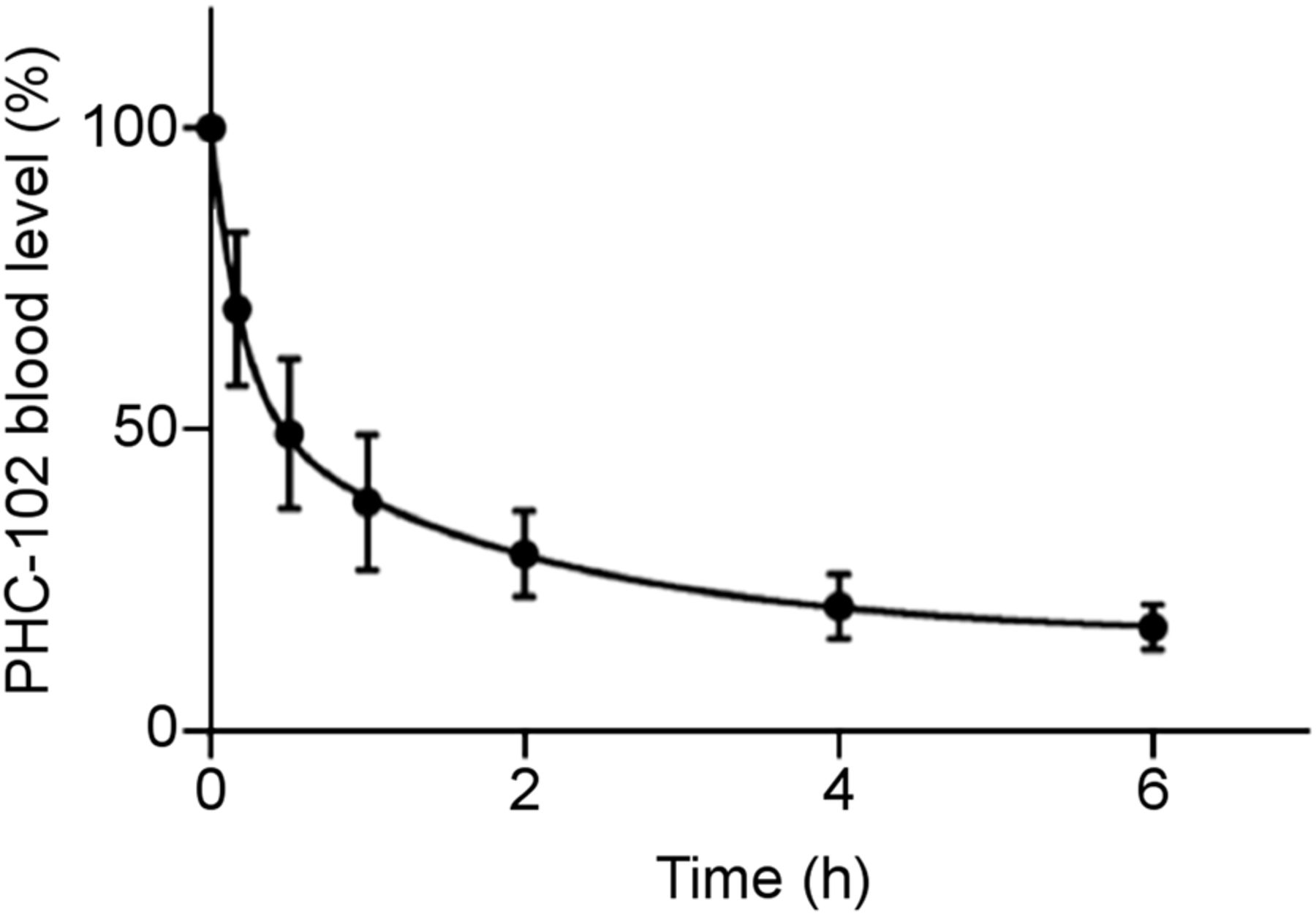
- FIGURE 1.
99mTc-PHC-102 blood levels. Blood clearance profiles were assessed by collecting blood samples at different time points and counting corresponding radioactivity values. Curve was calculated as average of values derived from individual patients. Variations in clearance rate were observed, possibly reflecting differences in kidney function.
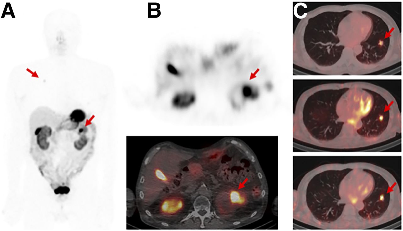
- FIGURE 2.
Anterior whole-body planar (A) and transverse SPECT (B) and 99mTc-PHC-102 fused SPECT/CT (C) images in 80-y-old patient with ccRCC (CAIX-positive). High uptake of radiotracer (arrows) in primary tumor (7.2 cm, upper pole of left kidney), in stomach, and in gallbladder was observed. Additional metastatic lesion (2.3 × 1.6 cm) in lung was also detected at all time points (0.5, 2, and 6 h).
- FIGURE 3.
Anterior SPECT/CT (A) and SPECT (B) scans obtained with 99mTc-PHC-102 in 49-y-old patient at (from left to right) 30 min, 2 h, and 6 h after administration of tracer. Primary neoplastic lesion (arrows) protruding from cortex of right kidney (1.6 cm) is visible at all time points. Renal excretion is appreciable from high signal observed in bladder at 30-min time point and from kidney uptake.
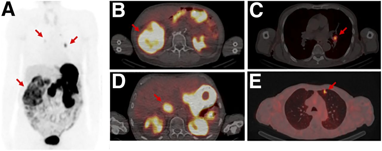
- FIGURE 4.
Anterior SPECT (arrows indicate RCC and metastases) (A) and transverse SPECT/CT (B) scans obtained with 99mTc-PHC-102 in 80-y-old patient at 6-h time point. Primary large tumor mass (A and B) in right kidney (7.2 cm), arrow in B indicates RCC) was detectable because of high tracer uptake. Tumor thrombus in vena cava (B and C); indicated with an arrow in C), as well as metastatic lesions in lymph node (D) indicated with an arrow) and in lung (E), indicated with an arrow), were also visible 6 h after 99mTc-PHC-102 injection because of high tracer uptake.


Tables
Subject Age (y) Sex Disease duration (y) Histology–CAIX 1 80 M <2 Positive 2 68 M <2 Positive 3 80 M <2 Positive 4 49 M <2 Negative 5 32 M <2 Unknown SUVmax SUVmean Organ 30 min 2 h 6 h 30 min 2 h 6 h Tumor 20.14 ± 3.94 18.43 ± 5.15 15.48 ± 4.74 14.02 ± 3.32 11.7 ± 3.08 10.38 ± 2.07 Kidney 35.04 ± 6.50 26.6 ± 4.05 17.50 ± 2.19 19.68 ± 4.49 16.75 ± 1.75 12.24 ± 1.33 Liver 4.26 ± 1.13 5.43 ± 1.11 6.06 ± 3.08 3.1 ± 1.16 4.00 ± 1.14 4.54 ± 2.57 Gallbladder 9.23 ± 3.66 17.2 ± 6.58 28.58 ± 11.80 6.55 ± 2.48 10.65 ± 2.77 15.28 ± 3.99 Intestine 6.78 ± 1.03 11.3 ± 4.57 10.80 ± 3.39 3.44 ± 1.12 3.65 ± 0.60 3.04 ± 0.93 Lung 0.94 ± 0.32 0.85 ± 0.30 0.56 ± 0.26 0.58 ± 0.26 0.40 ± 0.27 0.22 ± 0.13 Brain 0.32 ± 0.15 0.375 ± 0.22 0.28 ± 0.17 0.12 ± 0.12 0.11 ± 0.13 0.06 ± 0.05 Stomach 47.68 ± 15.63 58.5 ± 16.42 49.18 ± 28.54 21.22 ± 5.93 24.88 ± 6.12 20.96 ± 8.14 Spleen 1.28 ± 0.29 0.925 ± 0.34 0.78 ± 0.13 0.62 ± 0.26 0.35 ± 0.21 0.22 ± 0.08 Salivary gland 3.02 ± 2.52 1.32 ± 2.38 1.60 ± 0.94 2.3 ± 1.69 1.83 ± 1.31 1.08 ± 0.64 Average SUVmax and SUVmean are based on individual values from 5 patients imaged with microdosing 99mTc-PHC-102 SPECT/CT.
Supplemental Data
Files in this Data Supplement:


Jump to section
Related Articles
Cited By...
- Value of [68Ga]Ga-NYM046 PET/CT, in Comparison with 18F-FDG PET/CT, for Diagnosis of Clear Cell Renal Cell Carcinoma
- A deep learning approach for the discovery of tumor-targeting small organic ligands from DNA-Encoded Chemical Libraries
- Sortase-mediated site-specific modification of interleukin-2 for the generation of a tumor targeting acetazolamide-cytokine conjugate