


Article Figures & Data
Figures
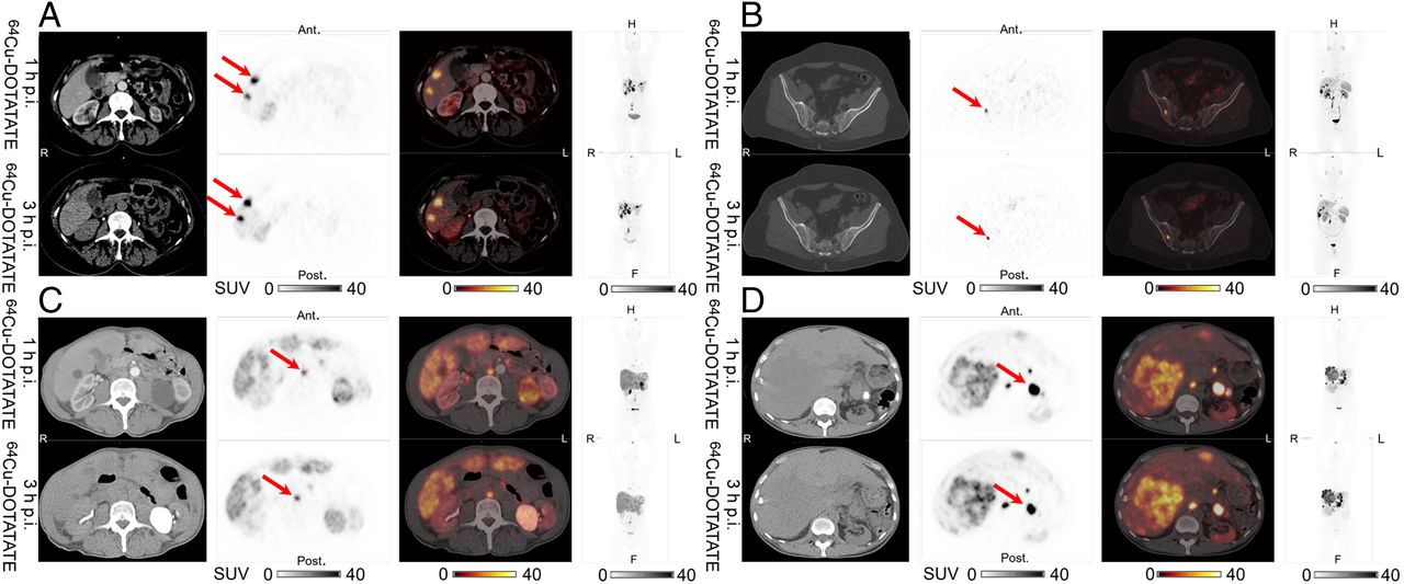
- FIGURE 1.
Representative examples of liver lesions (A), bone lesions (B), lymph node lesions (C), and pancreatic lesions (D) in same patients scanned at 1 and 3 h after injection. From left to right are shown CT, 64Cu-DOTATATE PET, 64Cu-DOTATATE PET/CT, and maximum-intensity projection, with corresponding SUV color bars below. All lesions were identified at both 1 h and 3 h on 64Cu-DOTATATE PET (arrows). Ant = anterior; F = feet; H = head; p.i. = after injection; post = posterior.
- FIGURE 2.
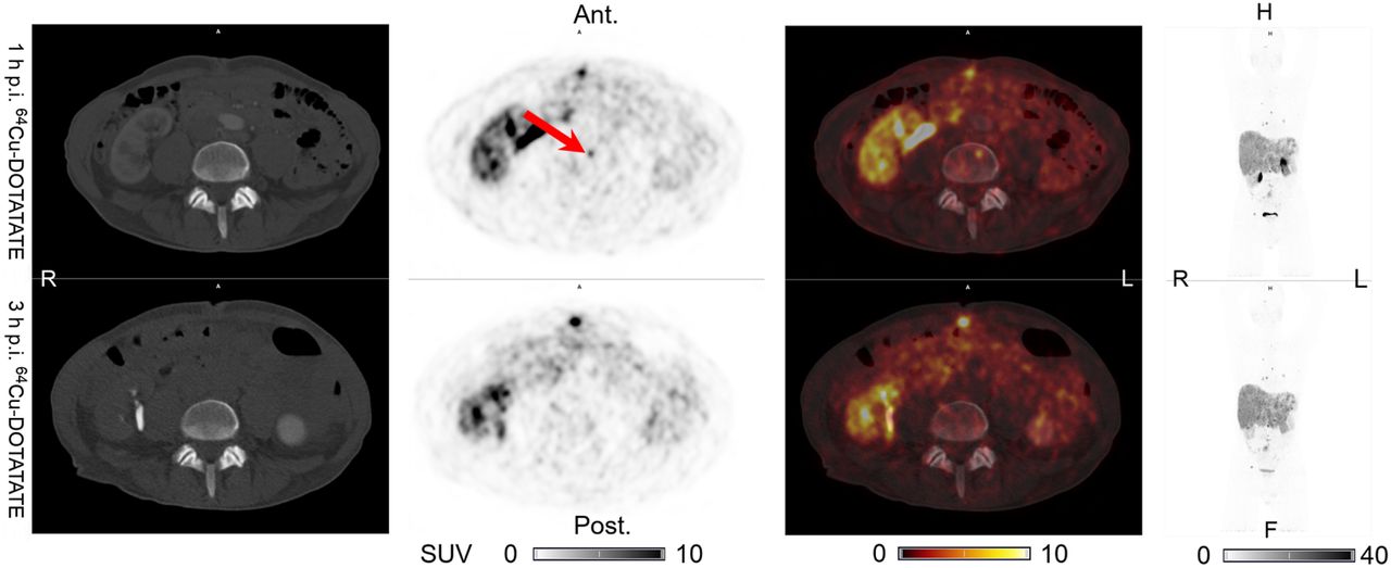
True-positive finding in patient 1: one additional lymph node lesion (arrows) visible on 64Cu-DOTATATE PET at 3 h after injection but not at 1 h, with visible CT correlate. Lesion was also visible on PET and CT at 1 h after injection on 64Cu-DOTATATE PET/CT performed 9 mo later. From left to right are shown CT, 64Cu-DOTATATE PET, 64Cu-DOTATATE PET/CT, and maximum-intensity projection, with corresponding SUV color bars below. Ant = anterior; F = feet; H = head; p.i. = after injection; post = posterior.
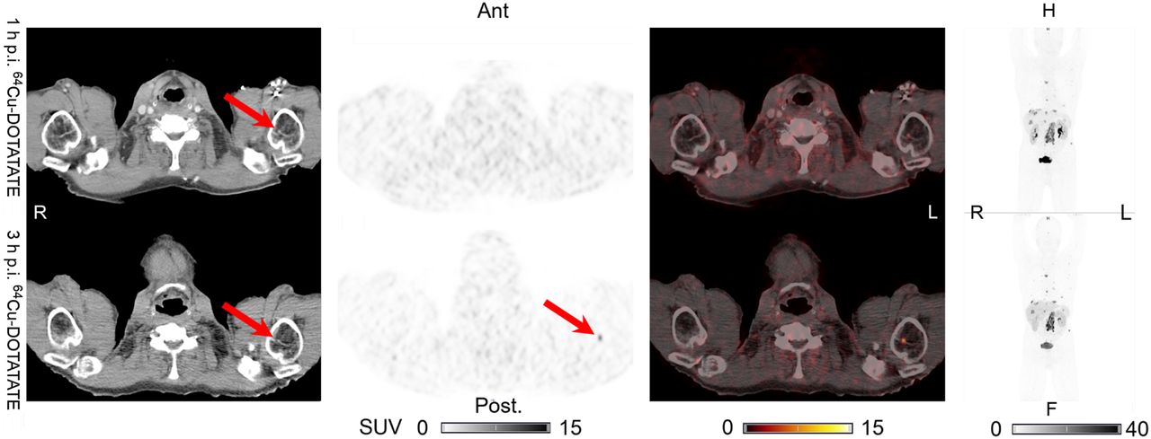
- FIGURE 3.
True-positive finding in patient 4: one additional bone lesion (arrows) visible on 64Cu-DOTATATE PET at 3 h after injection but not at 1 h, with visible CT correlate. No later SSTR PET images were available. From left to right are shown CT, 64Cu-DOTATATE PET, 64Cu-DOTATATE PET/CT, and maximum-intensity projection, with corresponding SUV color bars below. Ant = anterior; F = feet; H = head; p.i. = after injection; post = posterior.
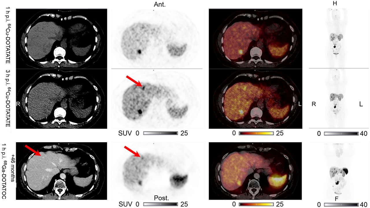
- FIGURE 4.
True-positive finding in patient 3: one additional liver lesion (arrows) visible on 64Cu-DOTATATE PET at 3 h after injection but not at 1 h, without CT correlate. This discordant lesion was also visible on PET and CT at 1 h after injection on 68Ga-DOTATOC PET performed 46 mo later. From left to right are shown CT, 64Cu-DOTATATE PET (68Ga-DOTATOC PET), 64Cu-DOTATATE PET/CT (68Ga-DOTATOC PET/CT), and maximum-intensity projection, with corresponding SUV color bars below. Ant = anterior; F = feet; H = head; p.i. = after injection; post = posterior.
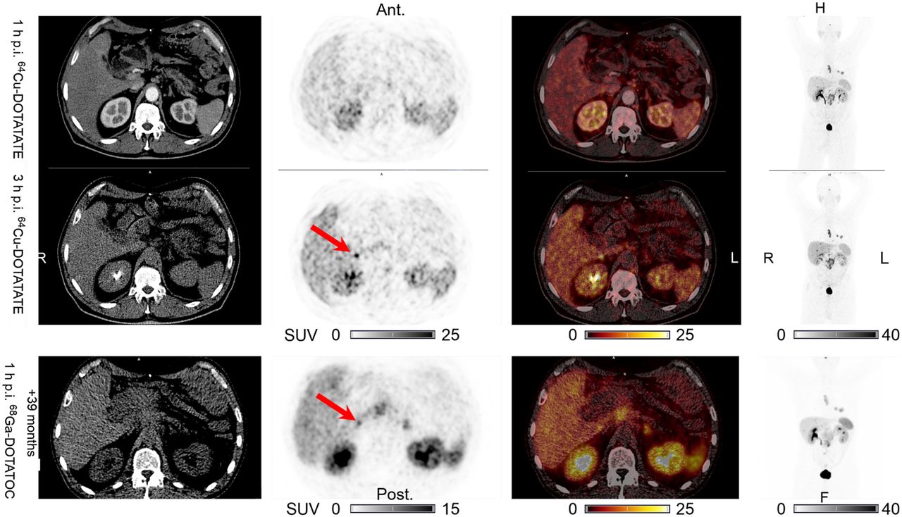
- FIGURE 5.
True-positive finding in patient 5: one additional liver lesion (arrows) visible on 64Cu-DOTATATE PET at 3 h after injection but not at 1 h, with no visible CT correlate. This discordant lesion was also visible on PET without CT correlate at 1 h after injection on 68Ga-DOTATOC PET/CT performed 39 mo later. From left to right are shown CT, 64Cu-DOTATATE PET (68Ga-DOTATOC PET), 64Cu-DOTATATE PET/CT(68Ga-DOTATOC PET/CT), and maximum-intensity projection, with corresponding SUV color bars below. Ant = anterior; F = feet; H = head; p.i. = after injection; post = posterior.
- FIGURE 6.
False-positive finding in patient 2: one additional bone lesion (arrow) visible on 64Cu-DOTATATE PET at 1 h after injection but not at 3 h, without CT correlate. No later SSTR PET images were available. No visible CT correlate was seen on latest CT scan, 8 mo later (not shown). From left to right are shown CT, 64Cu-DOTATATE PET, 64Cu-DOTATATE PET/CT, and maximum-intensity projection, with corresponding SUV color bars below. Ant = anterior; F = feet; H = head; p.i. = after injection; post = posterior.


Tables
Characteristic Data Sex Male 21 (60%) Female 14 (40%) Age (y) Mean 62 Range 40–81 Site of primary tumor Lung 3 (9%) Gastrointestinal 13 (37%) Pancreatic 5 (14%) Other 2 (6%) Unknown 12 (34%) Functional status Nonfunctioning 20 (57%) Functioning (carcinoid syndrome) 15 (43%) Grade Low (G1) 8 (23%) Intermediate (G2) 21 (60%) High (G3) 2 (6%) KI-67 proliferation index not available 4 (11%) Primary tumor removed No 20 (57%) Yes 15 (43%) Previous treatments* Surgery 15 (43%) Interferon α 19 (54%) Somatostatin analogs 14 (40%) Radiofrequency ablation (liver metastases) 3 (9%) External radiation therapy 1 (3%) Peptide receptor radionuclide therapy 12 (34%) ↵* Some patients received multiple treatments. Therefore, total number of treatments exceeded number of patients.
Data are n followed by percentage in parentheses, except for age.
Visible on 64Cu-DOTATATE PET Organ or tissue Concordant Only at 1 h after injection Only at 3 h after injection P† Lung 14 0 0 — Liver 298 0 2 0.98 Intestines 18 0 0 — Pancreas 12 0 0 — Intraabdominal carcinomatosis 7 0 0 — Bone 326 1 1 1.00 Lymph nodes 114 0 1 0.98 Other* 33 0 0 — Total 822 1 4 0.99 ↵* 22 soft-tissue lesions, 5 heart lesions, 1 prostate lesion, 1 adrenal lesion, 1 stomach lesion, 1 thyroid lesion, 1 brain lesion, and 1 spleen lesion.
↵† Testing for differences in number of lesions with negative binominal regression on 64Cu-DOTATATE PET between 1 and 3 h after injection. The analyses are performed per organ or region and on the total number of lesions.
Visible on 64Cu-DOTATATE PET Patient no. Concordant Only at 1 h after injection Only at 3 h after injection Status Months until follow-up Follow-up image modality Discordant organ system 1 Liver (11) LN (1) TP 0*/9 CT/Cu LN 2 Bone (15), LN (7), lung (1), carc (1), liver (43) Bone (1) FP 8 CT None 3 Liver (6), LN (8), int (1) Liver (1) TP 46/46 Ga/CT None 4 Bone (19), spleen (1), int (1), adrenal (1), heart (3), liver (5) Bone (1) TP 0* CT None 5 Liver (2), LN (19) heart (2), int (1) Liver (1) TP 39 Ga None ↵* Visible CT correlate on 64Cu-DOTATATE PET/CT.
LN = lymph nodes; TP = true-positive; Cu = follow-up 64Cu-DOTATATE PET at 1 h after injection; carc = intraabdominal carcinomatosis; FP = false-positive; int = intestines; Ga = 68Ga-DOTATOC PET.
Lesion SUVmax on 64Cu-DOTATATE PET Organ or tissue n* 1 h after injection 3 h after injection P† Lung 5 17.9 [3.0–32.9] 18.7 [5.3–32.1] 1.00 Liver 22 45.7 [37.2–54.3] 54.1 [44.3–64.0] <0.01 Intestines 12 64.4 [43.9–84.8] 77.8 [52.3–103.3] 0.19 Pancreas 8 79.0 [38.3–119.6] 85.9 [35.9–135.8] 1.00 Bone 12 44.2 [25.7–62.7] 50.1 [29.7–70.4] 0.46 Intraabdominal carcinomatosis 4 23.0 [7.7–38.3] 24.9 [9.0–40.7] 1.00 Lymph nodes 18‡ 40.9 [26.9–54.9] 43.5 [29.1–58.0] 0.76 ↵* Number of patients with lesions.
↵† Paired t testing with Bonferroni adjustment for multiple comparisons (n = 7) and capped at 1.00.
↵‡ n = 19 on 64Cu-DOTATATE PET at 3 h after injection (one additional lymph node lesion detected in 1 patient).
Data are mean values followed by 95% confidence interval in brackets.
Normal-organ SUVmean† on 64Cu-DOTATATE PET Organ or tissue n* 1 h after injection 3 h after injection P† Lung 35 0.27 [0.23–0.30] 0.15 [0.13–0.17] <0.01 Liver 32 4.0 [3.6–4.4] 5.7 [5.2–6.3] <0.01 Intestines 35 2.6 [2.1–3.1] 2.5 [2.0–3.1] 1.00 Uncinate process of pancreas 31 3.2 [2.7–3.6] 3.3 [2.7–3.9] 1.00 Cauda of pancreas 32 3.1 [2.8–3.5] 3.5 [3.2–3.9] 0.38 Bone 35 0.76 [0.66–0.85] 0.64 [0.56–0.73] <0.01 Muscle 35 0.63 [0.57–0.69] 0.49 [0.44–0.54] <0.01 Spleen 35 8.9 [7.8–10.0] 9.3 [8.2–10.4] 0.12 Pituitary gland 35 12.9 [10.8–14.9] 15.8 [13.4–18.2] <0.01 Adrenal gland 33 9.5 [8.0–11.0] 9.9 [8.2–11.6] 1.00 TTN† Organ or tissue n* 1 h after injection of 64Cu-DOTATATE 3 h after injection of 64Cu-DOTATATE P† Lung 5 87.9 [30.2–145.6] 160.9 [79.2–242.6] 0.04 Liver 19 12.6 [10.2–14.9] 11.0 [8.7–13.4] 0.03 Intestines 12 24.2 [14.9–33.4] 28.2 [16.5–40.0] 0.73 Pancreas 6 42.4 [12.3–72.5] 41.1 [8.7–73.4] 1.00 Bone 12 103.0 [38.6–167.4] 124.2 [57.1–191.2] 0.07 Intraabdominal carcinomatosis 4 14.0 [3.0–25.0] 22.5 [7.1–38.0] 0.50 Lymph nodes 18‡ 73.7 [43.0–104.4] 94.0 [61.6–126.4] 0.07 ↵* Number of patients with lesions and evaluable normal tissue.
↵† Paired t testing with Bonferroni adjustment for multiple comparisons (n = 7) and capped at 1.00.
↵‡ n = 19 on 64Cu-DOTATATE PET at 3 h after injection (one additional lymph node lesion detected in 1 patient).
Data are mean values followed by 95% confidence interval in brackets.


Jump to section
Related Articles
Cited By...
- Routine Use of [64Cu]Cu-DOTATATE PET/CT in a Neuroendocrine Tumor Center: Referral Patterns and Image Results of 2,249 Consecutive Scans
- Efficacy of [67Cu]Cu-EB-TATE Theranostic Against Somatostatin Receptor Subtype-2-Positive Neuroendocrine Tumors
- An Investigation of Lesion Detection Accuracy for Artificial Intelligence-Based Denoising of Low-Dose 64Cu-DOTATATE PET Imaging in Patients with Neuroendocrine Neoplasms
- An Investigation of Lesion Detection Accuracy for Artificial Intelligence-Based Denoising of Low-Dose 64Cu-DOTATATE PET Imaging in Patients with Neuroendocrine Neoplasms
- SNMMI Procedure Standard/EANM Practice Guideline for SSTR PET: Imaging Neuroendocrine Tumors
- Semiautomatic Tumor Delineation for Evaluation of 64Cu-DOTATATE PET/CT in Patients with Neuroendocrine Neoplasms: Prognostication Based on Lowest Lesion Uptake and Total Tumor Volume
- Somatostatin Receptor Imaging and Theranostics: Current Practice and Future Prospects