
Main menu

  • Home
  • Content
    • Current
    • Ahead of print
    • Past Issues
    • JNM Supplement
    • SNMMI Annual Meeting Abstracts
    • Continuing Education
    • JNM Podcasts
  • Subscriptions
    • Subscribers
    • Institutional and Non-member
    • Rates
    • Journal Claims
    • Corporate & Special Sales
  • Authors
    • Submit to JNM
    • Information for Authors
    • Assignment of Copyright
    • AQARA requirements
  • Info
    • Reviewers
    • Permissions
    • Advertisers
  • About
    • About Us
    • Editorial Board
    • Contact Information
  • More
    • Alerts
    • Feedback
    • Help
    • SNMMI Journals
  • SNMMI
    • JNM
    • JNMT
    • SNMMI Journals
    • SNMMI

User menu

  • Subscribe
  • My alerts
  • Log in
  • Log out
  • My Cart

Search

  • Advanced search
Journal of Nuclear Medicine
  • SNMMI
    • JNM
    • JNMT
    • SNMMI Journals
    • SNMMI
  • Subscribe
  • My alerts
  • Log in
  • Log out
  • My Cart
Journal of Nuclear Medicine

Advanced Search

  • Home
  • Content
    • Current
    • Ahead of print
    • Past Issues
    • JNM Supplement
    • SNMMI Annual Meeting Abstracts
    • Continuing Education
    • JNM Podcasts
  • Subscriptions
    • Subscribers
    • Institutional and Non-member
    • Rates
    • Journal Claims
    • Corporate & Special Sales
  • Authors
    • Submit to JNM
    • Information for Authors
    • Assignment of Copyright
    • AQARA requirements
  • Info
    • Reviewers
    • Permissions
    • Advertisers
  • About
    • About Us
    • Editorial Board
    • Contact Information
  • More
    • Alerts
    • Feedback
    • Help
    • SNMMI Journals
  • View or Listen to JNM Podcast
  • Visit JNM on Facebook
  • Join JNM on LinkedIn
  • Follow JNM on Twitter
  • Subscribe to our RSS feeds
Meeting ReportOncology: Clinical Therapy and Diagnosis

Automated Esophageal Gross Tumor Volume Segmentation in 18F-FDG PET and CT for Radiotherapy using Two-Stream 3D Deep Network Fusion

Tsung-Ying Ho, Dakai Jin, Dazhou Guo, CHEN-KAN TSENG, Jing Xiao, Le Lu and Tzu-Chen Yen
Journal of Nuclear Medicine May 2020, 61 (supplement 1) 498;
Tsung-Ying Ho
1Chang Gung Memorial Hospital Taipei Taiwan
  • Find this author on Google Scholar
  • Find this author on PubMed
  • Search for this author on this site
Dakai Jin
2PAII Inc. Bethesda MD United States
  • Find this author on Google Scholar
  • Find this author on PubMed
  • Search for this author on this site
Dazhou Guo
2PAII Inc. Bethesda MD United States
  • Find this author on Google Scholar
  • Find this author on PubMed
  • Search for this author on this site
CHEN-KAN TSENG
3Chang Gung Memorial Hospital At Linkou Taoyuan Taiwan
  • Find this author on Google Scholar
  • Find this author on PubMed
  • Search for this author on this site
Jing Xiao
4Ping An Technology Shenzhen China
  • Find this author on Google Scholar
  • Find this author on PubMed
  • Search for this author on this site
Le Lu
2PAII Inc. Bethesda MD United States
  • Find this author on Google Scholar
  • Find this author on PubMed
  • Search for this author on this site
Tzu-Chen Yen
5Chang-Gung Memorial Hospital Taoyuan Taiwan
  • Find this author on Google Scholar
  • Find this author on PubMed
  • Search for this author on this site
  • Article
  • Figures & Data
  • Info & Metrics
Loading

Abstract

498

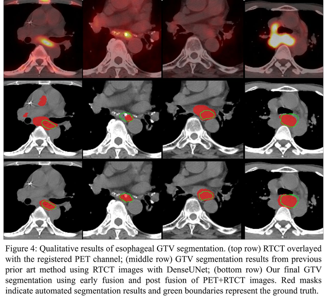
Objectives: Accurate delineation of gross tumor volume (GTV) is the decisive step in esophageal cancer radiotherapy. Physicians refer to high contrast from positron emission tomography/computed tomography (PET/CT) and manually delineate GTV on radiation therapy computed tomography (RTCT). However, 18F-FDG PET/CT has not been well-explored for computer-based automated esophageal GTV delineation. In this work, we aim to utilize the complementary information of 18F-FDG PET and RTCT to facilitate the automated GTV delineation task in the esophageal cancer treatment.

Methods: We curated a dataset of diagnostic 18F-FDG PET/CT, RTCT images and 3D GTV masks from 110 patients with stage II or greater esophageal cancer and undergoing radiotherapy. We propose a two-stream 3D deep network fusion pipeline to segment esophageal GTV using 18F-FDG PET and RTCT. First, we align the PET image to RTCT through the registration of PETCT to RTCT using the B-spline based deformable registration algorithm with a robust anatomy-based initialization. Next, we trained a two-stream pipeline that combines and merges predictions from two independent sub-networks, one only trained using the RTCT and one trained using both RTCT and aligned PET images. The former exploits the anatomical contextual information in CT, while the latter takes advantage of PET sensitivity. The predictions of these two streams are then deeply fused together with the original RTCT to provide a final GTV prediction. Furthermore, a 3D progressive holistically nested network (PHNN) model is adopted for effective fusion and segmentation. Five-fold cross-validation is used to fully evaluate our method.

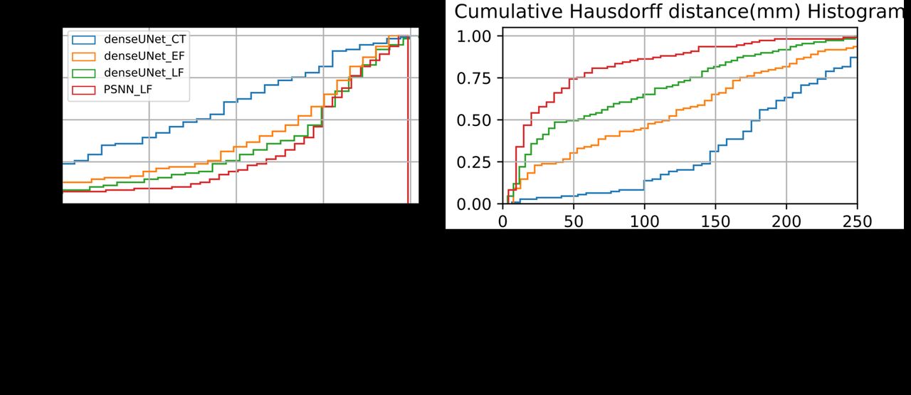
Results: The proposed two-stream 3D deep network fusion method using 18F-FDG PET and RTCT modalities has achieved 0.755±0.148 Dice score (DSC) and 4.7±5.2 cm Hausdorff distance (HD). It significantly outperforms the previous leading RTCT based esophageal GTV segmentation method, i.e. DenseUnet by 10% DSC increase and 8.2 cm HD reduction.

Conclusions: Our work demonstrates that the 18F-FDG PET images can be effectively integrated with RTCT to better segment the esophageal GTV using a two-stream 3D deep network fusion method. We provide significant and tangible improvements compared to the recent RTCT-based representative work, which represents a step forward toward reliable and automated esophageal GTV segmentation.

Figure
  • Download figure
  • Open in new tab
  • Download powerpoint
Figure
  • Download figure
  • Open in new tab
  • Download powerpoint
Figure
  • Download figure
  • Open in new tab
  • Download powerpoint
Figure
  • Download figure
  • Open in new tab
  • Download powerpoint
Figure
  • Download figure
  • Open in new tab
  • Download powerpoint
Previous
Back to top

In this issue

Journal of Nuclear Medicine
Vol. 61, Issue supplement 1
May 1, 2020
  • Table of Contents
  • Index by author
Article Alerts
Sign In to Email Alerts with your Email Address
Email Article

Thank you for your interest in spreading the word on Journal of Nuclear Medicine.

NOTE: We only request your email address so that the person you are recommending the page to knows that you wanted them to see it, and that it is not junk mail. We do not capture any email address.

Enter multiple addresses on separate lines or separate them with commas.
Automated Esophageal Gross Tumor Volume Segmentation in 18F-FDG PET and CT for Radiotherapy using Two-Stream 3D Deep Network Fusion
(Your Name) has sent you a message from Journal of Nuclear Medicine
(Your Name) thought you would like to see the Journal of Nuclear Medicine web site.
Citation Tools
Automated Esophageal Gross Tumor Volume Segmentation in 18F-FDG PET and CT for Radiotherapy using Two-Stream 3D Deep Network Fusion
Tsung-Ying Ho, Dakai Jin, Dazhou Guo, CHEN-KAN TSENG, Jing Xiao, Le Lu, Tzu-Chen Yen
Journal of Nuclear Medicine May 2020, 61 (supplement 1) 498;

Citation Manager Formats

  • BibTeX
  • Bookends
  • EasyBib
  • EndNote (tagged)
  • EndNote 8 (xml)
  • Medlars
  • Mendeley
  • Papers
  • RefWorks Tagged
  • Ref Manager
  • RIS
  • Zotero
Share
Automated Esophageal Gross Tumor Volume Segmentation in 18F-FDG PET and CT for Radiotherapy using Two-Stream 3D Deep Network Fusion
Tsung-Ying Ho, Dakai Jin, Dazhou Guo, CHEN-KAN TSENG, Jing Xiao, Le Lu, Tzu-Chen Yen
Journal of Nuclear Medicine May 2020, 61 (supplement 1) 498;
Twitter logo Facebook logo LinkedIn logo Mendeley logo
  • Tweet Widget
  • Facebook Like
  • Google Plus One
Bookmark this article

Jump to section

  • Article
  • Figures & Data
  • Info & Metrics

Related Articles

  • No related articles found.
  • Google Scholar

Cited By...

  • No citing articles found.
  • Google Scholar

More in this TOC Section

Oncology: Clinical Therapy and Diagnosis

  • Added value of single photon emission computed tomography (SPECT) / Computed Tomography (CT) with radioiodine whole body scan in follow up of patients with differentiated thyroid cancer
  • Assessment of incidental pituitary uptake on FDG PET/CT scan: Digital vs. conventional PET/CT
  • Thyroglobulin measurement in fine-needle aspiration optimize lateral neck dissection in patients with papillary thyroid cancer
Show more Oncology: Clinical Therapy and Diagnosis

Miscellaneous

  • The Diagnostic Contribution of 18F-FDG PET/CT SCAN in Cancer of BMUO
  • Role of 18F-FDG PET-CT in Epitheloid Hemangioendothelioma
Show more Miscellaneous

Similar Articles

SNMMI

© 2025 SNMMI

Powered by HighWire