
Main menu

  • Home
  • Content
    • Current
    • Ahead of print
    • Past Issues
    • JNM Supplement
    • SNMMI Annual Meeting Abstracts
    • Continuing Education
    • JNM Podcasts
  • Subscriptions
    • Subscribers
    • Institutional and Non-member
    • Rates
    • Journal Claims
    • Corporate & Special Sales
  • Authors
    • Submit to JNM
    • Information for Authors
    • Assignment of Copyright
    • AQARA requirements
  • Info
    • Reviewers
    • Permissions
    • Advertisers
  • About
    • About Us
    • Editorial Board
    • Contact Information
  • More
    • Alerts
    • Feedback
    • Help
    • SNMMI Journals
  • SNMMI
    • JNM
    • JNMT
    • SNMMI Journals
    • SNMMI

User menu

  • Subscribe
  • My alerts
  • Log in
  • Log out
  • My Cart

Search

  • Advanced search
Journal of Nuclear Medicine
  • SNMMI
    • JNM
    • JNMT
    • SNMMI Journals
    • SNMMI
  • Subscribe
  • My alerts
  • Log in
  • Log out
  • My Cart
Journal of Nuclear Medicine

Advanced Search

  • Home
  • Content
    • Current
    • Ahead of print
    • Past Issues
    • JNM Supplement
    • SNMMI Annual Meeting Abstracts
    • Continuing Education
    • JNM Podcasts
  • Subscriptions
    • Subscribers
    • Institutional and Non-member
    • Rates
    • Journal Claims
    • Corporate & Special Sales
  • Authors
    • Submit to JNM
    • Information for Authors
    • Assignment of Copyright
    • AQARA requirements
  • Info
    • Reviewers
    • Permissions
    • Advertisers
  • About
    • About Us
    • Editorial Board
    • Contact Information
  • More
    • Alerts
    • Feedback
    • Help
    • SNMMI Journals
  • View or Listen to JNM Podcast
  • Visit JNM on Facebook
  • Join JNM on LinkedIn
  • Follow JNM on Twitter
  • Subscribe to our RSS feeds
Meeting ReportOncology: Clinical Therapy and Diagnosis

Imaging characteristics and diagnostic performance of 18F-FDG PET/CT for melanoma patients who demonstrate hyperprogressive disease when treated with immunotherapy

Ryusuke Nakamoto, Lisa Zaba, Jarrett Rosenberg, Sunil Reddy, Tomomi Nobashi, Guido Davidzon, Carina Aparici, Judy Nguyen, Farshad Moradi, Andrei Iagaru and Benjamin Franc
Journal of Nuclear Medicine May 2020, 61 (supplement 1) 484;
Ryusuke Nakamoto
1Radiology Stanford University Stanford CA United States
  • Find this author on Google Scholar
  • Find this author on PubMed
  • Search for this author on this site
Lisa Zaba
2Dermatology Stanford University Stanford CA United States
  • Find this author on Google Scholar
  • Find this author on PubMed
  • Search for this author on this site
Jarrett Rosenberg
1Radiology Stanford University Stanford CA United States
  • Find this author on Google Scholar
  • Find this author on PubMed
  • Search for this author on this site
Sunil Reddy
3Oncology Stanford University Stanford CA United States
  • Find this author on Google Scholar
  • Find this author on PubMed
  • Search for this author on this site
Tomomi Nobashi
4Kyoto University Kyoto-city Japan
  • Find this author on Google Scholar
  • Find this author on PubMed
  • Search for this author on this site
Guido Davidzon
1Radiology Stanford University Stanford CA United States
  • Find this author on Google Scholar
  • Find this author on PubMed
  • Search for this author on this site
Carina Aparici
1Radiology Stanford University Stanford CA United States
  • Find this author on Google Scholar
  • Find this author on PubMed
  • Search for this author on this site
Judy Nguyen
1Radiology Stanford University Stanford CA United States
  • Find this author on Google Scholar
  • Find this author on PubMed
  • Search for this author on this site
Farshad Moradi
1Radiology Stanford University Stanford CA United States
  • Find this author on Google Scholar
  • Find this author on PubMed
  • Search for this author on this site
Andrei Iagaru
1Radiology Stanford University Stanford CA United States
  • Find this author on Google Scholar
  • Find this author on PubMed
  • Search for this author on this site
Benjamin Franc
1Radiology Stanford University Stanford CA United States
  • Find this author on Google Scholar
  • Find this author on PubMed
  • Search for this author on this site
  • Article
  • Figures & Data
  • Info & Metrics
Loading

Abstract

484

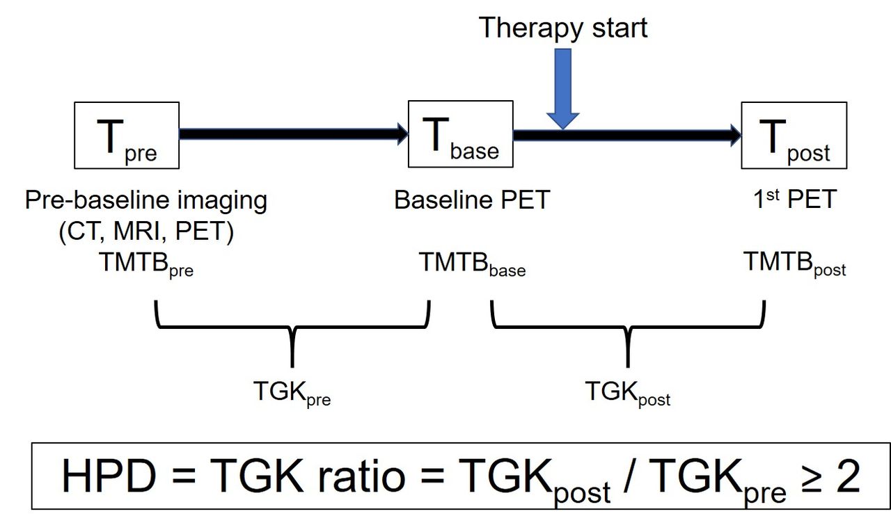
Objectives: Immune checkpoint inhibitors (ICIs) have recently emerged as one of the most important advances in cancer treatment. On the other hand, cancer patients treated with ICIs sometimes experience very poor prognosis called “hyperprogressive disease (HPD)”. HPD is usually defined as a RECIST progression at the first evaluation and as a ≥ 2-fold increase of the tumor growth kinetic ratio (TGKR) compared with pre-immunotherapy. Previous studies reported that HPD correlated with shorter progression-free survival (PFS) and overall survival (OS). At this moment, no useful method to predict the risk of HPD accurately before immunotherapy has been established, because its mechanism has not been elucidated. This retrospective study investigated 18F-FDG PET/CT findings of melanoma patients who demonstrated HPD after the start of ICIs. Then, we investigated the ability of baseline PET/CT parameters, acquired before the start of immunotherapy, to predict the risk of HPD. In addition, we evaluated whether we could diagnose HPD accurately based only on quantitative values obtained from baseline and first restaging PET/CT without information of TGR, a parameter that requires pre-baseline imaging before baseline imaging (3 time-point imaging).

Methods: Seventy-six patients (M, 49; F, 27) who underwent PET/CT scans before and approximately 3 months after immunotherapy from July 2010 to November 2018 were retrospectively enrolled. PET/CT parameters, including the sums of MTV, TLG, and the longest diameters of target lesions (total measured tumor burden, TMTB) based on irRECIST were measured from baseline PET/CT (MTVbase, TLGbase, and TMTBbase, respectively) and first restaging PET/CT (MTVpost, TLGpost, and TMTBpost, respectively). The ratio of MTV (MTVpost / MTVbase, MTVr), TLG (TLGpost / TLGbase, TLGr), and TMTB (TMTBpost / TMTBbase, TMTBr) before and approximately 3 months after immunotherapy were also calculated. These PET/CT parameters were compared between HPD and other patients. The area under the curves (AUCs) were used to assess the diagnostic performances of baseline PET/CT parameters to predict the risk of HPD, and those of MTVr, TLGr, and TMTBr to diagnose HPD without information of TGR. Kaplan-Meier survival analysis, and log-rank test were used for statistical analysis.

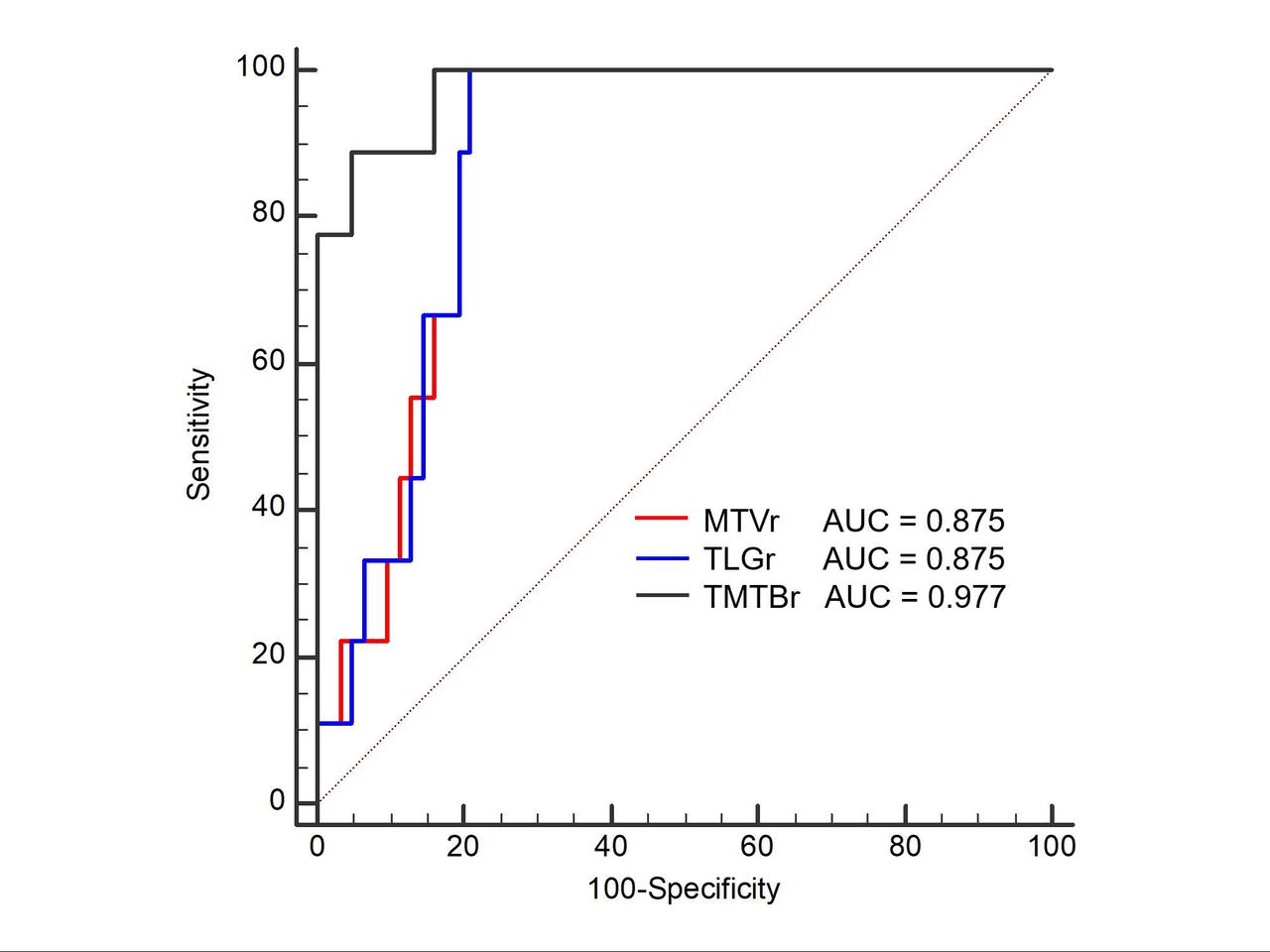
Results: Nine patients (M, 8; F, 1) met the criteria of HPD (11.8%) (TGKR ≥ 2, Fig. 1). MTVbase of the HPD patients was higher than that of non-HPD (TGKR < 2) patients (P = 0.037) and OS of HPD patients was worse than that of non-HPD patients (7 vs. more than 60 months, P = 0.0001) (Table). The AUC of MTVbase (> 155.45ml) to predict the risk of HPD was 0.703 (95% CI 0.585 to 0.804, P = 0.043), with a sensitivity of 66.7% and specificity of 81.2% (Fig. 2). The AUCs of MTVr (≥ 1.25), TLGr (≥ 1.15), and TMTBr (≥ 1.27) to diagnose HPD without information of TGKR were 0.875 (95% CI 0.775 to 0.941, P < 0.0001), 0.875 (95% CI 0.775 to 0.941, P < 0.0001), and 0.977 (95% CI 0.909 to 0.998, P < 0.0001), respectively, with all sensitivities of 100%, and specificities of 79%, 79%, and 83.9%, respectively (Fig. 3). One pseudoprogrssion case (1.3%) was seen in this population. MTVr, TLGr, and TMTBr of this case were 2.31 (> 1.25), 2.10 (> 1.15), and 1.62 (> 1.27), respectively, misdiagnosed as HPD when utilizing the ratio of tumor burden. TGKR of this pseudoprogression case was 1.29 (< 2.0).

Conclusions: Whole-body MTV of HPD patients at baseline PET was higher than other patients, but patients at high risk of developing HPD could not be accurately identified based on baseline PET/CT metabolic parameters prior to initiating immunotherapy. The ratio of PET/CT parameters before and approximately 3 months after the start of immunotherapy may be helpful to diagnose HPD, when patients do not undergo pre-baseline imaging, although distinguishing pseudoprogression from HPD must take into account the clinical assessment in addition to imaging.

View this table:
  • View inline
  • View popup

Comparison of PET/CT findings between HPD and non-HPD patients

Figure
  • Download figure
  • Open in new tab
  • Download powerpoint

Figure
  • Download figure
  • Open in new tab
  • Download powerpoint
Figure
  • Download figure
  • Open in new tab
  • Download powerpoint
Previous
Back to top

In this issue

Journal of Nuclear Medicine
Vol. 61, Issue supplement 1
May 1, 2020
  • Table of Contents
  • Index by author
Article Alerts
Sign In to Email Alerts with your Email Address
Email Article

Thank you for your interest in spreading the word on Journal of Nuclear Medicine.

NOTE: We only request your email address so that the person you are recommending the page to knows that you wanted them to see it, and that it is not junk mail. We do not capture any email address.

Enter multiple addresses on separate lines or separate them with commas.
Imaging characteristics and diagnostic performance of 18F-FDG PET/CT for melanoma patients who demonstrate hyperprogressive disease when treated with immunotherapy
(Your Name) has sent you a message from Journal of Nuclear Medicine
(Your Name) thought you would like to see the Journal of Nuclear Medicine web site.
Citation Tools
Imaging characteristics and diagnostic performance of 18F-FDG PET/CT for melanoma patients who demonstrate hyperprogressive disease when treated with immunotherapy
Ryusuke Nakamoto, Lisa Zaba, Jarrett Rosenberg, Sunil Reddy, Tomomi Nobashi, Guido Davidzon, Carina Aparici, Judy Nguyen, Farshad Moradi, Andrei Iagaru, Benjamin Franc
Journal of Nuclear Medicine May 2020, 61 (supplement 1) 484;

Citation Manager Formats

  • BibTeX
  • Bookends
  • EasyBib
  • EndNote (tagged)
  • EndNote 8 (xml)
  • Medlars
  • Mendeley
  • Papers
  • RefWorks Tagged
  • Ref Manager
  • RIS
  • Zotero
Share
Imaging characteristics and diagnostic performance of 18F-FDG PET/CT for melanoma patients who demonstrate hyperprogressive disease when treated with immunotherapy
Ryusuke Nakamoto, Lisa Zaba, Jarrett Rosenberg, Sunil Reddy, Tomomi Nobashi, Guido Davidzon, Carina Aparici, Judy Nguyen, Farshad Moradi, Andrei Iagaru, Benjamin Franc
Journal of Nuclear Medicine May 2020, 61 (supplement 1) 484;
Twitter logo Facebook logo LinkedIn logo Mendeley logo
  • Tweet Widget
  • Facebook Like
  • Google Plus One
Bookmark this article

Jump to section

  • Article
  • Figures & Data
  • Info & Metrics

Related Articles

  • No related articles found.
  • Google Scholar

Cited By...

  • No citing articles found.
  • Google Scholar

More in this TOC Section

Oncology: Clinical Therapy and Diagnosis

  • Added value of single photon emission computed tomography (SPECT) / Computed Tomography (CT) with radioiodine whole body scan in follow up of patients with differentiated thyroid cancer
  • Assessment of incidental pituitary uptake on FDG PET/CT scan: Digital vs. conventional PET/CT
  • Thyroglobulin measurement in fine-needle aspiration optimize lateral neck dissection in patients with papillary thyroid cancer
Show more Oncology: Clinical Therapy and Diagnosis

Sarcoma, Melanoma & Others

  • Role of 18F-FDG PET/CT Imaging in Cardiac Masses
  • Prognostic value of volumetric PET parameters at early response evaluation in melanoma patients treated with immunotherapy
  • 18F-FDG PET/CT in prognostication of malignant peripheral nerve sheath tumors.
Show more Sarcoma, Melanoma & Others

Similar Articles

SNMMI

© 2025 SNMMI

Powered by HighWire