


Abstract
1179
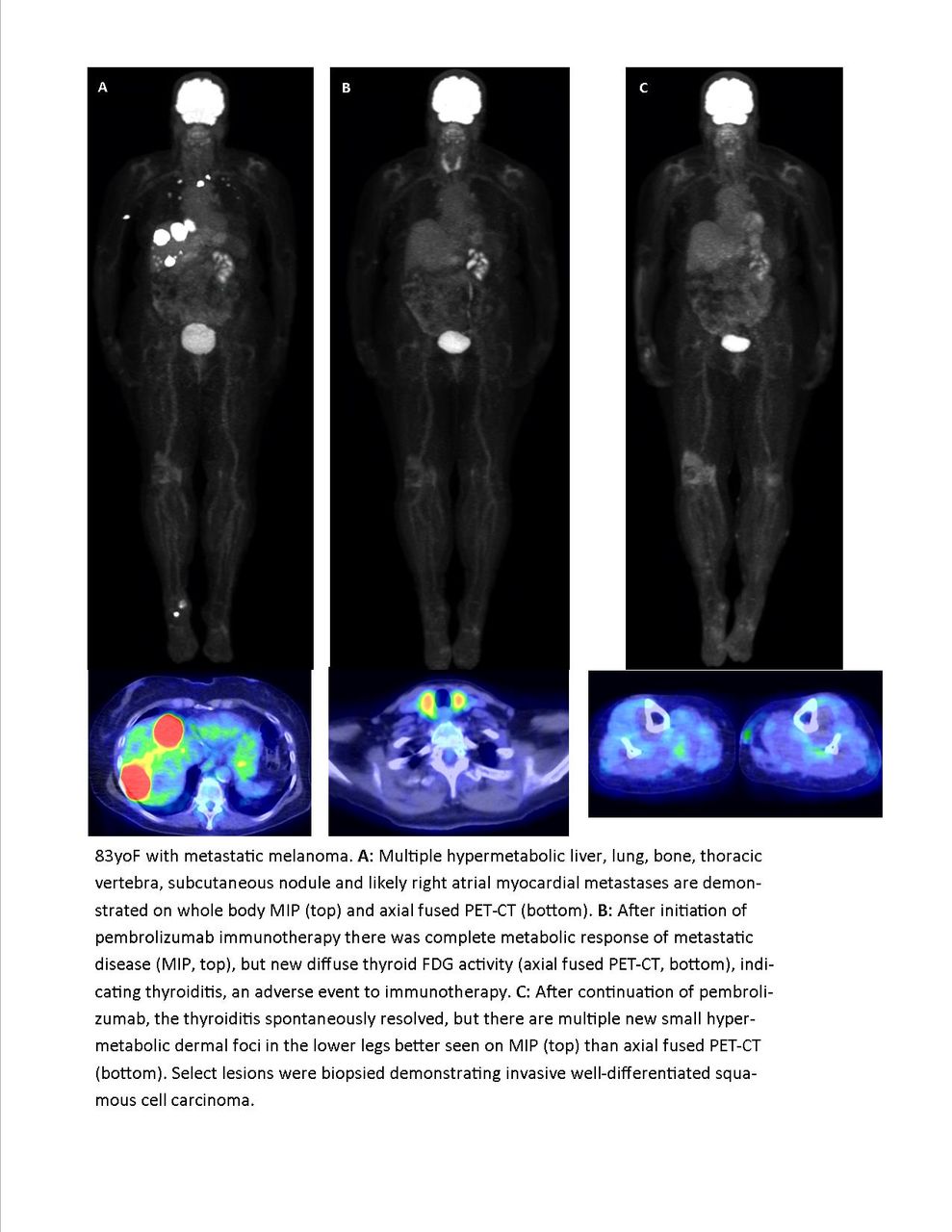
Objectives: Immunotherapy is an emerging tool in the treatment of a variety of otherwise drug resistant cancers. Immunotherapy agents, also known as immune checkpoint inhibitors, act on one of several critical normal checkpoints in the immune system: programmed cell death protein 1 (PD-1), programmed cell death ligand 1 (PD-L1) or cytotoxic T lymphocyte-associated antigen 4 (CTLA-4). In doing so, these agents effectively stimulate the immune system to target the cancer cells. More well-known agents include nivolumab (PD-1), pembrolizumab (PD-1) and ipilimumab (CTLA-4), but others exist. Initially approved for treatment refractory metastatic melanoma, there are now many malignancies for which immunotherapy are indicated. Unfortunately, these immunotherapy agents have also been associated with a variety of immune related adverse events. These adverse events most commonly affect the skin and colon but appear to be able to involve any organ system, most commonly presenting as an inflammatory process at the affected site, but more unique and organ specific manifestation have been recognized. Severity of the adverse event ranges greatly. Many of these adverse events manifest on serial PET examinations obtained over a treatment interval. Familiarity with these potential immunotherapy related adverse events is critical for the PET reader to recognize and become familiar in order to not mistakenly diagnose recurrent or worsening malignancy when in fact a positive treatment response may be present, but accompanied by an treatment related adverse event. When such an adverse event is suspected, it is critical to relay this to the referring clinician so that the benefits of any positive treatment response can be considered against the risks continuing the immunotherapy. This educational exhibit contains many image-rich examples of these immunotherapy related adverse events. Pertinent patient and treatment history is provided. Additionally, a case of “pseudoprogression”, a potentially confusing scenario associated with immune checkpoint inhibitors where sites of malignancy demonstrate apparent worsening on FDG PET imaging shortly after initiation of immunotherapy only to spontaneously improve on subsequent follow up, is featured. The Figure depicts an 83 year old female with metastatic melanoma. The patient demonstrates near complete metabolic response of widely metastatic disease after initiation of pembrolizumab immunotherapy. The patient experience immunotherapy-related thyroiditis, but symptoms were mild so the treatment was continued. On subsequent follow-up PET, the spontaneously resolved, but multiple new small hypermetabolic dermal foci appeared in the lower legs. Select lesions were biopsied demonstrating invasive well-differentiated squamous cell carcinoma. In this particular case, multispecialty consensus deemed the appearance of squamous cell carcinoma as unlikely related to the pembrolizumab. However, there are case reports in the literature of squamous cell carcinomas developing in a similar clinical context. It is important to be aware of the possible association in order to avoid confusion over the more common and similar appearing immunotherapy related panniculitis.

In this issue


Jump to section
Related Articles
Cited By...
- No citing articles found.