


Article Figures & Data
Figures
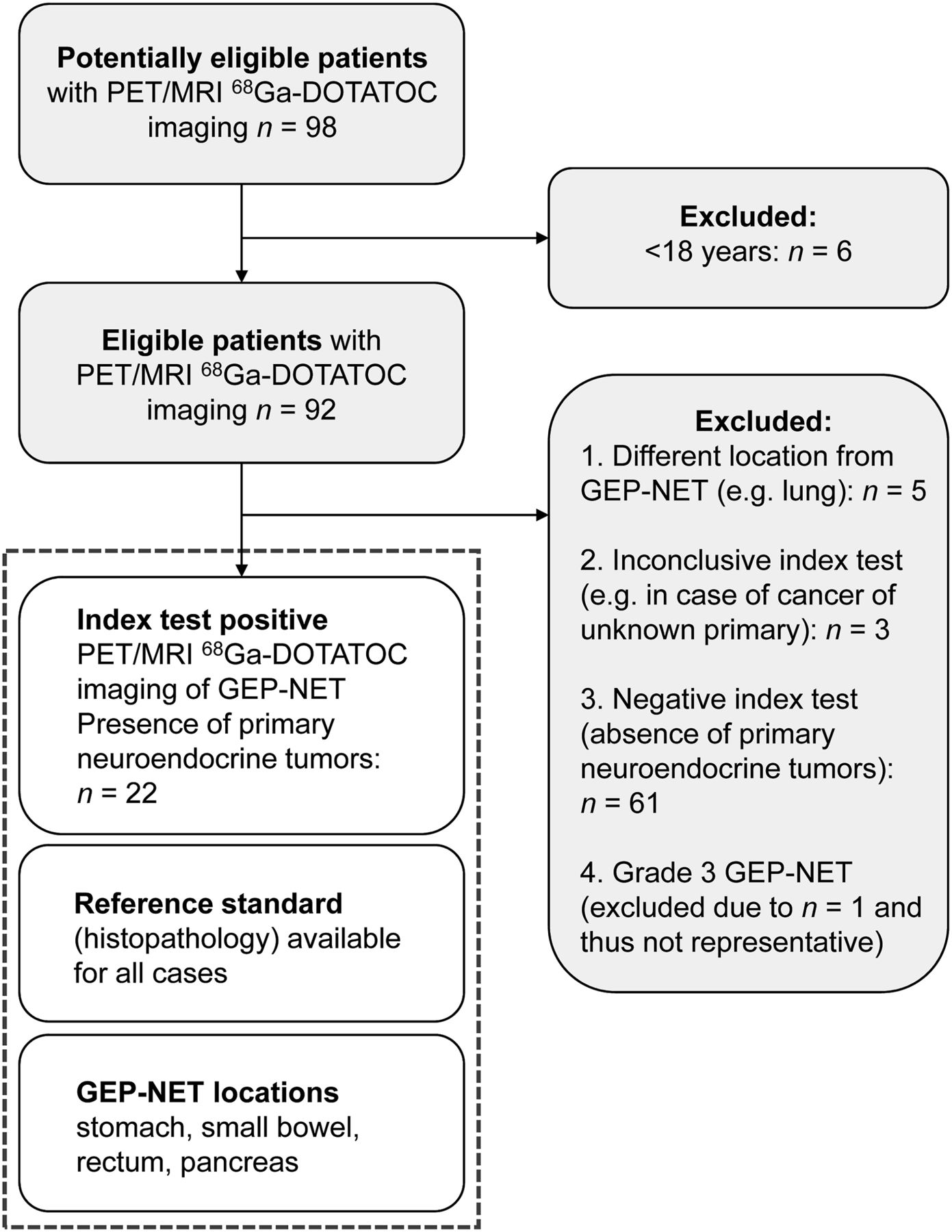
- FIGURE 1.
Study flowchart.
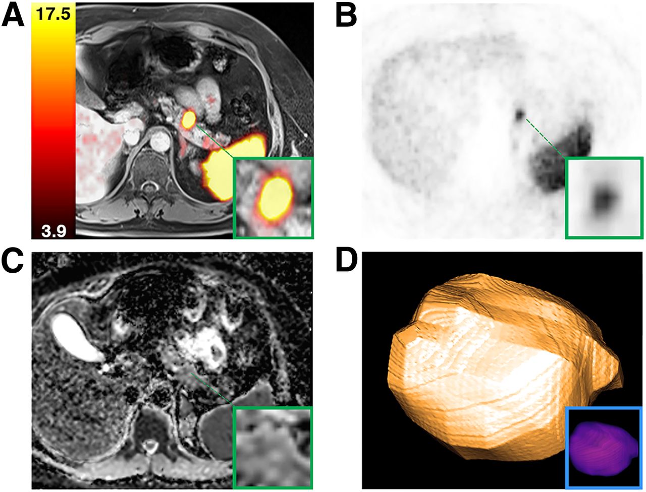
- FIGURE 2.
Example of 3D VOI lesion analysis in 50-y-old patient with grade 1 pancreas NET (SUVmean of 15 and ADCmin of 900 mm2/s; combined SUVmean/ADCmin ratio, 0.02). Shown are fusion of enhanced T1-weighted VIBE (volumetric interpolated breath-hold sequence) MRI with 68Ga-DOTATOC PET (A), 68Ga-DOTATOC PET (B), ADC map (C), and 3D lesion model (D).
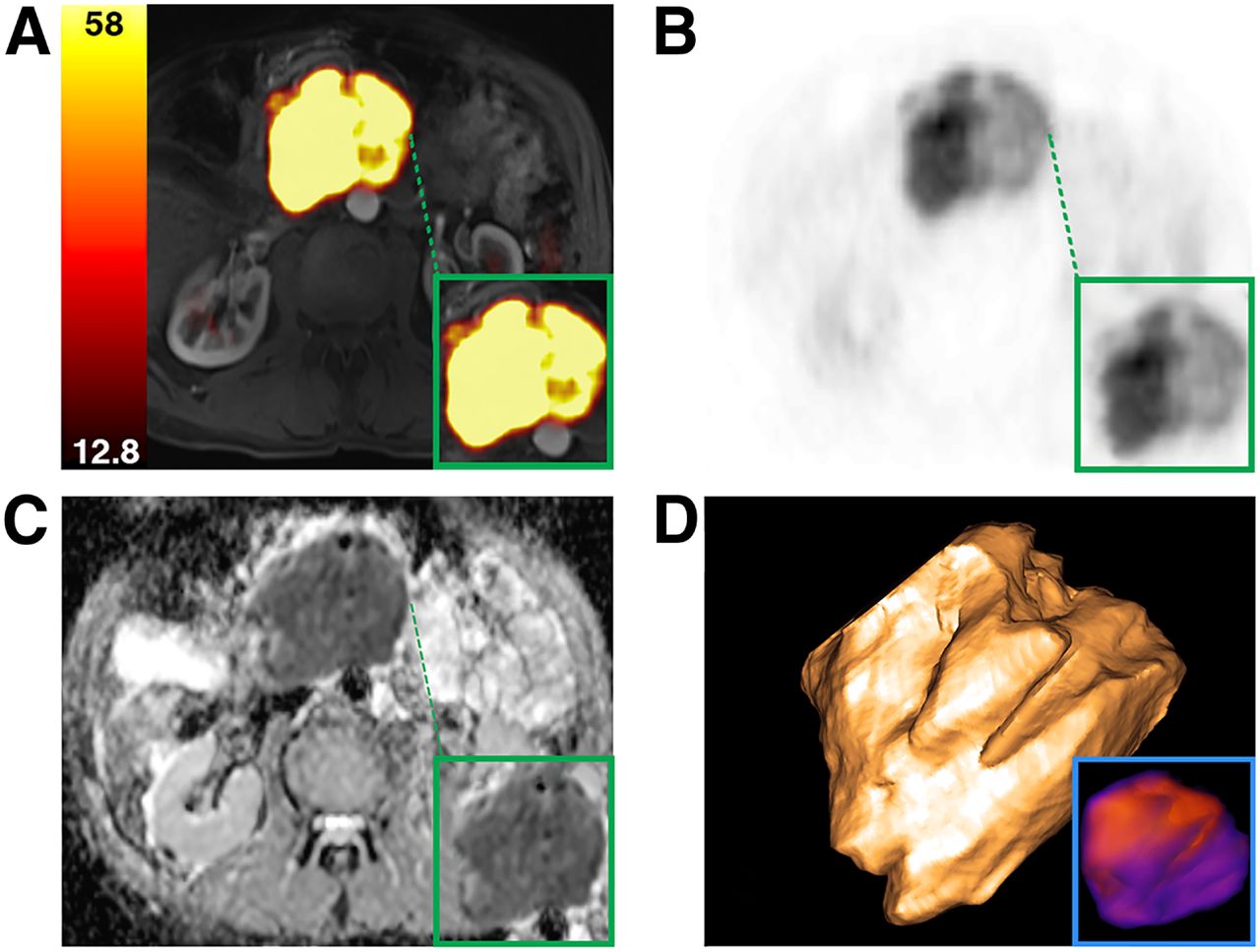
- FIGURE 3.
Example of 3D VOI lesion analysis in 64-y-old patient with grade 2 pancreas NET (SUVmean of 45 and ADCmin of 490 mm2/s; combined SUVmean/ADCmin ratio, 0.09). Shown are fusion of enhanced T1-weighted VIBE (volumetric interpolated breath-hold sequence) MRI with 68Ga-DOTATOC PET (A), 68Ga-DOTATOC PET (B), ADC map (C), and 3D lesion model (D).
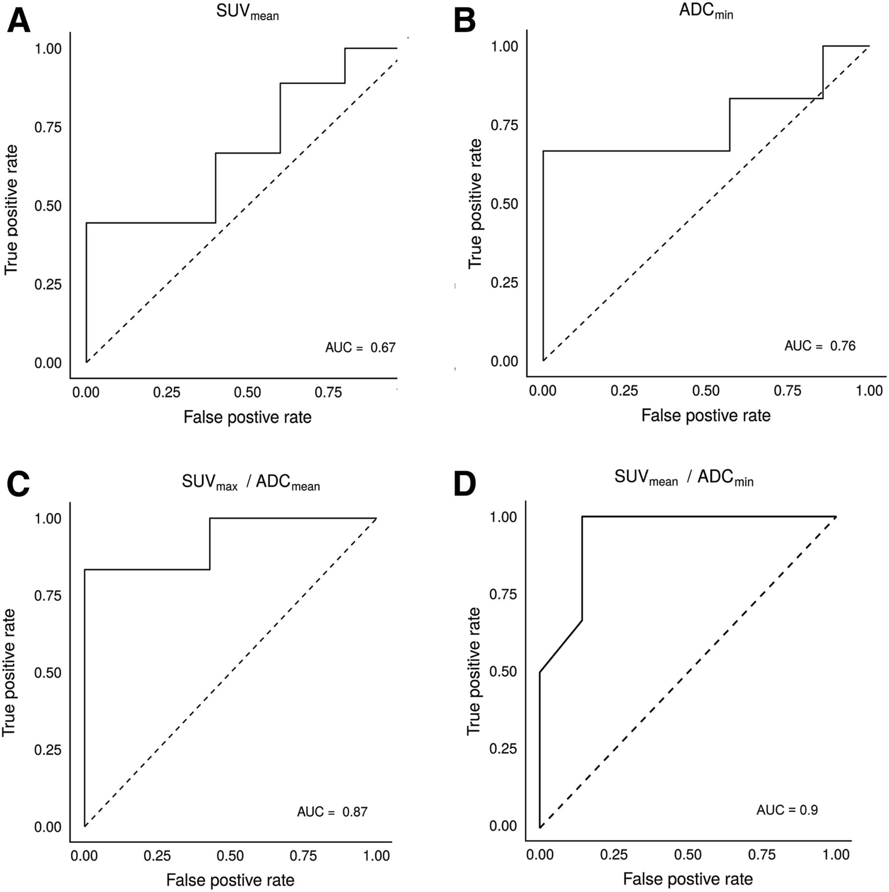
- FIGURE 4.
Receiver-operating-characteristic curves from 68Ga-DOTATOC PET and MRI ADC parameters. SUVmean demonstrates poor to moderate discriminative test performance (A), whereas ADCmin shows fair discriminative ability (B). Of combined ratios and parameters, SUVmean/ADCmin (D) demonstrates better discriminative test performance than SUVmax/ADCmean (C), with AUC of 0.90, sensitivity of 86%, and specificity of 100%. AUC = area under the curve.


Tables
Characteristic Data Age (y) 61 (43–81) Sex Male 15 (68.2%) Female 7 (31.8%) Histologic tumor grade (24 lesions) Grade 1 12 (50%) Grade 2 12 (50%) Tumor location Stomach 1 (4.2%) Small bowel 9 (37.5%) Rectum 1 (4.2%) Pancreas 13 (54.2%) Presence of metastases 20 (80.0%) Qualitative data are expressed as numbers followed by percentages in parentheses; continuous data are expressed as mean ± SD followed by range in parentheses.
Sequence Orientation Bandwidth (Hz/Px) TR/TE (ms) Matrix FOV (mm) Voxel size (mm3) TA (s) T2-weighted HASTE Axial 710 1,400/95 320 400 1.3 × 1.3 × 5.0 68 T2-weighted TIRM Coronal 300 4,390/53 256 450 1.8 × 1.8 × 4.0 142 T1-weighted fs VIBE Axial 450 3.9/1.86 320 400 1.3 × 1.3 × 3.0 17 T2-weighted fs TSE Axial 243 2,200/100 448 400 0.9 × 0.9 × 5.0 230 EPI DWI Axial 2,232 5,600/55 134 380 1.4 × 1.4 × 5.0 204 T1-weighted fs VIBE (dynamic) Axial 450 3.95/1.92 320 360 1.1 × 1.1 × 3.0 17 (per phase) T1-weighted fs STARVIBE (Siemens) Axial 870 3.05/1.44 320 380 12.x1.2 × 1.2 278 TR = repetition time; TE = echo time; FOV = field of view; TA = time of acquisition; HASTE = half-Fourier acquisition single-shot turbo spin echo; TIRM = turbo inversion recovery magnitude; fs = fat saturation; VIBE = volumetric interpolated breath-hold sequence; EPI = echo planar imaging.
- TABLE 3
Comparison of Imaging Parameters Between Different World Health Organization Grade GEP NETs
Parameter Grade 1 (n = 12) Grade 2 (n = 12) Diameter (mm) 19.7 ± 9.6 (12.0–95.0) 40.7 ± 30.4 (11.0–40.0) TFTV (cm3) 6.4 ± 9.9 (1.2–34.2) 70.6 ± 112.2 (1.5–351.1) Ki-67 proliferation index (%) 1.6 ± 0.6 (0.9–2.0) 5.3 ± 2.6 (2.3–10) TBR 6.6 ± 1.9 (3.1–10.1) 12.7 ± 9.3 (3.9–33.2) SUVmean 14.7 ± 7.0 (4.0–23.2) 23.1 ± 12.3 (8.0–45.0) SUVmax 34.9 ± 16.9 (14.5–62.5) 42.3 ± 26.6 (14.8–89.5) ADCmean (×10−3 mm2/s) 1.24 ± 0.18 (1.05–1.49) 0.96 ± 0.26 (0.50–1.22) ADCmin (×10−3 mm2/s) 0.67 ± 0.14 (0.51–0.87) 0.49 ± 0.24 (0.23–0.85) Enhancement ratio 1.2 ± 0.8 (0.6–2.9) 1.0 ± 0.4 (0.4 to 1.6) Data are mean ± SD followed by range in parentheses.
Supplemental Data
Files in this Data Supplement:


Jump to section
Related Articles
Cited By...
- No citing articles found.