


Article Figures & Data
Figures
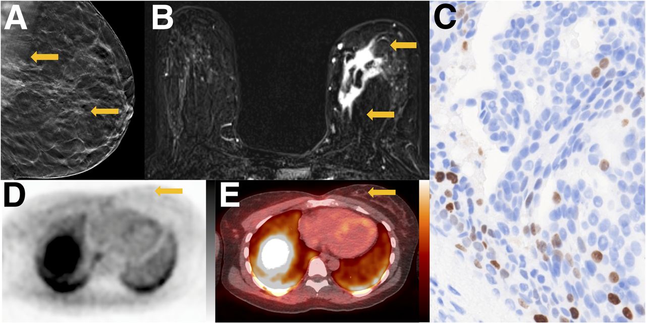
- FIGURE 1.
Tumor with low proliferative status. A 42-y-old woman with ER-positive/human epidermal growth factor receptor 2–negative primary breast cancer. (A) Tomosynthesis mediolateral oblique projection demonstrates irregular mass with spiculated margins and associated calcifications (arrows). (B) Axial contrast-enhanced T1-weighted subtraction image demonstrates irregular mass in medial breast with heterogeneous enhancement (arrows). (C) Ki-67 staining demonstrates low percentage of actively dividing cells (11%) (×20). (D) Axial 18F-ISO-1 image demonstrates no qualitative uptake in medial breast (arrow, SUVmax of 1.5 g/mL). (E) Corresponding 18F-ISO-1 PET/CT demonstrates biopsy clip marking site of malignancy (arrow). PET and PET/CT images are scaled to 0–5 g/mL SUV, −160 to +240 HU.
- FIGURE 2.
Tumor with high proliferative status. A 40-y-old woman with triple-negative breast cancer. (A) Mammographic craniocaudal projection demonstrates high-density irregular mass (arrow) with overlying palpable marker. (B) Axial contrast-enhanced T1-weighted image demonstrates that mass (arrow) is irregular with heterogeneous enhancement, with central signal dropout from biopsy marker. (C) Ki-67 staining demonstrates high percentage of actively dividing cells (74%) (×20). (D) Axial 18F-ISO-1 image demonstrates qualitative uptake at site of malignancy (arrow; SUVmax of 2.6 g/mL) (E) Corresponding CT image demonstrates irregular mass (arrow). PET and CT images are scaled to 0–5 g/mL SUV, −160 to +240 HU.
- FIGURE 3.
Plot of SUVmax in groups stratified by Ki-67 below or above 20. (A) SUVmax shows significant difference between patient tumors stratified by low (n = 15) and high (n = 14) Ki-67 values in all 29 tumors. (B) SUVmax stratified by low (n = 8) and high (n = 13) Ki-67 values restricted to IDC (n = 21) show significant differences based on Ki-67 threshold. Center line of each distribution indicates median value; error bars show 95% confidence interval of median. *P < 0.05.
- FIGURE 4.
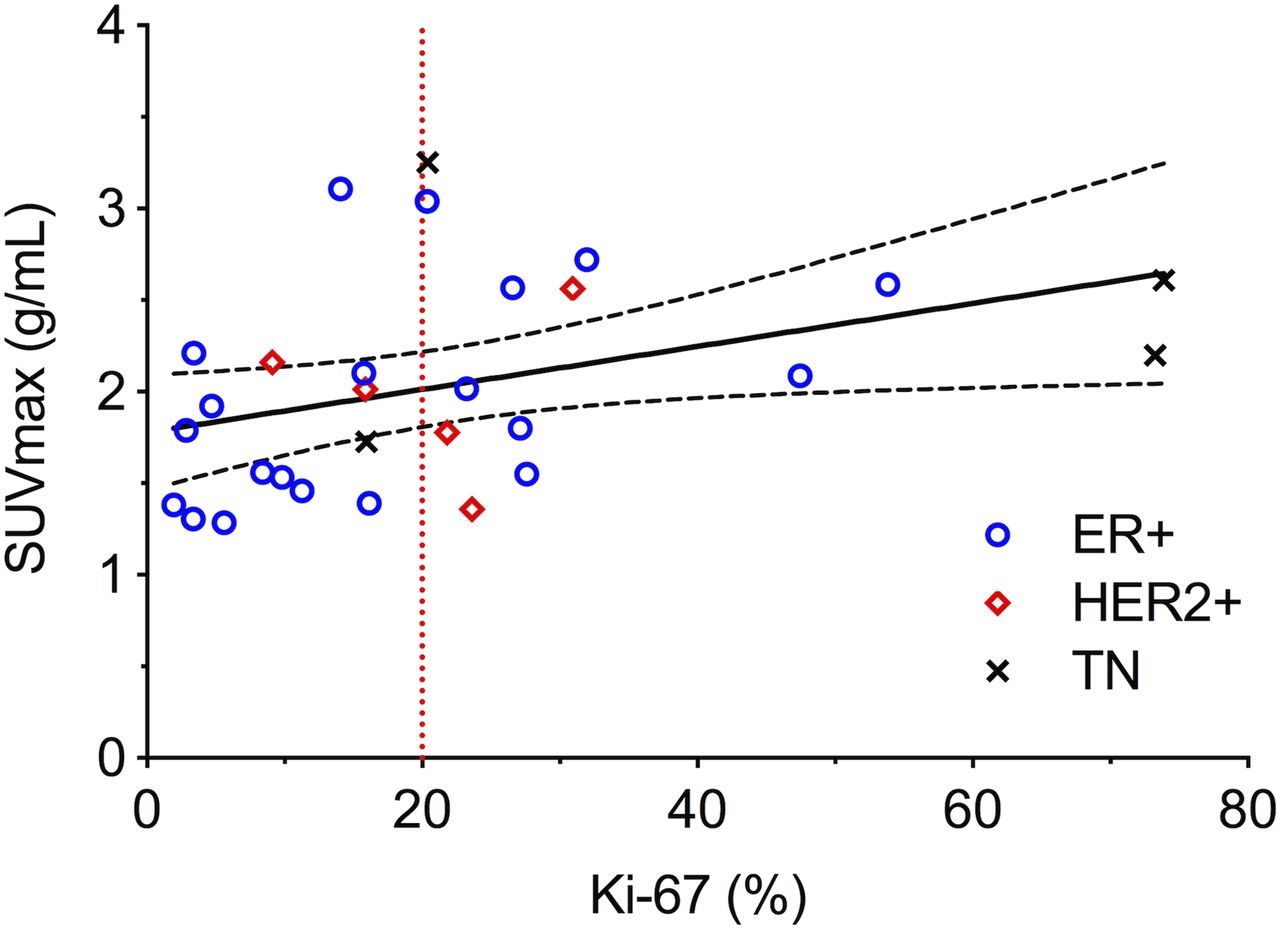
Scatterplots of SUVmax vs. Ki-67 for all 29 tumors. Spearman tests found significant correlations with Ki-67 (ρ = 0.46, P = 0.01). Solid linear regression trend line and dashed 95% confidence intervals are included for reference. TN = triple-negative.


Tables
Characteristic Data Age range (y) 32–79 (median, 55) Female (n) 28 (28/28 = 100%) Race (n) Caucasian 17 (17/28 = 61%) Black 9 (9/28 = 32%) Asian 1 (1/28 = 4%) Hispanic 1 (1/28 = 4%) Histology (n) IDC 21 (21/29 = 72%) ILC 4 (4/29 = 14%) Mixed (IDC and ILC) 4 (4/29 = 14%) Histologic grade (n) 1 2 (2/29 = 7%) 2 15 (15/29 = 52%) 3 11 (11/29 = 38%) Not graded 1 (1/29 = 3%) AJCC tumor anatomic stage group* (n) 1A 9 (9/29 = 31%) 2A 9 (9/29 = 31%) 2B 7 (7/29 = 25%) 3A 2 (2/29 = 7%) 3B 1 (1/29 = 4%) IV 1 (1/29 = 4%) Receptor status (n) ER+ or PR+/HER2− 20 (20/29 = 69%) HER2+ 5 (5/29 = 17%) Triple-negative (ER−/PR−/HER2−) 4 (4/29 = 14%) ↵* According to AJCC Cancer Staging Manual, 8th ed.
AJCC = American Joint Committee on Cancer; PR = progesterone receptor; HER2 = human epidermal growth factor receptor 2.
Supplemental Data
Files in this Data Supplement:


