


Article Figures & Data
Figures
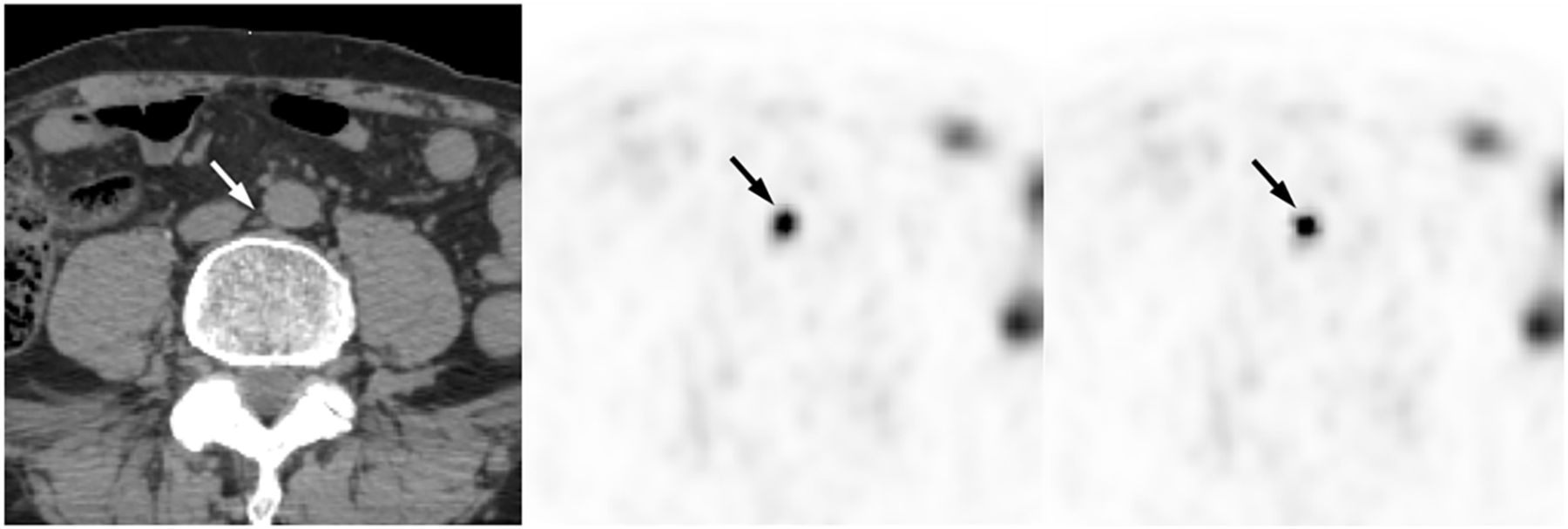
- FIGURE 1.
A 75-y-old man with history of pT3a Gleason 7 (3 + 4) prostate cancer after radical prostatectomy 11 y ago, and salvage radiotherapy for biochemical recurrence 7 y ago, now with slowly rising PSA (2.1 ng/mL). Interaortocaval lymph node (left panel, arrow) had volume of 118.3 cm3 on CT. SUVmax measured on PET was 7.0 with miPSMA score of 1 (middle panel, arrow). After correction, SUVmax was 22.6 with miPSMA score of 3 (right panel, arrow).
- FIGURE 2.
CF per lesion volume. (A) Distribution of CFs (y-axis) is graphed by lesion volume in mm3 (x-axis). (B) CF is graphed by volume and pixel size.
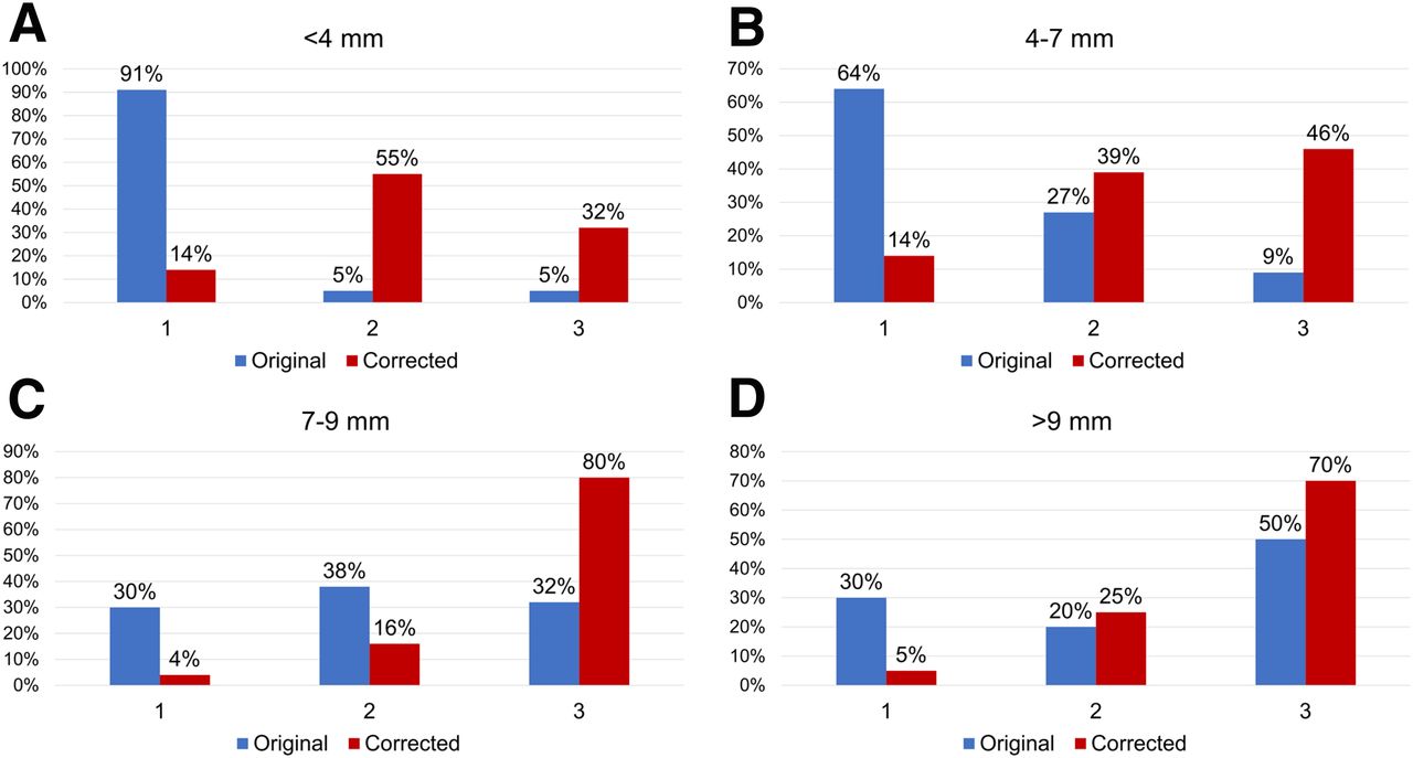
- FIGURE 3.
Distribution of PSMA scores as function of lesion size before and after smoothing filter and PVC for lesions below 4 mm (A), between 4 and 7 mm (B), between 7 and 9 mm (C), and between 9 and 12 mm (D).


Tables
Score PSMA expression Uptake 0 None <Blood-pool activity 1 Low ≥Blood pool and <liver 2 Intermediate ≥Liver and <parotid gland 3 High ≥Parotid gland Parameter Original Corrected Mean SUVmax (P < 0.00001) 11.0 ± 9.3 (1.8–57.1) 28.5 ± 22.8 (5.1–116.3) Mean SUVmean (P < 0.00001) 6.7 ± 5.7 (1–36.3) 14.1 ± 11.3 (2.2–54.8) Mean miPSMA score (P < 0.00001) 1.6 ± 0.76 (1–3) 2.28 ± 0.77 (1–3) Data are mean ± SD, followed by range in parentheses.
Lesion size CF <4 mm (n = 22) 4 (2.5–6.4 ± 1.1) 4–7 mm (n = 140) 2.8 (1.6–4.9 ± 0.64) 7–9 mm (n = 50) 2.3 (1.6–3.3 ± 0.43) 9–12 mm (n = 20) 1.8 (1.4–2.4 ± 0.64) Data are mean followed by range ± SD in parentheses.
Lesion size Original Corrected SUVmax miPSMA SUVmax miPSMA <4 mm 5.3 ± 5.2 (2.1–25.6) 1.1 ± 0.5 21.5 ± 21.2 (5.6–96.3), P = 0.00072 2.1 ± 0.8, P < 0.00001 4–7 mm 8.4 ± 6.0 (1.8–34.3) 1.4 ± 0.6 24.4 ± 19.7 (5.1–116.3), P < 0.00001 2.2 ± 0.8, P < 0.00001 7–9 mm 16.4 ± 9.2 (3.7–45.8) 2.0 ± 0.8 39.0 ± 25.5 (5.8–102.9), P < 0.00001 2.6 ± 0.7, P < 0.00001 9–12 mm 22.2 ± 15.6 (3.8–57.1) 2.2 ± 0.9 39.2 ± 27.1 (8.4–102.0), P = 0.00004 2.5 ± 0.8, P = 0.114 Data are mean ± SD with or without range in parentheses. P values are for original vs. corrected data.
Lesion size Corrected RoT SUVmax miPSMA SUVmax miPSMA <4 mm 21.5 ± 21.2 (5.6–96.3) 2.1 ± 0.8 21.0 ± 20.4 (8.3–101.4), P = 0.681 2.1 ± 0.6, P = 0.616 4–7 mm 24.4 ± 19.7 (5.1–116.3) 2.2 ± 0.8 23.7 ± 16.8 (5.1–96.7), P = 0.184 2.3 ± 0.8, P = 0.665 7–9 mm 39.0 ± 25.5 (5.8–102.9) 2.6 ± 0.7 37.4 ± 21.0 (8.4–104.5), P = 0.206 2.6 ± 0.7, P = 0.905 9–12 mm 39.2 ± 27.1 (8.4–102.0) 2.5 ± 0.8 40.1 ± 28.1 (6.9–103.0), P = 0.595 2.5 ± 0.8, P = 0.867 Data are mean ± SD. Numbers in parentheses represent range; range for miPSMA is fixed (1–3) and therefore not included in this table. P values are for corrected vs. RoT data.
Lesion size Original Corrected RoT Score 1 Score 2 Score 3 Score 1 Score 2 Score 3 Score 1 Score 2 Score 3 <4 mm 20 (91) 1 (4.5) 1 (4.5) 5 (22.7) 10 (45.5) 7 (31.8) 3 (13.6) 13 (59.1) 6 (27.3) 4–7 mm 90 (64.3) 38 (27.1) 12 (8.6) 31 (22.1) 51 (36.4) 58 (41.4) 25 (17.9) 53 (37.8) 62 (44.3) 7–9 mm 15 (30) 19 (38) 16 (32) 6 (12) 10 (20) 34 (68) 5 (10) 9 (18) 36 (76) 9–12 mm 6 (30) 4 (20) 10 (50) 4 (20) 3 (15) 13 (65) 3 (10) 4 (25) 13 (65) Data are numbers followed by percentages in parentheses.


Jump to section
Related Articles
Cited By...
- No citing articles found.