


Johannes Czernin, MD, editor in chief of The Journal of Nuclear Medicine, talked with David W. Townsend, PhD, and Thomas Beyer, PhD, MBA, about their pioneering work in the development of PET/CT technology.
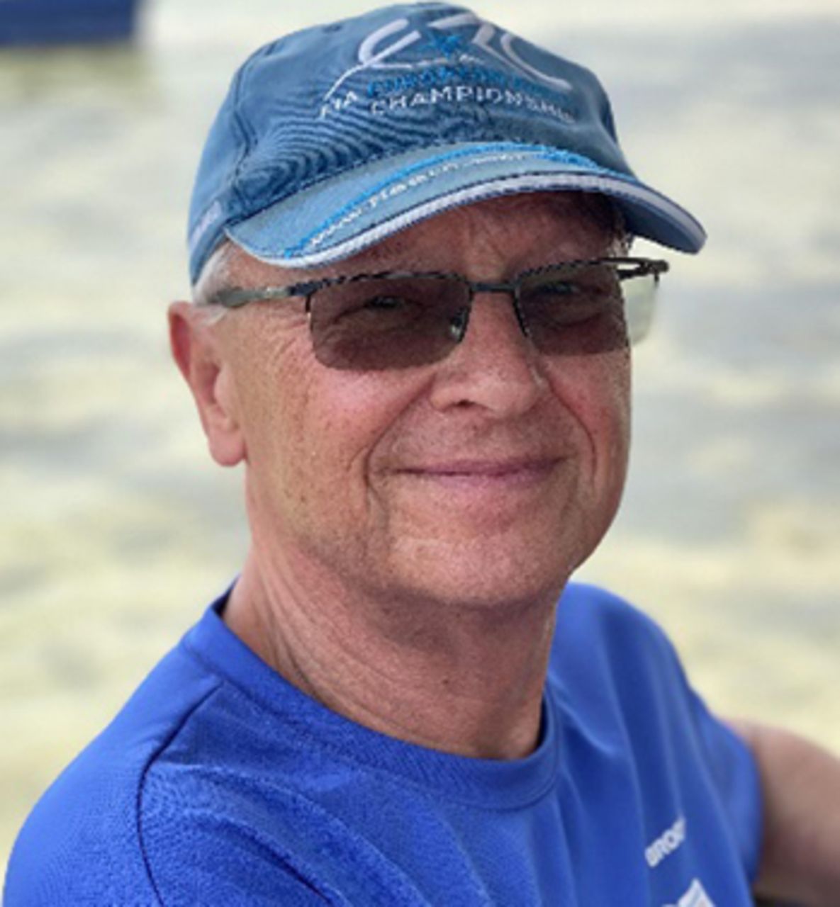
Townsend, whose PhD was in particle physics, was a staff member for 8 years at the European Centre for Nuclear Research in Geneva, Switzerland. In 1980, he joined the faculty of the University of Geneva Hospital. In 1993, he moved to the University of Pittsburgh Medical Center (PA) as an associate professor of radiology and senior PET physicist. He was the codirector of the Pittsburgh PET Facility from 1996 to 2002 and became a professor of radiology in 2000. The PET/CT scanner, developed by Townsend and Ronald Nutt, PhD, was named by TIME Magazine as the medical invention of the year 2000. From 2003 to 2009, Townsend was director of the Molecular Imaging and Translational Research Program at the University of Tennessee, Knoxville. In 2009, he became head of PET and SPECT development for the Singapore Bioimaging Consortium and a professor of radiology at the National University of Singapore. There he was appointed director of the A*STAR-NUS Clinical Imaging Research Centre in 2010, a position from which he retired in 2018. Townsend received the 2004 Distinguished Clinical Scientist Award from the Academy of Molecular Imaging and the 2008 Austrian Nuclear Medicine Pioneer Award. He shared with Nutt the 2010 Institute of Electrical and Electronics Engineers Medal for Innovations in Health Care Technology. Among numerous other awards, he received the Paul C. Aebersold Award from SNMMI and the Edward J. Hoffman Medical Imaging Scientist Award from IEEE.
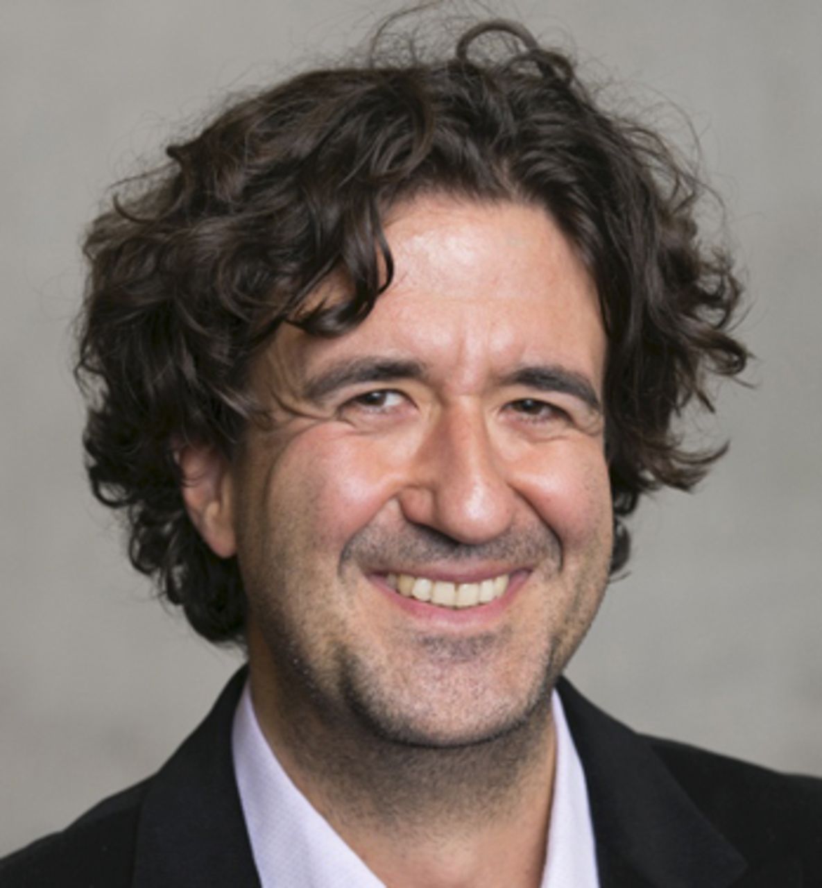
Beyer is a professor of physics in medical imaging at the Medical University of Vienna (Austria). He is also the founder of cmi-experts GmbH (Zurich, Switzerland), a cross-modality imaging consulting company. He earned his physics degree in 2000 from the University of Surrey (Guildford, U.K.). He first worked with Townsend in Geneva, and from 1994 to 1999 he was a member of the University of Pittsburgh Medical Center team developing the first integrated PET/CT system. From 1999 to 2002 he worked with Siemens/CTI PET Systems GmbH (Münster, Germany) as an international PET/CT specialist. He was an assistant and later associate professor at the University of Duisburg–Essen (Germany) before working in various positions in health-care industries. He was appointed to his current academic position in 2013. In 2018 he cofounded a university spinoff (Dediciad GmbH) building artificial intelligence prediction models using molecular imaging information.
Dr. Czernin: This discussion marks the 20th anniversary of your landmark paper introducing PET/CT that was published in JNM in 2000 (J Nucl Med. 2000;41:1369–1379). It is fitting that this paper has been cited exactly 2,000 times today. This makes it the second most frequently cited paper in the history of JNM, second only to Richard Wahl’s introduction of the PET Response Criteria in Solid Tumors (J Nucl Med. 2009;50[suppl 1]:122S–150S), underscoring the extraordinary impact that your idea had not only on progress in imaging instrumentation but, even more important, on the practice of medicine. I want to take you back in time to tell us about the origin of the idea.
Dr. Townsend: I guess the story would have begun back in the 1980s, when Alan Jeavons and I worked on a rotating PET scanner based on modified wire chamber detectors. To overcome the low sensitivity of these detectors, we were required to develop 3D image reconstruction algorithms and implement fully 3D acquisition. In 1987, I was invited by Terry Jones for a sabbatical at Hammersmith Hospital (London, U.K.) to improve PET sensitivity by applying our 3D reconstruction algorithms to his bismuth germanate block–based ring PET scanner from CTI. After that was successful, Terry suggested removing about half the block detectors and rotating the remainder to simulate a full-ring PET system that would be much lower in cost than a full ring, a proposal that was supported by Ron Nutt from CTI. Our team, including Martin Wensveen, Henri Tochon-Danguy, Peter Frey, and Anne Christin, together with Larry Byars (CTI), built such a device (the PRT [partial-ring tomograph]) and evaluated it clinically at the University Hospital of Geneva (Switzerland) in 1989 in the department of nuclear medicine under the direction of Prof. Alfred Donath. The PRT PET system was eventually commercialized by CTI as the ECAT ART [advanced rotating tomograph] scanner. One of the physicians we worked with in Geneva was Rudi Egli, an oncology surgeon.
Dr. Beyer: David, is it true that Dr. Egli had watched the PRT scanner with the covers off and noticed lots of free space and suggested fitting a CT into the gaps?
Dr. Townsend: This is indeed true! Around 1990, he suggested that, with the gaps between the PET detectors, we could add another imaging modality such as CT. We discussed the idea with Ron Nutt and Terry Jones and decided to try to develop such a device: a combined PET/CT scanner. At the time, I was joined by Michel Defrise from Brussels, who spent a sabbatical with us in Geneva in 1993 to work on 3D reconstruction.
Dr. Beyer: I joined your group in Geneva that same year after I had just completed a summer internship at the Paul Scherrer Institute (Villigen, Switzerland). You had asked me to look into ways of using the CT for attenuation correction of the PET, which we felt was an asset to putting 2 machines together but not the driving factor for a PET/CT. A few months later I left to finish my studies in Leipzig. You had moved to Pittsburgh and invited me to do my master’s thesis with you and Paul Kinahan. And this is how I really got involved for several years.
“PET/CT is the death of nuclear medicine.”
Dr. Townsend: I was recruited to the University of Pittsburgh in late 1993, by which time Ron had convinced Siemens to contribute a CT scanner at no cost if I could obtain grant funding to build a PET/CT prototype. I was joined at the University of Pittsburgh by Paul Kinahan, and in 1994 we submitted an R01 with the support of Mark Mintun, PET director, and Rich Baron, chair of radiology. The first submission was rejected but the grant was funded on resubmission. We began the PET/CT project in 1995 with NIH funding and the support of CTI PET Systems in Knoxville, TN.
Dr. Beyer: I recall our first PowerPoint design when we thought of a concept to merge the CT and ECAT ART PET detector blocks in-plane, only to realize how much space the CT components required, again after having seen a CT scanner with its covers off. This ultimately led to a coaxially displaced design concept for the early SMART [Somatom (CT) ART] PET/CT, with the PET and CT parts placed back to back on a single rotating metal annulus. In retrospect, I think that perhaps the biggest asset in the whole development process was that you had the right people at the right time at the right place who were all geared up to create something new.
Dr. Czernin: What was the initial response to your PET/CT concept by the major industry partners CTI and Siemens? And what were the responses by nuclear medicine physicians and radiologists?
Dr. Townsend: We first needed to generate funds, which we did with the NIH R01. Interestingly, the study section gave it a terrible score the first time round. It was declared a complete waste of time and money to tie up a CT scanner to just do attenuation correction for PET. Paul and I were very frustrated but then resubmitted it. It received the highest score in that review cycle and was funded in 1995. This allowed us to build the prototype and financially support some of the people involved, including Thomas.
Dr. Czernin: When you resubmitted the grant did you focus on attenuation correction or did you already propose diagnostic CT quality?
Dr. Townsend: We tried to correct the reviewers’ misunderstanding that this was focused on attenuation correction. As Thomas said, CT-based attenuation correction was sort of a byproduct of putting the 2 machines together. But the idea was to produce an integrated PET/CT with a clinical-grade PET and a clinical-grade CT system.
Dr. Beyer: At the time, the state of the art in PET-only was the ECAT EXACT and the ECAT EXACT HR+. Much longer acquisition times were needed to get the same quality data with the rotating ECAT ART. For the early commercialization of PET/CT we felt strongly that the PET components should have maximum sensitivity.
Dr. Czernin: Thomas, when you joined the group to work with David, who provided feedback to aim for specific performance characteristics? What was the vision for a fully integrated, high-performance clinical system?
Dr. Beyer: Well, that’s a complicated question, because when I joined the team during the Geneva times, my exposure to medical doctors was limited. It was mainly the bunch of physicists that David rounded up for pizza and hardcore physics. Later, during the Pittsburgh times, we engaged with many more clinicians at the University of Pittsburgh Medical Center. Our sparring partners were, for example, Martin Charron, who was a pediatric nuclear medicine physician, and Carolyn Meltzer and Todd Blodgett from the PET Center. We should, of course, mention an earlier pioneer of multimodality imaging, the late Bruce Hasegawa from the University of California, San Francisco. Bruce developed the first combined SPECT/CT device and introduced us to Willi Kalender from Erlangen (Germany), the developer of spiral CT. Willi pushed the team to incorporate as high-performance a CT as possible in the first commercial PET/CT design from Siemens.
Dr. Townsend: And don’t forget, we were not the first in the context of hybrid imaging. In 2004, after I had just given a talk in Japan, Yuji Nakamoto approached me and told me of earlier developments toward a dual-modality PET/CT at Gunma University under the direction of a neurosurgeon. The system was built with the support of Hitachi and had been used successfully on many neurooncology patients already in the 1980s. Because the group had not published on it, this pioneering development had not received the attention it deserved.
Dr. Czernin: When you started presenting the concept and early data, what was the initial response of the community? Who were the naysayers?
Dr. Townsend: Around 1999 I was invited to Guys’ and St. Thomas’ Hospitals in London (U.K.) to give a PET/CT talk. A senior nuclear medicine physician there at that time felt this was all nonsense and that precise anatomy with the PET scan was not necessary in nuclear medicine, except maybe in 10% of cases—and that even then software could be used, so that PET/CT was a complete nonstarter. Later, Dale Bailey and Paul Marsden took me out to dinner and apologized for the negative reaction from the audience.
Dr. Beyer: In 1999 I gave a talk at the annual German nuclear medicine meeting, and a guy stood up and said, “This is the death of nuclear medicine.” After I moved to Essen in 2002 and we took delivery of the first commercial PET/CT in Europe, I gave a seminar at our university hospital for about 20 people. In the room was the head of radiation therapy, who said to me during my presentation: “Mr. Beyer, before you tell us about PET/CT, you have to convince us of the power of PET.” And he literally left the room.
Dr. Townsend: Similarly, when I presented the abstract chosen for the Image of the Year at the SNM meeting in Los Angeles (CA) in 1999 the only comment came from a person with a heavy German accent who said, “You may be able to combine the 2 imaging modalities, but you will never combine radiology and nuclear medicine; your device will fail.”
Dr. Beyer: Well, he was right on the professional combination, wasn’t he? I recall that in 2003 a nuclear medicine physician from The Netherlands published a paper in which he stated that only 20% of cases needed a CT and the rest can be read with PET only. Funny to see that he immediately stopped these inconsiderate remarks as soon as he took delivery of his own PET/CT.
Dr. Czernin: Let’s look forward now, as the rest is history. Thousands of PET/CT scanners have been installed, combining the highest-end PET, CT, and MR systems. Simon Cherry, Terry Jones, Ramsey Badawi, and Joel Karp led teams that came up with the total-body PET. Until recently, many of us had the feeling that we had reached the limits of PET technology. They proved us wrong. What is the future of PET, PET/CT, and PET/MR technology?
Dr. Townsend: One of the biggest technical impacts of PET/MR has been to push the silicon photomultipliers into the PET/CT to improve performance. So whatever else happens with PET/MR from an instrumentation point of view, the PET components in PET/CT are much higher-performance now, thanks to the underlying instrumentation development in PET/MR.
Dr. Beyer: I conceive of a single detector that works both in PET/CT and PET/MR. Perhaps in 5 y, people will compose their PET/CT or PET/MR system following a LEGO-type design approach using the same PET detector. I hope that with this economy of scale, systems will become a bit less expensive. I also believe that many recent publications have helped renew interest in truly quantitative PET. The new PET/CT systems make it easier to perform dynamic imaging. And I hope that we exercise rigor in assessing the value of the temporal domain above and beyond the spatial domain. We will need to think about combining imaging data from PET/CT with nonimaging data. I am a firm believer in artificial intelligence–driven clinical decision support systems and their potential for more accurate and efficient treatment decisions.
Overall, there is immense momentum in the field with these latest technical developments, both in the existing commercial PET/CT arena and the EXPLORER domain, that make people aware of the beauty and the power as well as the potential of whole- or total-body PET. I have really high hopes, much like you, it seems, that the limits of PET technology have not yet been reached. To me, total-body PET/CT is a quantum leap, opening up completely new opportunities for quantitative and longitudinal imaging and addressing the challenges of systems biology with completely novel approaches. This is so exciting that I would love to start over in this field again.
Dr. Czernin: David, do you want to comment on that?
Dr. Townsend: All the ideas that Terry and the inspiring PET group at Hammersmith Hospital used to have in the 1980s have finally come together. Terry Jones, as well as Mike Phelps (and the late Ed Hoffman) at the University of California at Los Angeles, were the guys who pushed the limits of the technology. For Terry and Mike to see it come together after all those years, I think is a tremendous achievement. To be able to look at multiorgan interactions, to be able to look at the whole body in subsecond time frames, is something that Terry basically always dreamed about when we were working together at Hammersmith.
Dr. Czernin: Your work has had a substantial impact on the practice of medicine and patient care. What do you think is the most important contribution of PET/CT to patient care relative to the time before PET/CT?
Dr. Townsend: Having been in Pittsburgh when we were just getting started with the prototype, we scanned 300 patients. There was still a lot of opposition as to why we were combining PET and CT. But a few clinical cases made us realize that doing PET and CT together was quite different from doing them separately. It took just a few clinical cases to show how patient management could be changed. And when we started presenting those few cases at different meetings, people started to realize that there was something different here.
Dr. Beyer: I agree entirely with David regarding the power of truly fused images and that a few cases were enough for an eye opener. We were fortunate enough to add to the benefits of an integrated diagnostic approach, sometimes referred to as a “1-stop shop.” Already with the prototype PET/CT, the total examination time was less than the sum of the 2 exams. Then and now, we must understand that patients want an accurate diagnosis with as few diagnostic tests that take as little time as possible. This calls for protocols that are optimized primarily for patients and only secondarily for radiation safety officers.
Dr. Czernin: PET/CT use will continue to increase. Radiology training now often includes body PET/CT fellowships. The newer generation of imaging specialists is trained in PET and CT. This creates an avalanche of knowledge that serves as a multiplier of PET use. PET is now 100% mainstream and completely established. It is amazing what has happened over the last 20 y with technologic advances, the emergence of theranostics, and an ever-expanding portfolio of PET imaging probes. David and Thomas, thank you for your remarkable contributions, and thank you for taking the time to talk with me and our readers.
- © 2020 by the Society of Nuclear Medicine and Molecular Imaging.


Jump to section
Related Articles
Cited By...
- No citing articles found.