
Main menu

  • Home
  • Content
    • Current
    • Ahead of print
    • Past Issues
    • JNM Supplement
    • SNMMI Annual Meeting Abstracts
    • Continuing Education
    • JNM Podcasts
  • Subscriptions
    • Subscribers
    • Institutional and Non-member
    • Rates
    • Journal Claims
    • Corporate & Special Sales
  • Authors
    • Submit to JNM
    • Information for Authors
    • Assignment of Copyright
    • AQARA requirements
  • Info
    • Reviewers
    • Permissions
    • Advertisers
  • About
    • About Us
    • Editorial Board
    • Contact Information
  • More
    • Alerts
    • Feedback
    • Help
    • SNMMI Journals
  • SNMMI
    • JNM
    • JNMT
    • SNMMI Journals
    • SNMMI

User menu

  • Subscribe
  • My alerts
  • Log in
  • My Cart

Search

  • Advanced search
Journal of Nuclear Medicine
  • SNMMI
    • JNM
    • JNMT
    • SNMMI Journals
    • SNMMI
  • Subscribe
  • My alerts
  • Log in
  • My Cart
Journal of Nuclear Medicine

Advanced Search

  • Home
  • Content
    • Current
    • Ahead of print
    • Past Issues
    • JNM Supplement
    • SNMMI Annual Meeting Abstracts
    • Continuing Education
    • JNM Podcasts
  • Subscriptions
    • Subscribers
    • Institutional and Non-member
    • Rates
    • Journal Claims
    • Corporate & Special Sales
  • Authors
    • Submit to JNM
    • Information for Authors
    • Assignment of Copyright
    • AQARA requirements
  • Info
    • Reviewers
    • Permissions
    • Advertisers
  • About
    • About Us
    • Editorial Board
    • Contact Information
  • More
    • Alerts
    • Feedback
    • Help
    • SNMMI Journals
  • View or Listen to JNM Podcast
  • Visit JNM on Facebook
  • Join JNM on LinkedIn
  • Follow JNM on Twitter
  • Subscribe to our RSS feeds
Research ArticleFocus on Molecular Imaging

Quo Vadis, Molecular Imaging?

Jan Grimm, Fabian Kiessling and Bernd J. Pichler
Journal of Nuclear Medicine October 2020, 61 (10) 1428-1434; DOI: https://doi.org/10.2967/jnumed.120.241984
Jan Grimm
1Molecular Pharmacology Program and Department of Radiology, Memorial Sloan Kettering Cancer Center, and Pharmacology Program and Department of Radiology, Weil Cornell Medical College, New York, New York
  • Find this author on Google Scholar
  • Find this author on PubMed
  • Search for this author on this site
Fabian Kiessling
2Center for Biohybrid Medical Systems, Institute for Experimental Molecular Imaging, University Hospital Aachen, RWTH Aachen University, Aachen, Germany, and Fraunhofer Institute for Digital Medicine, Bremen, Germany; and
  • Find this author on Google Scholar
  • Find this author on PubMed
  • Search for this author on this site
Bernd J. Pichler
3Werner Siemens Imaging Center, Department of Preclinical Imaging and Radiopharmacy, Eberhard Karls University, Tuebingen, Germany, and Cluster of Excellence iFIT (EXC 2180) “Image-Guided and Functionally Instructed Tumor Therapies,” University of Tübingen, Tübingen, Germany
  • Find this author on Google Scholar
  • Find this author on PubMed
  • Search for this author on this site
  • Article
  • Figures & Data
  • Info & Metrics
  • PDF
Loading

Article Figures & Data

Figures

  • FIGURE 1.
    • Download figure
    • Open in new tab
    • Download powerpoint
    FIGURE 1.

    Imaging without probes. (A) High-resolution optoacoustic imaging using raster-scanning optoascoustic mesoscopy of subcutaneously implanted CT26 tumor in mouse. (Courtesy of Katja Haedicke and Jan Grimm, Memorial Sloan Kettering Cancer Center.) (B) Mid-infrared optoacoustic microscopy of excised pancreatic mouse tissue, with overlay of lipid (CH2 [yellow]) and protein (amide [blue]) maps showing clusters of pancreatic acinar glands embedded in protein. (Reprinted with permission of (5).) (C) Spatial correlation between pH-weighted molecular MRI, 18F-FDOPA PET, and MR spectroscopy (MRS) in 2 patients with anaplastic astrocytomas showing CEST asymmetry consistent with low pH in regions with confirmed elevated 18F-FDOPA uptake on PET and elevated lactate on MRS. From left to right are shown T2-weighted fluid-attenuated inversion recovery (FLAIR), pH-weighted MRI using amine CEST, 18F-FDOPA PET, and MRS from area shown in red box in FLAIR images. a.u. = arbitrary units; BG = background; Cho = choline; Cr = creatine; FDOPA = 6-fluoro-L-dopa; Lip/Lac = lipids/lactates; NAA = N-acetylaspartate; NMR = nuclear magnetic resonance. (Reprinted with permission of (8).)
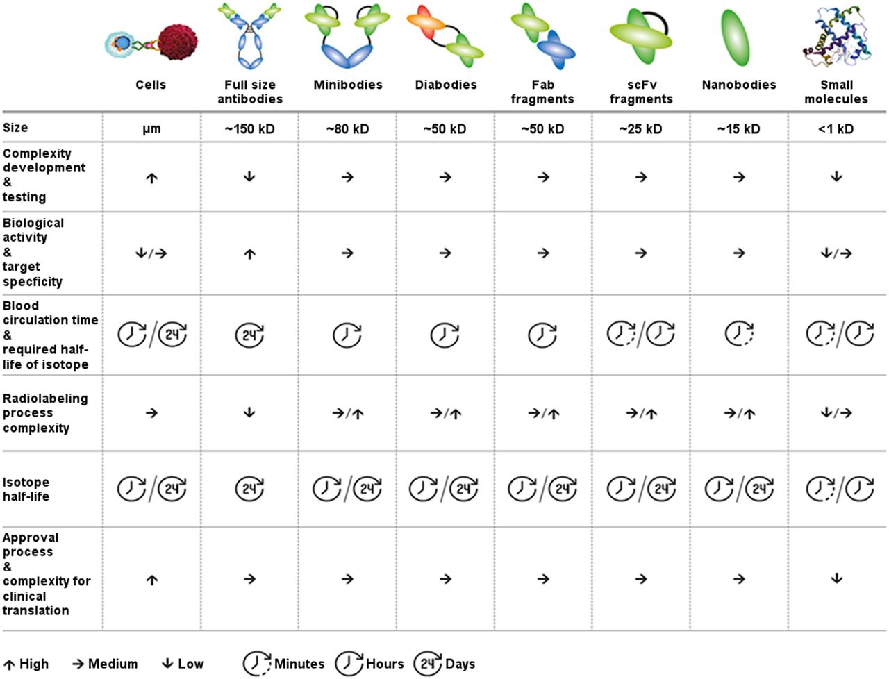
  • FIGURE 2.
    • Download figure
    • Open in new tab
    • Download powerpoint
    FIGURE 2.

    MI probes based on biologicals: format defines and impacts not only pharmacokinetics but also development time, costs, and level of effort for clinical translation. Fab fragments = antigen-binding fragments; scFv = single-chain variable fragment.

  • FIGURE 3.
    • Download figure
    • Open in new tab
    • Download powerpoint
    FIGURE 3.

    Scheme illustrating main sites of accumulation and elimination of diagnostic nanomaterials. This is simplistic view; composition of materials and their interaction with body components may cause different properties in individual cases. Color intensities of human shapes (top row) indicate overall body distribution. (Left) Diagnostic agents smaller than albumin can be renally eliminated and thus usually have short blood half-life and intracorporal persistence. They rapidly overcome biologic barriers, leading to low unspecific accumulation and low background for targeted imaging applications (bottom row). (Middle) Some materials can leave vasculature but are not renally eliminated. They usually show smaller distribution volume but longer body persistence and MPS uptake (liver, spleen, and lymph nodes are shown as an example) and can have long blood half-lives. Diagnostically, these materials can be used for EPR prediction, for MPS staining, and as theranostic drug delivery systems. Antibodies, as considerably small nanosystems (∼12 nm for IgG), also fall into this class. Because of their still-sufficient tissue penetration capabilities, they are frequently used as molecular diagnostic agents, which is reasonable if long intervals between injection and imaging are acceptable in clinical workflow. (Right) Microbubbles can be used as ultrasound contrast agents. Microbubbles remain strictly intravascular and favorably enable vascular visualization and intravascular MI. They are being explored for their capability of locally promoting drug delivery and as carriers of drugs and genes. Gray underlays in bottom row indicate distribution in and penetration of materials into tissue compartments. BV = blood vessel; IS = interstitial space; SPION = superparamagnetic iron oxide nanoparticles; TC = target cell. (Modified clip art from Servier medical art database [https://smart.servier.com/].)
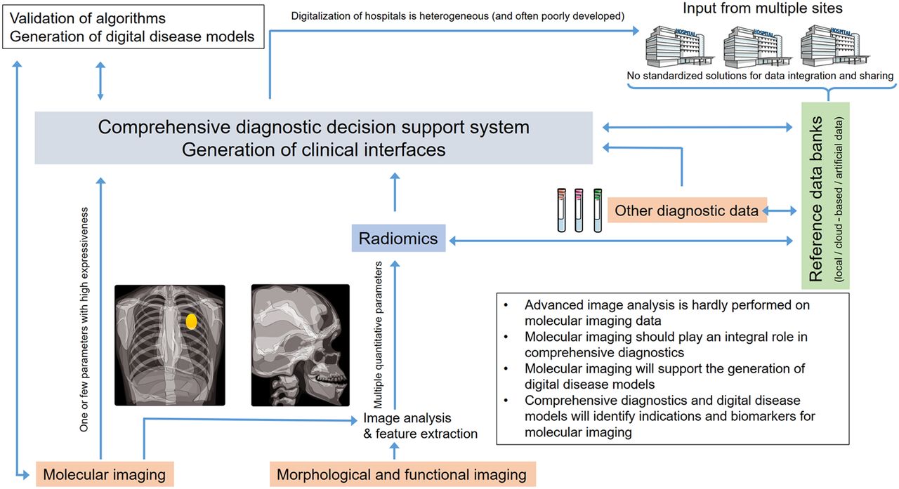
  • FIGURE 4.
    • Download figure
    • Open in new tab
    • Download powerpoint
    FIGURE 4.

    Currently, MI aims mostly at extracting one or a few key biomarkers, whereas radiomics aims to achieve diagnostic precision from integrating multiple features extracted from functional and morphologic imaging data. However, multiple features could be extracted from MI data to enhance its diagnostic accuracy. Integrating imaging features with other diagnostic data (e.g., from physical examinations, electrophysiology, clinical chemistry, and omics) leads to comprehensive diagnostics, where artificial intelligence supports decision making based on learning from large data collections. There is a great need to understand and validate output of artificial intelligence–based decision support systems. Systems biology modeling and generation of virtual disease models will become important. These models will profit from MI of key parameters of pathologic processes and inversely provide indications for developing new MI probes. (Modified clip art from Servier medical art database [https://smart.servier.com/].)

  • FIGURE 5.
    • Download figure
    • Open in new tab
    • Download powerpoint
    FIGURE 5.

    (A) Three-dimensional computer-aided design sketch of modularized mini-panel PET detection system. (B) Photograph of system. (C) Core setup of microfluidics system (a), continuous flow of radiotracer medium (b), and representative image from PET detector during acquisition (c). Red boxes denote analyzed areas (40). (D) Example of comprehensive data integration that connects workflows of in vivo MI and in vitro postmortem tissue metabolomics. (a) Flowchart of procedure for image-guided tissue extraction. (b–d) Temperature measured with sensors in stomach, in rectum, and embedded under skin during freezing and embedding (b), during time that mouse was kept at room temperature and covered with dry ice (c), and during milling (d). (e) Example of nuclear magnetic resonance spectra from samples obtained with image-guided milling machine. CIMR = continuously infused microfluidic radioassay; LYSO/SiPM = lutetium yttrium oxyorthosilicate/silicon photomultiplier; PEEK = polyether ether ketone; VOI = volume of interest. (Reprinted with permission of (43).)

PreviousNext
Back to top

In this issue

Journal of Nuclear Medicine: 61 (10)
Journal of Nuclear Medicine
Vol. 61, Issue 10
October 1, 2020
  • Table of Contents
  • Table of Contents (PDF)
  • About the Cover
  • Index by author
Print
Download PDF
Article Alerts
Sign In to Email Alerts with your Email Address
Email Article

Thank you for your interest in spreading the word on Journal of Nuclear Medicine.

NOTE: We only request your email address so that the person you are recommending the page to knows that you wanted them to see it, and that it is not junk mail. We do not capture any email address.

Enter multiple addresses on separate lines or separate them with commas.
Quo Vadis, Molecular Imaging?
(Your Name) has sent you a message from Journal of Nuclear Medicine
(Your Name) thought you would like to see the Journal of Nuclear Medicine web site.
Citation Tools
Quo Vadis, Molecular Imaging?
Jan Grimm, Fabian Kiessling, Bernd J. Pichler
Journal of Nuclear Medicine Oct 2020, 61 (10) 1428-1434; DOI: 10.2967/jnumed.120.241984

Citation Manager Formats

  • BibTeX
  • Bookends
  • EasyBib
  • EndNote (tagged)
  • EndNote 8 (xml)
  • Medlars
  • Mendeley
  • Papers
  • RefWorks Tagged
  • Ref Manager
  • RIS
  • Zotero
Share
Quo Vadis, Molecular Imaging?
Jan Grimm, Fabian Kiessling, Bernd J. Pichler
Journal of Nuclear Medicine Oct 2020, 61 (10) 1428-1434; DOI: 10.2967/jnumed.120.241984
Twitter logo Facebook logo LinkedIn logo Mendeley logo
  • Tweet Widget
  • Facebook Like
  • Google Plus One
Bookmark this article

Jump to section

  • Article
    • Abstract
    • MI WITHOUT PROBES
    • RADIOTRACERS AND SMALL MOLECULES
    • NANO- AND MICROSYSTEMS
    • COMPREHENSIVE DIAGNOSTICS
    • NEW INDICATIONS FOR MI
    • OUTLOOK
    • DISCLOSURE
    • Footnotes
    • REFERENCES
  • Figures & Data
  • Info & Metrics
  • PDF

Related Articles

  • This Month in JNM
  • PubMed
  • Google Scholar

Cited By...

  • No citing articles found.
  • Google Scholar

More in this TOC Section

  • From Genome to Phenome: Opportunities and Challenges of Molecular Imaging
  • Imaging the Activity of Efflux Transporters at the Blood–Brain Barrier in Neurologic Diseases: Radiotracer Selection Criteria
  • Molecular Imaging of Pulmonary Fibrosis
Show more Focus on Molecular Imaging

Similar Articles

Keywords

  • molecular imaging
  • diagnostic probe
  • radiotracer
  • radiomics
  • Multiparametric imaging
SNMMI

© 2025 SNMMI

Powered by HighWire