


Article Figures & Data
Figures
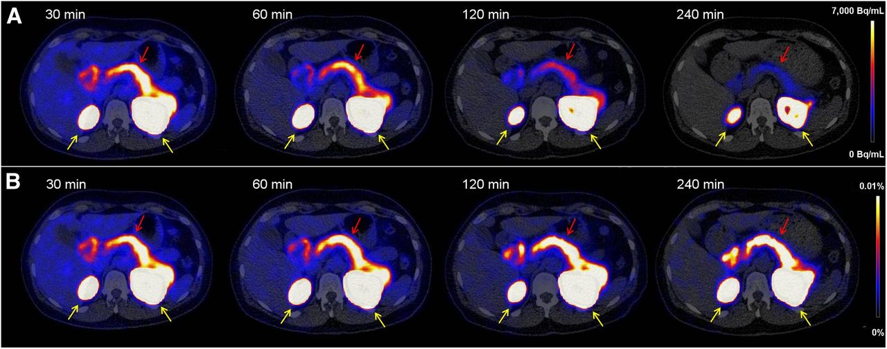
- FIGURE 1.
Transversal PET/CT images of abdomen showing biodistribution of 68Ga-NODAGA-exendin-4 at 30, 60, 120, and 240 min after injection. (A) Tracer accumulation in Bq/mL, showing washout as well as physical decay. (B) Tracer accumulation in percentage injected dose/g of tissue, showing only washout of tracer. Kidneys are indicated by yellow arrows and pancreas by red arrows.
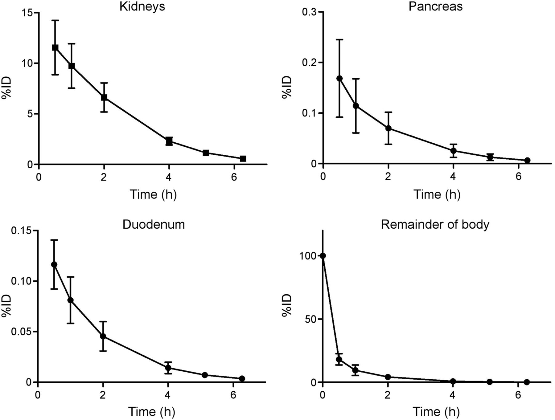
- FIGURE 2.
Time–activity curves of kidneys, pancreas, duodenum, and remainder of body (n = 6). Data are mean ± SD. %ID = percentage injected dose.


Tables
- TABLE 1
Patient Characteristics, Injected Activities, and Time After Injection of Performed PET/CT Scans
Time after injection (min) Patient no. Sex Age (y) Weight (kg) Injected activity (MBq) Scan 1 Scan 2 Scan 3 Scan 4 1 F 24 72.0 106.1 32 66 121 239 2 F 53 58.0 107.5 32 62 120 238 3 F 55 75.6 108.0 30 83 119 235 4 M 65 83.6 105.0 30 61 120 234 5 F 64 88.8 105.1 30 56 115 235 6 M 63 84.5 101.6 30 62 118 236 - TABLE 2
Organ-Absorbed Doses (mGy/MBq) and Effective Doses (mSv/MBq) Acquired Using Reference Adult Models in OLINDA/EXM 1.1
Site (n = 6) Mean SD Adrenals 0.012 0.002 Brain 0.005 0.0006 Breasts 0.005 0.0006 Gallbladder wall 0.008 0.0008 Stomach wall 0.007 0.0007 Heart wall 0.006 0.0006 Kidneys 0.472 0.1019 Intestine, lower large: wall 0.006 0.0007 Intestine, upper large: wall 0.007 0.0007 Intestine, small 0.008 0.0010 Liver 0.008 0.0008 Lungs 0.006 0.0006 Muscle 0.006 0.0006 Ovaries 0.006 0.0008 Pancreas 0.023 0.0083 Red marrow 0.006 0.0005 Osteogenic cells 0.008 0.0010 Skin 0.005 0.0005 Spleen 0.001 0.0001 Testes 0.002 0.0003 Thymus 0.005 0.0006 Thyroid 0.005 0.0006 Urinary bladder wall 0.005 0.0007 Uterus 0.004 0.0003 Total body 0.008 0.0009 Effective dose (mSv/MBq) 0.007 0.0007 - TABLE 3
Estimated Absorbed Doses (mGy/MBq) and Effective Doses (mSv/MBq) in Children Acquired Using Newborn, 1-Year-Old, and 5-Year-Old Models in OLINDA/EXM 1.1
Site Newborn 1-y-old 5-y-old Kidneys 5.430 ± 1.086 2.037 ± 0.408 1.125 ± 0.225 Pancreas 0.530 ± 0.217 0.160 ± 0.062 0.082 ± 0.021 Adrenals 0.129 ± 0.015 0.059 ± 0.007 0.033 ± 0.004 Spleen 0.112 ± 0.017 0.050 ± 0.006 0.028 ± 0.003 Small intestine 0.114 ± 0.017 0.047 ± 0.007 0.047 ± 0.007 Whole body 0.117 ± 0.014 0.046 ± 0.006 0.023 ± 0.003 Effective dose (mSv/MBq) 0.116 ± 0.016 0.038 ± 0.005 0.019 ± 0.003 Data are mean ± SD.
Supplemental Data
Files in this Data Supplement:


Jump to section
Related Articles
Cited By...
- Glucagonlike Peptide-1 Receptor Imaging in Individuals with Type 2 Diabetes
- Glucagonlike Peptide-1 Receptor Imaging in Individuals with Type 2 Diabetes
- 68Ga-NODAGA-Exendin-4 PET Scanning for Focal Congenital Hyperinsulinism: Need for Replication
- 68Ga-NODAGA-Exendin-4 PET/CT Improves the Detection of Focal Congenital Hyperinsulinism