
Main menu

  • Home
  • Content
    • Current
    • Ahead of print
    • Past Issues
    • JNM Supplement
    • SNMMI Annual Meeting Abstracts
  • Subscriptions
    • Subscribers
    • Institutional and Non-member
    • Rates
    • Corporate & Special Sales
    • Journal Claims
  • Authors
    • Submit to JNM
    • Information for Authors
    • Assignment of Copyright
    • AQARA requirements
  • Info
    • Permissions
    • Advertisers
    • Continuing Education
  • About
    • About Us
    • Editorial Board
    • Contact Information
  • More
    • Alerts
    • Feedback
    • Help
    • SNMMI Journals
  • SNMMI
    • JNM
    • JNMT
    • SNMMI Journals
    • SNMMI

User menu

  • Subscribe
  • My alerts
  • Log in
  • Log out
  • My Cart

Search

  • Advanced search
Journal of Nuclear Medicine
  • SNMMI
    • JNM
    • JNMT
    • SNMMI Journals
    • SNMMI
  • Subscribe
  • My alerts
  • Log in
  • Log out
  • My Cart
Journal of Nuclear Medicine

Advanced Search

  • Home
  • Content
    • Current
    • Ahead of print
    • Past Issues
    • JNM Supplement
    • SNMMI Annual Meeting Abstracts
  • Subscriptions
    • Subscribers
    • Institutional and Non-member
    • Rates
    • Corporate & Special Sales
    • Journal Claims
  • Authors
    • Submit to JNM
    • Information for Authors
    • Assignment of Copyright
    • AQARA requirements
  • Info
    • Permissions
    • Advertisers
    • Continuing Education
  • About
    • About Us
    • Editorial Board
    • Contact Information
  • More
    • Alerts
    • Feedback
    • Help
    • SNMMI Journals
  • Follow JNM on Twitter
  • Visit JNM on Facebook
  • Join JNM on LinkedIn
  • Subscribe to our RSS feeds
Meeting ReportNeurosciences

Evaluation of [18F]MNI-1126, an18F-labeled SV2A PET tracer in healthy, Parkinson disease and Alzheimer’s disease subjects

Cristian Constantinescu, Vincent Carroll, Alexandra Gouasmat, Cedric Tresse, David Russell, Roger Gunn, Olivier Barret, Eugenii Rabiner and Kenneth Marek
Journal of Nuclear Medicine May 2019, 60 (supplement 1) 320;
Cristian Constantinescu
3Invicro New Haven CT United States
  • Find this author on Google Scholar
  • Find this author on PubMed
  • Search for this author on this site
Vincent Carroll
3Invicro New Haven CT United States
  • Find this author on Google Scholar
  • Find this author on PubMed
  • Search for this author on this site
Alexandra Gouasmat
3Invicro New Haven CT United States
  • Find this author on Google Scholar
  • Find this author on PubMed
  • Search for this author on this site
Cedric Tresse
3Invicro New Haven CT United States
  • Find this author on Google Scholar
  • Find this author on PubMed
  • Search for this author on this site
David Russell
3Invicro New Haven CT United States
  • Find this author on Google Scholar
  • Find this author on PubMed
  • Search for this author on this site
Roger Gunn
2Invicro London United Kingdom
  • Find this author on Google Scholar
  • Find this author on PubMed
  • Search for this author on this site
Olivier Barret
3Invicro New Haven CT United States
  • Find this author on Google Scholar
  • Find this author on PubMed
  • Search for this author on this site
Eugenii Rabiner
2Invicro London United Kingdom
  • Find this author on Google Scholar
  • Find this author on PubMed
  • Search for this author on this site
Kenneth Marek
3Invicro New Haven CT United States
1Institute for Neurodegenerative Disorders New Haven CT United States
  • Find this author on Google Scholar
  • Find this author on PubMed
  • Search for this author on this site
  • Article
  • Figures & Data
  • Info & Metrics
Loading

Abstract

320

Introduction: We have recently developed [18F]MNI-1126, as an 18F analog of the established [11C]UCB-J PET radioligand, for imaging synaptic vesicle glycoprotein 2A (SV2A). The tracer was previously tested in non-human primates, which demonstrated its specificity for SV2A as well as comparable imaging characteristics with [11C]UCB-J. We herein report a pilot study with [18F]MNI-1126 performed to further assess its in vivo characteristics in human brain, and to examine its capacity to measure differences in SV2A density between healthy subjects and those diagnosed with Parkinson disease (PD), and Alzheimer disease (AD).

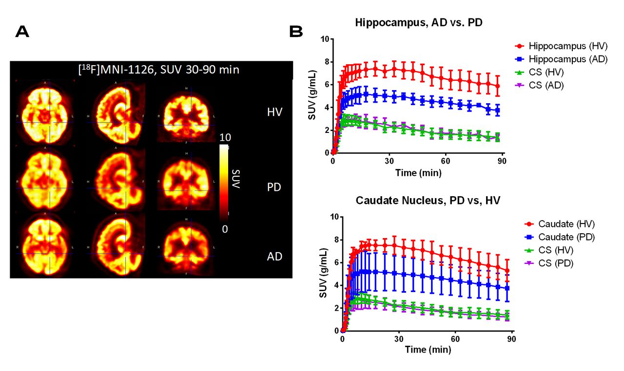
Methods: Brain PET imaging was performed in 11 human subjects, 4 HV (males, 53 ± 3 y), 4 PD (3 males, 1 female, 70 ± 7 y) and 3 AD (1 male, 2 females, 67 ± 10 y, MMSE 3,14 and 22). Dynamic image data were acquired for up to 180 min on a Siemens ECAT HR+ scanner following a 3 min bolus injection of [18F]MNI-1126 (297.7 ± 58.3 MBq). Arterial blood sampling with associated metabolite assays was acquired and enabled the generation of an arterial input function for kinetic modeling. Dynamic images were motion corrected and normalized into MNI space. Regions of interest were defined by Hammers, N30R83 atlas and intersected with gray and white matter segments. Regional brain time-activity curves (TACs) were generated and analyzed by one-tissue (1T), two-tissue (2T) compartment models, and Logan graphical analysis (LGA) to estimate regional volumes of distribution (VT). A binding potential (BPND) was estimated by assuming that the VT of centrum semiovale (CS) was reflective of the non-displaceable signal. Non-invasive LGA (NI-LGA) and simplified reference tissue model (SRTM), with CS as reference region were also explored. Time stability of both VT and BPND outcome measures was evaluated. Cross-sectional analysis of VT and BPND were performed across the healthy, PD and AD subject groups. Results: [18F]MNI-1126 readily entered the brain and its distribution was consistent with SV2A similar to [11C]UCB-J - high brain uptake (max SUV 9-12) reached at 15-30 min after injection, moderate washout, and high specific binding (max BPND ~5.0). Unmetabolized parent fraction was 52.4 ± 15.5 %, 24.9 ± 8.5 %, and 22.6 ± 7.3 %, at 15 min, 60 min, and 120 min post injection. Fraction of unbound tracer to plasma proteins (i.e., free fraction) was 32% ± 2%. VT and BPND estimates were relatively stable for scan durations of 40 min and 60 min, respectively (~5% different from 120 min). The 1T model produced better (lower AIC) and more reliable (low SE) fits than 2T. Across methods regional BPND computed with 1T model was highly correlated (R2~ 0.99) with LGA (no bias) and NI-LGA. NI-LGA estimates presented an increasing negative bias with BPND (slope ~0.85). Lower global VT (6%, 14%) and BPND (20%, 29%) were found in PD and AD subjects, respectively, compared to HV. In AD, the largest reductions in BPND were found in hippocampus (51%), and superior lateral temporal cortex (39%). These results indicate that reductions in SV2A density in AD can be measured in brain regions besides hippocampus, which have been previously reported with [11C]UCB-J. In PD, the largest reductions in BPND were found in caudate nucleus (38%), hippocampus (33%), and thalamus (30%).

Conclusions: These first-in-human studies with [18F]MNI-1126 have confirmed its favorable in vivo imaging properties. Disease specific decreases in regional SV2A density were detected and quantified in both PD and AD subjects. These encouraging proof-of-concept data provide a rationale for future multicenter clinical trials to validate the utility of SV2A PET imaging in AD, PD and other neurodegenerative disorders.

Figure
  • Download figure
  • Open in new tab
  • Download powerpoint
Previous
Back to top

In this issue

Journal of Nuclear Medicine
Vol. 60, Issue supplement 1
May 1, 2019
  • Table of Contents
  • Index by author
Article Alerts
Sign In to Email Alerts with your Email Address
Email Article

Thank you for your interest in spreading the word on Journal of Nuclear Medicine.

NOTE: We only request your email address so that the person you are recommending the page to knows that you wanted them to see it, and that it is not junk mail. We do not capture any email address.

Enter multiple addresses on separate lines or separate them with commas.
Evaluation of [18F]MNI-1126, an18F-labeled SV2A PET tracer in healthy, Parkinson disease and Alzheimer’s disease subjects
(Your Name) has sent you a message from Journal of Nuclear Medicine
(Your Name) thought you would like to see the Journal of Nuclear Medicine web site.
Citation Tools
Evaluation of [18F]MNI-1126, an18F-labeled SV2A PET tracer in healthy, Parkinson disease and Alzheimer’s disease subjects
Cristian Constantinescu, Vincent Carroll, Alexandra Gouasmat, Cedric Tresse, David Russell, Roger Gunn, Olivier Barret, Eugenii Rabiner, Kenneth Marek
Journal of Nuclear Medicine May 2019, 60 (supplement 1) 320;

Citation Manager Formats

  • BibTeX
  • Bookends
  • EasyBib
  • EndNote (tagged)
  • EndNote 8 (xml)
  • Medlars
  • Mendeley
  • Papers
  • RefWorks Tagged
  • Ref Manager
  • RIS
  • Zotero
Share
Evaluation of [18F]MNI-1126, an18F-labeled SV2A PET tracer in healthy, Parkinson disease and Alzheimer’s disease subjects
Cristian Constantinescu, Vincent Carroll, Alexandra Gouasmat, Cedric Tresse, David Russell, Roger Gunn, Olivier Barret, Eugenii Rabiner, Kenneth Marek
Journal of Nuclear Medicine May 2019, 60 (supplement 1) 320;
del.icio.us logo Digg logo Reddit logo Twitter logo Facebook logo Google logo Mendeley logo
  • Tweet Widget
  • Facebook Like
  • Google Plus One
Bookmark this article

Jump to section

  • Article
  • Figures & Data
  • Info & Metrics

Related Articles

  • No related articles found.
  • Google Scholar

Cited By...

  • No citing articles found.
  • Google Scholar

More in this TOC Section

Neurosciences

  • Brain-related complications of COVID-19 and the role of FDG-PET in detecting vascular and non-vascular defects throughout the body
  • A critical review of the utility of PET imaging in the diagnosis and management of psychosis
  • The radiological study of traumatic brain injury: an emphasis upon molecular imaging
Show more Neurosciences

Novel Brain Imaging Targets

  • Clinical Protocol to Quantify AMPA Receptors Using Novel 11C-labeled PET Tracer of K2
  • Initial experience with 18F-Flouroglutamine PET/CT imaging in brain tumor response assessment
  • First-in-human study using [11C]MDTC and positron emission tomography for imaging the cannabinoid receptor type 2
Show more Novel Brain Imaging Targets

Similar Articles

SNMMI

© 2022 Journal of Nuclear Medicine

Powered by HighWire