
Main menu

  • Home
  • Content
    • Current
    • Ahead of print
    • Past Issues
    • JNM Supplement
    • SNMMI Annual Meeting Abstracts
    • Continuing Education
    • JNM Podcasts
  • Subscriptions
    • Subscribers
    • Institutional and Non-member
    • Rates
    • Journal Claims
    • Corporate & Special Sales
  • Authors
    • Submit to JNM
    • Information for Authors
    • Assignment of Copyright
    • AQARA requirements
  • Info
    • Reviewers
    • Permissions
    • Advertisers
  • About
    • About Us
    • Editorial Board
    • Contact Information
  • More
    • Alerts
    • Feedback
    • Help
    • SNMMI Journals
  • SNMMI
    • JNM
    • JNMT
    • SNMMI Journals
    • SNMMI

User menu

  • Subscribe
  • My alerts
  • Log in
  • My Cart

Search

  • Advanced search
Journal of Nuclear Medicine
  • SNMMI
    • JNM
    • JNMT
    • SNMMI Journals
    • SNMMI
  • Subscribe
  • My alerts
  • Log in
  • My Cart
Journal of Nuclear Medicine

Advanced Search

  • Home
  • Content
    • Current
    • Ahead of print
    • Past Issues
    • JNM Supplement
    • SNMMI Annual Meeting Abstracts
    • Continuing Education
    • JNM Podcasts
  • Subscriptions
    • Subscribers
    • Institutional and Non-member
    • Rates
    • Journal Claims
    • Corporate & Special Sales
  • Authors
    • Submit to JNM
    • Information for Authors
    • Assignment of Copyright
    • AQARA requirements
  • Info
    • Reviewers
    • Permissions
    • Advertisers
  • About
    • About Us
    • Editorial Board
    • Contact Information
  • More
    • Alerts
    • Feedback
    • Help
    • SNMMI Journals
  • View or Listen to JNM Podcast
  • Visit JNM on Facebook
  • Join JNM on LinkedIn
  • Follow JNM on Twitter
  • Subscribe to our RSS feeds
Meeting ReportNeurosciences

Tmem59 deletion activates microglia and reduces phagocytic capacity

Yanyan Kong, Jiao Wang, Jie Li, Fengchun Hua, Bizeng Zhao, Tieqiao Wen and Yihui Guan
Journal of Nuclear Medicine May 2019, 60 (supplement 1) 1506a;
Yanyan Kong
3PET Center, Huashan Hospital Fudan University Shanghai China
  • Find this author on Google Scholar
  • Find this author on PubMed
  • Search for this author on this site
Jiao Wang
1Laboratory of Molecular Neural Biology, School of Life Sciences, Shanghai University Shanghai China
  • Find this author on Google Scholar
  • Find this author on PubMed
  • Search for this author on this site
Jie Li
1Laboratory of Molecular Neural Biology, School of Life Sciences, Shanghai University Shanghai China
  • Find this author on Google Scholar
  • Find this author on PubMed
  • Search for this author on this site
Fengchun Hua
3PET Center, Huashan Hospital Fudan University Shanghai China
  • Find this author on Google Scholar
  • Find this author on PubMed
  • Search for this author on this site
Bizeng Zhao
2No.6 Shanghai Peoples Hospital, Jiaotong Universit Shanghai China
  • Find this author on Google Scholar
  • Find this author on PubMed
  • Search for this author on this site
Tieqiao Wen
1Laboratory of Molecular Neural Biology, School of Life Sciences, Shanghai University Shanghai China
  • Find this author on Google Scholar
  • Find this author on PubMed
  • Search for this author on this site
Yihui Guan
3PET Center, Huashan Hospital Fudan University Shanghai China
  • Find this author on Google Scholar
  • Find this author on PubMed
  • Search for this author on this site
  • Article
  • Figures & Data
  • Info & Metrics
Loading

Abstract

1506a

Introduction: Neuroinflammation is widely regarded as a chronic innate immune response in the brain and apotentially pathogenic factor in a number of neurodegenerative diseases such as Alzheimer’s disease (AD), as well as traumatic brain injury. Recently, due to its key signaling steps in the initiation of immune activation, greater attention has been paid to the potential of neuroinflammation as a therapeutic target. Microglia, which comprise approximately 20% of all glial cells, serve as the principal immune cells and play crucial roles in the central nervous system, responding to neuroinflammation via migration and the execution of phagocytosis. The activation of microglia is characterized by an increase in the expression level of TSPO, which can be detected by PET.Transmembrane protein 59 (Tmem59) is a membrane protein that plays an important role in neural stem cell differentiation, glioma apoptosis, dendritic spine formation, and Alzheimer’s disease (AD), nevertheless, the involvement of the Tmem59 gene in the brain immune response has not yet been reported. In the current work, we investigated the effect of Tmem59 in neuroinflammation.

Methods: 18F-DPA-714 imaging in WT and KO mice were performed using a micro PET/CT. Tmem59 down-expression models and vector controls of BV2 microglia cells were established by psiRNA-Tmem59 and Vehicle plasmid transfection, respectively. The change of gene expression was detected by western blotting analysis. Cell phagocytosis assay was used to study the effect of Dcf1 downregulation on the phagocytic ability of LPS-activated microglia

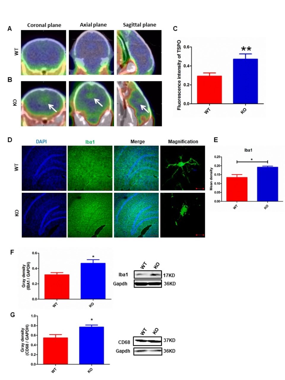
Results: FIGURE 1 | Tmem59 deletion induces activation of microglial cells in vivo. 18F-DPA-714 (green) was used to trace TSPO (a biomarker of microglia) by PET to observe the activity of microglia in vivo. (A,B) Brain observation by PET in WT (A) and Tmem59-KO (B) mice. White arrowhead denotes activated microglia. (C) Quantitation of the TSPO in WT and Tmem59-KO mouse brain (Supplementary Table S1). (D) Immunohistochemical observation of microglia from WT and Tmem59-KO mouse brain sections. Microglial cells were detected by the Iba1 biomarker (green), and the nuclei were counterstained with DAPI (blue). Scale bars represent 200 µm. Higher magnification of confocal images were shown in right panel. Scale bars, 10 µm. (E) Quantitation of the mean density of Iba1 staining in WT and Tmem59-KO mouse brain sections (mean±SEM). n = 4. ∗p < 0.05. Protein expression of Iba1 (F) and CD68 (G) in WT and Tmem59-KO mouse brain tissue . Quantification of protein expression levels normalized to Gapdh. Data are expressed as the mean±SEM. n = 3. ∗p < 0.05; ∗∗p < 0.01.FIGURE 2 | Tmem59 deletion suppresses the phagocytic ability of BV2 microglia cells. BV2 microglial cells were transfected with the psiRNA-hH1neo plasmid or the psiRNA-Tmem59 plasmid. 24 h post-transfection, BV2 microglia were stimulated with LPS (1000 ng/ml) and incubated for 12 h. (A) Image showing the phagocytic ability of BV2 microglia cells. Quanta were spontaneous green, and the cell skeleton was detected by ActinRed (red). Scale bars represent 20 µm. (B) Analysis of the average quantum absorption in each cell to assess the phagocytic activity of BV2 microglia. Data are expressed as the mean±SEM. n = 4. ∗p < 0.05 (Supplementary Table S12).

Conclusions: In conclusion, our data indicates that Tmem59-deficient microglia induced aberrant proinflammatory cytokines release and subsequent microglial dysfunction, which blocked the phagocytic abilities of activated microglia. Taken together, these observations provide novel insight into the role of Tmem59 in activated microglial cells during the neuroimmune response, and further lay the foundation for the elucidation of the mechanism underlying neuroinflammatory-related diseases.

Figure
  • Download figure
  • Open in new tab
  • Download powerpoint
Figure
  • Download figure
  • Open in new tab
  • Download powerpoint
Previous
Back to top

In this issue

Journal of Nuclear Medicine
Vol. 60, Issue supplement 1
May 1, 2019
  • Table of Contents
  • Index by author
Article Alerts
Sign In to Email Alerts with your Email Address
Email Article

Thank you for your interest in spreading the word on Journal of Nuclear Medicine.

NOTE: We only request your email address so that the person you are recommending the page to knows that you wanted them to see it, and that it is not junk mail. We do not capture any email address.

Enter multiple addresses on separate lines or separate them with commas.
Tmem59 deletion activates microglia and reduces phagocytic capacity
(Your Name) has sent you a message from Journal of Nuclear Medicine
(Your Name) thought you would like to see the Journal of Nuclear Medicine web site.
Citation Tools
Tmem59 deletion activates microglia and reduces phagocytic capacity
Yanyan Kong, Jiao Wang, Jie Li, Fengchun Hua, Bizeng Zhao, Tieqiao Wen, Yihui Guan
Journal of Nuclear Medicine May 2019, 60 (supplement 1) 1506a;

Citation Manager Formats

  • BibTeX
  • Bookends
  • EasyBib
  • EndNote (tagged)
  • EndNote 8 (xml)
  • Medlars
  • Mendeley
  • Papers
  • RefWorks Tagged
  • Ref Manager
  • RIS
  • Zotero
Share
Tmem59 deletion activates microglia and reduces phagocytic capacity
Yanyan Kong, Jiao Wang, Jie Li, Fengchun Hua, Bizeng Zhao, Tieqiao Wen, Yihui Guan
Journal of Nuclear Medicine May 2019, 60 (supplement 1) 1506a;
Twitter logo Facebook logo LinkedIn logo Mendeley logo
  • Tweet Widget
  • Facebook Like
  • Google Plus One
Bookmark this article

Jump to section

  • Article
  • Figures & Data
  • Info & Metrics

Related Articles

  • No related articles found.
  • Google Scholar

Cited By...

  • No citing articles found.
  • Google Scholar

More in this TOC Section

Neurosciences

  • PET CT Quantification to direct Alzheimer’s patients to anti-amyloid therapy
  • Multicenter trial study for usefulness of physical parameters toward the standardization of brain SPECT image: relation to visual analysis
  • Assessment of Brain Perfusion 123I-IMP SPECT imaging Using Low Energy High Resolution Collimator image reconstructed by Flash 3D
Show more Neurosciences

Neurology & Psychiatry Imaging Posters

  • comparison study of F Choline PET CT with MRI in intracranial space occupying lesions and evaluation of diagnostic accuracy for differentiating neoplastic versus non neoplastic lesions
  • Radioactive iodine labelled MIBG cardiac sympathetic imaging is less promising in differentiating PD from other parkinsonism for early onset patients
  • Dr.
Show more Neurology & Psychiatry Imaging Posters

Similar Articles

SNMMI

© 2025 SNMMI

Powered by HighWire