
Main menu

  • Home
  • Content
    • Current
    • Ahead of print
    • Past Issues
    • JNM Supplement
    • SNMMI Annual Meeting Abstracts
    • Continuing Education
    • JNM Podcasts
  • Subscriptions
    • Subscribers
    • Institutional and Non-member
    • Rates
    • Journal Claims
    • Corporate & Special Sales
  • Authors
    • Submit to JNM
    • Information for Authors
    • Assignment of Copyright
    • AQARA requirements
  • Info
    • Reviewers
    • Permissions
    • Advertisers
  • About
    • About Us
    • Editorial Board
    • Contact Information
  • More
    • Alerts
    • Feedback
    • Help
    • SNMMI Journals
  • SNMMI
    • JNM
    • JNMT
    • SNMMI Journals
    • SNMMI

User menu

  • Subscribe
  • My alerts
  • Log in
  • Log out
  • My Cart

Search

  • Advanced search
Journal of Nuclear Medicine
  • SNMMI
    • JNM
    • JNMT
    • SNMMI Journals
    • SNMMI
  • Subscribe
  • My alerts
  • Log in
  • Log out
  • My Cart
Journal of Nuclear Medicine

Advanced Search

  • Home
  • Content
    • Current
    • Ahead of print
    • Past Issues
    • JNM Supplement
    • SNMMI Annual Meeting Abstracts
    • Continuing Education
    • JNM Podcasts
  • Subscriptions
    • Subscribers
    • Institutional and Non-member
    • Rates
    • Journal Claims
    • Corporate & Special Sales
  • Authors
    • Submit to JNM
    • Information for Authors
    • Assignment of Copyright
    • AQARA requirements
  • Info
    • Reviewers
    • Permissions
    • Advertisers
  • About
    • About Us
    • Editorial Board
    • Contact Information
  • More
    • Alerts
    • Feedback
    • Help
    • SNMMI Journals
  • View or Listen to JNM Podcast
  • Visit JNM on Facebook
  • Join JNM on LinkedIn
  • Follow JNM on Twitter
  • Subscribe to our RSS feeds
Meeting ReportNeurosciences

Brain F18 FDG PET CT in patients with Anti-LGI1 encephalitis

Mohammad Salehi Sadaghiani, Leah Dickstein, John Probasco and Lilja Solnes
Journal of Nuclear Medicine May 2019, 60 (supplement 1) 1474;
Mohammad Salehi Sadaghiani
4Radiology Johns Hopkins Hospital Baltimore MD United States
  • Find this author on Google Scholar
  • Find this author on PubMed
  • Search for this author on this site
Leah Dickstein
2Neurology Johns Hopkins Hospital Baltimore MD United States
  • Find this author on Google Scholar
  • Find this author on PubMed
  • Search for this author on this site
John Probasco
3Johns Hopkins Dept. of Neurology Baltimore MD United States
  • Find this author on Google Scholar
  • Find this author on PubMed
  • Search for this author on this site
Lilja Solnes
1Johns Hopkins Garrison MD United States
  • Find this author on Google Scholar
  • Find this author on PubMed
  • Search for this author on this site
  • Article
  • Figures & Data
  • Info & Metrics
Loading

Abstract

1474

Objectives: To evaluate metabolic activity on brain F18 FDG PET CT in patients with Anti-LGI1 encephalitis.

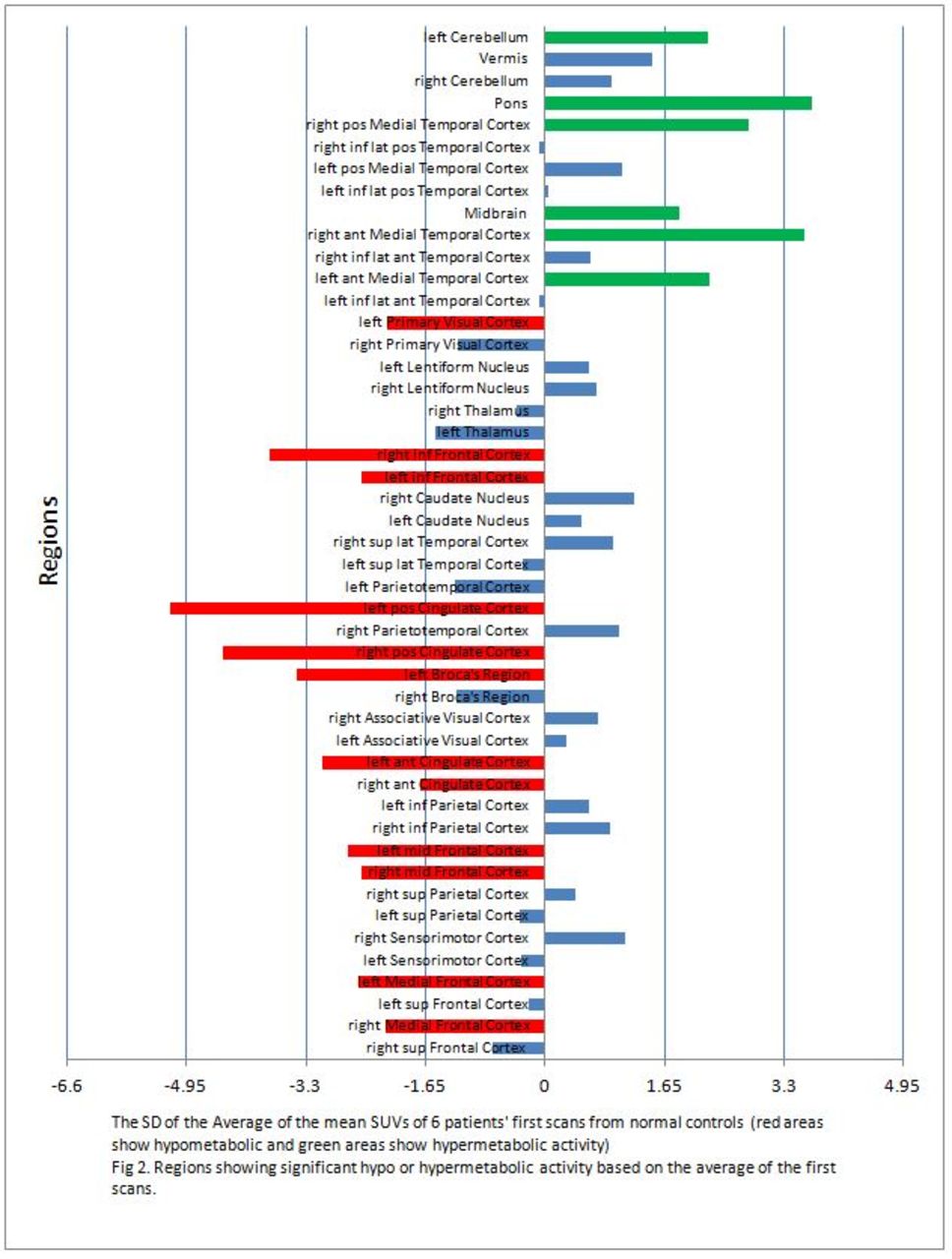
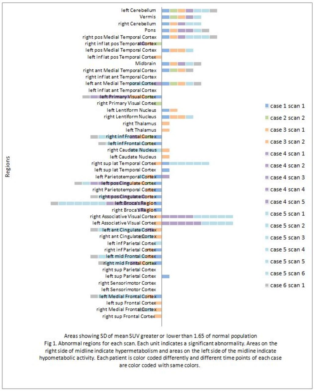
Methods: In this retrospective study, 6 patients with confirmed diagnosis of Anti-LGI1 encephalitis who had total of 16 F18 FDG PET CT scans of the brain were included in the study. Quantitative analysis for PET imaging was done using NeuroQ™ version 3.75. NeuroQ™ provides regional assessment of human brain scans based on automated quantification of mean pixel values lying within standardized regions of interests (ROIs) and compares the brain regional activity to normal activity values of 50 normal controls. 47 areas in the brain were segmented and analyzed. Abnormal regions are determined based on having internally normalized ROI radiotracer uptake values falling more than 1.65 standard deviations below the mean value (in the Hypometabolic operation) and more than 1.65 standard deviations above the mean value (in the Hypermetabolic operation) for asymptomatic control group. Besides each individual scan results (Fig1) we also calculated the SD of average of the first scans of the 6 patients from the normal controls (Fig 2).

Results: The resultant SD of the averages of the first scans from normal controls showed that pons, midbrain, right anterior medial temporal cortex, left anterior medial temporal cortex and right posterior medial temporal cortex show hypermetabolic activity, while left primary visual cortex, right inferior frontal cortex, left inferior frontal cortex, left posterior cingulated cortex, right posterior cingulated cortex, left broca region, left anterior cingulated cortex, right anterior cingulated cortex, left mid frontal cortex, right mid frontal cortex, left medial frontal cortex and right medial frontal cortex showed significant hypometabolic activity (Fig 1). The significant hyper and hypometabolic areas for each scan are shown in Fig 2.

Conclusions: Anti-LGI1 encephalitis is a subtype of autoimmune encephalitis that results from antibodies targeted at leucine rich glioma inactivated 1 (LGI1) protein. This autoantigen is secreted from neuronal proteins and acts as a ligand for ADAM22 and ADAM23 which are epilepsy-related proteins (1). Early intervention improves long term outcomes (2). Patients may show cognitive abnormality and seizures (3). MRI results usually reveal medial temporal hyperintensity (4). Less is known regarding the F18 FDG PET evaluation of the patients. Herein, we present the results of 47 brain clusters based on FDG PET scans compared to 50 normal controls. Considering the abnormal signal in medial temporal areas on MRI, our results show that these areas are hypermetabolic. On the other hand frontal and cingulated areas show hypometabolic activity. These results provide better understanding of the nature of this neurologic disease and may help us diagnose patients at onset of disease which will allow us to intervene earlier.

Figure
  • Download figure
  • Open in new tab
  • Download powerpoint
Figure
  • Download figure
  • Open in new tab
  • Download powerpoint
Previous
Back to top

In this issue

Journal of Nuclear Medicine
Vol. 60, Issue supplement 1
May 1, 2019
  • Table of Contents
  • Index by author
Article Alerts
Sign In to Email Alerts with your Email Address
Email Article

Thank you for your interest in spreading the word on Journal of Nuclear Medicine.

NOTE: We only request your email address so that the person you are recommending the page to knows that you wanted them to see it, and that it is not junk mail. We do not capture any email address.

Enter multiple addresses on separate lines or separate them with commas.
Brain F18 FDG PET CT in patients with Anti-LGI1 encephalitis
(Your Name) has sent you a message from Journal of Nuclear Medicine
(Your Name) thought you would like to see the Journal of Nuclear Medicine web site.
Citation Tools
Brain F18 FDG PET CT in patients with Anti-LGI1 encephalitis
Mohammad Salehi Sadaghiani, Leah Dickstein, John Probasco, Lilja Solnes
Journal of Nuclear Medicine May 2019, 60 (supplement 1) 1474;

Citation Manager Formats

  • BibTeX
  • Bookends
  • EasyBib
  • EndNote (tagged)
  • EndNote 8 (xml)
  • Medlars
  • Mendeley
  • Papers
  • RefWorks Tagged
  • Ref Manager
  • RIS
  • Zotero
Share
Brain F18 FDG PET CT in patients with Anti-LGI1 encephalitis
Mohammad Salehi Sadaghiani, Leah Dickstein, John Probasco, Lilja Solnes
Journal of Nuclear Medicine May 2019, 60 (supplement 1) 1474;
Twitter logo Facebook logo LinkedIn logo Mendeley logo
  • Tweet Widget
  • Facebook Like
  • Google Plus One
Bookmark this article

Jump to section

  • Article
  • Figures & Data
  • Info & Metrics

Related Articles

  • No related articles found.
  • Google Scholar

Cited By...

  • No citing articles found.
  • Google Scholar

More in this TOC Section

Neurosciences

  • PET CT Quantification to direct Alzheimer’s patients to anti-amyloid therapy
  • Multicenter trial study for usefulness of physical parameters toward the standardization of brain SPECT image: relation to visual analysis
  • Assessment of Brain Perfusion 123I-IMP SPECT imaging Using Low Energy High Resolution Collimator image reconstructed by Flash 3D
Show more Neurosciences

Neurology & Psychiatry Imaging Posters

  • comparison study of F Choline PET CT with MRI in intracranial space occupying lesions and evaluation of diagnostic accuracy for differentiating neoplastic versus non neoplastic lesions
  • Radioactive iodine labelled MIBG cardiac sympathetic imaging is less promising in differentiating PD from other parkinsonism for early onset patients
  • Dr.
Show more Neurology & Psychiatry Imaging Posters

Similar Articles

SNMMI

© 2025 SNMMI

Powered by HighWire