
Main menu

  • Home
  • Content
    • Current
    • Ahead of print
    • Past Issues
    • JNM Supplement
    • SNMMI Annual Meeting Abstracts
    • Continuing Education
    • JNM Podcasts
  • Subscriptions
    • Subscribers
    • Institutional and Non-member
    • Rates
    • Journal Claims
    • Corporate & Special Sales
  • Authors
    • Submit to JNM
    • Information for Authors
    • Assignment of Copyright
    • AQARA requirements
  • Info
    • Reviewers
    • Permissions
    • Advertisers
  • About
    • About Us
    • Editorial Board
    • Contact Information
  • More
    • Alerts
    • Feedback
    • Help
    • SNMMI Journals
  • SNMMI
    • JNM
    • JNMT
    • SNMMI Journals
    • SNMMI

User menu

  • Subscribe
  • My alerts
  • Log in
  • My Cart

Search

  • Advanced search
Journal of Nuclear Medicine
  • SNMMI
    • JNM
    • JNMT
    • SNMMI Journals
    • SNMMI
  • Subscribe
  • My alerts
  • Log in
  • My Cart
Journal of Nuclear Medicine

Advanced Search

  • Home
  • Content
    • Current
    • Ahead of print
    • Past Issues
    • JNM Supplement
    • SNMMI Annual Meeting Abstracts
    • Continuing Education
    • JNM Podcasts
  • Subscriptions
    • Subscribers
    • Institutional and Non-member
    • Rates
    • Journal Claims
    • Corporate & Special Sales
  • Authors
    • Submit to JNM
    • Information for Authors
    • Assignment of Copyright
    • AQARA requirements
  • Info
    • Reviewers
    • Permissions
    • Advertisers
  • About
    • About Us
    • Editorial Board
    • Contact Information
  • More
    • Alerts
    • Feedback
    • Help
    • SNMMI Journals
  • View or Listen to JNM Podcast
  • Visit JNM on Facebook
  • Join JNM on LinkedIn
  • Follow JNM on Twitter
  • Subscribe to our RSS feeds
Meeting ReportNeurosciences

In vivo imaging of Tau deposition using a new tracer-18F-S23 in comparison with 18F-THK5317 in Alzheimer’s disease

Liping Fu, Kaixiang Zhou, Xiaojun Zhang, Fang Yi, Mengchao Cui, Jinming Zhang, Baixuan Xu and Jiahe Tian
Journal of Nuclear Medicine May 2019, 60 (supplement 1) 1462;
Liping Fu
1Department of Nuclear Medicine, Chinese PLA General Hospital Beijing China
  • Find this author on Google Scholar
  • Find this author on PubMed
  • Search for this author on this site
Kaixiang Zhou
2Key Laboratory of Radiopharmaceuticals, Ministry of Education, Beijing Normal University Beijing China
  • Find this author on Google Scholar
  • Find this author on PubMed
  • Search for this author on this site
Xiaojun Zhang
1Department of Nuclear Medicine, Chinese PLA General Hospital Beijing China
  • Find this author on Google Scholar
  • Find this author on PubMed
  • Search for this author on this site
Fang Yi
3Lishilu Outpatient, PLA Rocket Force General Hospital Beijing China
  • Find this author on Google Scholar
  • Find this author on PubMed
  • Search for this author on this site
Mengchao Cui
2Key Laboratory of Radiopharmaceuticals, Ministry of Education, Beijing Normal University Beijing China
  • Find this author on Google Scholar
  • Find this author on PubMed
  • Search for this author on this site
Jinming Zhang
1Department of Nuclear Medicine, Chinese PLA General Hospital Beijing China
  • Find this author on Google Scholar
  • Find this author on PubMed
  • Search for this author on this site
Baixuan Xu
1Department of Nuclear Medicine, Chinese PLA General Hospital Beijing China
  • Find this author on Google Scholar
  • Find this author on PubMed
  • Search for this author on this site
Jiahe Tian
1Department of Nuclear Medicine, Chinese PLA General Hospital Beijing China
  • Find this author on Google Scholar
  • Find this author on PubMed
  • Search for this author on this site
  • Article
  • Figures & Data
  • Info & Metrics
Loading

Abstract

1462

Purpose: To evaluate the effectiveness of a new tau tracer ((S)-1-(4-(6-(dimethylamino) naphthalen-2-yl)phenoxy)-3-fluoropropan-2-ol,18F-S23) in identifying AD patients from healthy controls (HCs) as compared with 18F-THK5317.

Methods: The study cohort consisted of 6 AD, 1 FTLD, 1 PSP, 1 DLB, 6 HCs and 2 young volunteers. Dynamic PET was performed over a period of 90 min after injection of tracers in 1 AD patient, 6 HCs and 2 young volunteers. Static data acquisitions with a duration of 20-min from 40 min to 60 min after tracers’ administration were performed in all patients to analyze the standardized uptake value (SUV) and cortical-to-cerebellum SUV ratios (SUVRs). Voxel-based analysis of both tracers was employed to assess their uptake differences between populations.

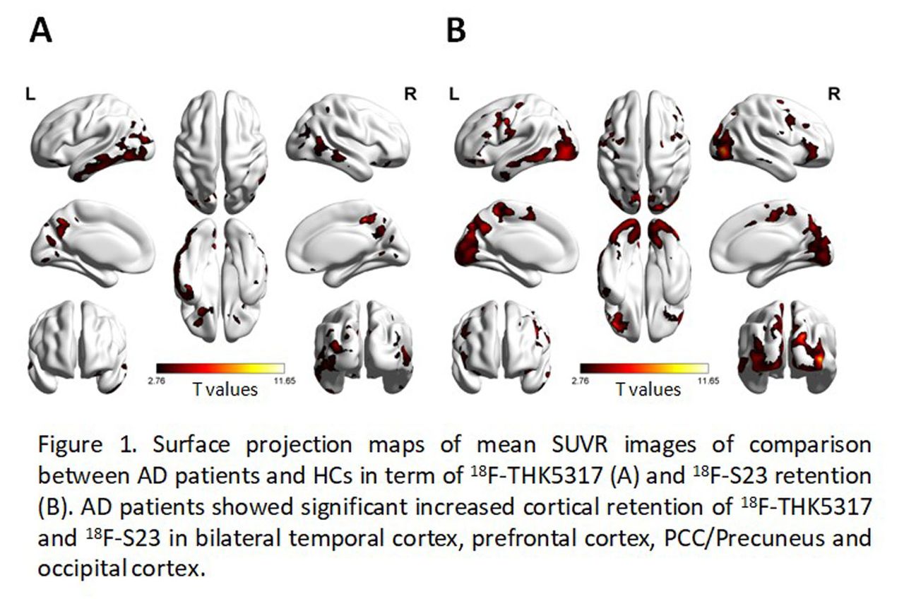
Results: Both tracers were distributed to the brain of subjects rapidly but the clearance of 18F-S23 was slower than 18F-THK5317 within 90 min acquisition. The AD patient showed increased cortical 18F-S23 binding (e.g., the basal temporal cortex and posterior cingulated cortex); minimal accumulation of tracers was seen in young volunteers. Nonspecific uptake was observed in both tracers in basal ganglia and brain stem, but the radioactive uptake of eyeballs was observed in 18F-THK5317 only. In comparative studies, the cortical average SUVR of 18F-THK5317 was 1.32 ± 0.06 for AD versus 1.21 ± 0.03 for HCs (F = 5.73, P < 0.05), and for 18F-S23, that was 1.20 ± 0.10 versus 1.15 ± 0.06 (F = 3.17, P < 0.05) (Table 1.). In voxelwised analysis, AD patients showed significant increased cortical 18F-S23 binding in the bilateral basal temporal cortex, hippocampus, prefrontal cortex, insular, ACC and lateral parietal cortex; 18F-THK5317 displayed increased abnormal radioactive retention in bilateral basal temporal cortex, lateral temporal cortex, prefrontal cortex, PCC/precuneus and lateral parietal cortex (Figure 1.). The patterns of uptake of both tracers were similar in FLTD and PSP patients, which were observed increased radioactive retention in the anterior temporal cortex and in the globus pallidus and midbrain, respectively. No obvious abnormal uptake was found in the DLB patient.

Conclusions: Although the binding intensity and distribution of 18F-S23 were slightly different with that of 18F-THK5317 in controls, the significant difference between AD and HCs in cortical regions indicates that 18F-S23 might be a promising Tau-tracer for PET imaging.

View this table:
  • View inline
  • View popup

SUVRs of Cortical Brain Region Relative to Cerebellum by Diagnostic Group

Figure
  • Download figure
  • Open in new tab
  • Download powerpoint
Previous
Back to top

In this issue

Journal of Nuclear Medicine
Vol. 60, Issue supplement 1
May 1, 2019
  • Table of Contents
  • Index by author
Article Alerts
Sign In to Email Alerts with your Email Address
Email Article

Thank you for your interest in spreading the word on Journal of Nuclear Medicine.

NOTE: We only request your email address so that the person you are recommending the page to knows that you wanted them to see it, and that it is not junk mail. We do not capture any email address.

Enter multiple addresses on separate lines or separate them with commas.
In vivo imaging of Tau deposition using a new tracer-18F-S23 in comparison with 18F-THK5317 in Alzheimer’s disease
(Your Name) has sent you a message from Journal of Nuclear Medicine
(Your Name) thought you would like to see the Journal of Nuclear Medicine web site.
Citation Tools
In vivo imaging of Tau deposition using a new tracer-18F-S23 in comparison with 18F-THK5317 in Alzheimer’s disease
Liping Fu, Kaixiang Zhou, Xiaojun Zhang, Fang Yi, Mengchao Cui, Jinming Zhang, Baixuan Xu, Jiahe Tian
Journal of Nuclear Medicine May 2019, 60 (supplement 1) 1462;

Citation Manager Formats

  • BibTeX
  • Bookends
  • EasyBib
  • EndNote (tagged)
  • EndNote 8 (xml)
  • Medlars
  • Mendeley
  • Papers
  • RefWorks Tagged
  • Ref Manager
  • RIS
  • Zotero
Share
In vivo imaging of Tau deposition using a new tracer-18F-S23 in comparison with 18F-THK5317 in Alzheimer’s disease
Liping Fu, Kaixiang Zhou, Xiaojun Zhang, Fang Yi, Mengchao Cui, Jinming Zhang, Baixuan Xu, Jiahe Tian
Journal of Nuclear Medicine May 2019, 60 (supplement 1) 1462;
Twitter logo Facebook logo LinkedIn logo Mendeley logo
  • Tweet Widget
  • Facebook Like
  • Google Plus One
Bookmark this article

Jump to section

  • Article
  • Figures & Data
  • Info & Metrics

Related Articles

  • No related articles found.
  • Google Scholar

Cited By...

  • No citing articles found.
  • Google Scholar

More in this TOC Section

Neurosciences

  • PET CT Quantification to direct Alzheimer’s patients to anti-amyloid therapy
  • Multicenter trial study for usefulness of physical parameters toward the standardization of brain SPECT image: relation to visual analysis
  • Assessment of Brain Perfusion 123I-IMP SPECT imaging Using Low Energy High Resolution Collimator image reconstructed by Flash 3D
Show more Neurosciences

Neurology & Psychiatry Imaging Posters

  • comparison study of F Choline PET CT with MRI in intracranial space occupying lesions and evaluation of diagnostic accuracy for differentiating neoplastic versus non neoplastic lesions
  • Radioactive iodine labelled MIBG cardiac sympathetic imaging is less promising in differentiating PD from other parkinsonism for early onset patients
  • Dr.
Show more Neurology & Psychiatry Imaging Posters

Similar Articles

SNMMI

© 2025 SNMMI

Powered by HighWire